1371
Revisiting outer volume subtraction with deep-learning tools for highly-accelerated real-time cine CMR1University of Minnesota, Saint Paul, MN, United States, 2National Heart-Lung and Blood Institute, Bethesda, MD, United States, 3University of Minnesota, Minneapolis, MN, United States
Synopsis
Keywords: Image Reconstruction, Cardiovascular, real-time, cardiac cine, heart, outer volume subtraction
Motivation: Real-time cine CMR provides a free-breathing ECG-free approach for heart function assessment. Nevertheless, commercially available real-time cine CMR methods without temporal regularization have limited acceleration and spatio-temporal resolutions.
Goal(s): Use deep learning (DL) to remove extra-cardiac volume that aliases into the heart and improve acceleration rates for real-time cine CMR using only spatial regularization.
Approach: We characterize pseudo-periodic ghosting artifacts arising from cardiac motion in time-interleaved sequences, then use DL to detect and remove them. This is followed by self-supervised physics-driven DL reconstruction.
Results: Proposed technique effectively estimates and removes background signal, leading to substantial image quality improvement.
Impact: We characterize and use deep learning (DL) to estimate pseudo-periodic ghosting artifacts arising from cardiac motion in time-interleaved real-time cine sequences. Background removal followed by physics-driven DL reconstruction substantially improves reconstruction at nominal R=8 for higher spatio-temporal resolution acquisitions.
Introduction
Real-time cine CMR is a free-breathing ECG-free alternative to breath-hold cine CMR for functional assessment of the heart1. These acquisitions currently use parallel imaging2,3, which limits their spatio-temporal resolution. Spatio-temporal regularization4,5,6,7,8 is used for higher accelerations, but these risk temporal blurring9 while also incurring high computational complexity. Thus, improved reconstruction methods without temporal regularization are desirable. The main impediment for high acceleration rates in parallel imaging reconstruction in this setting is the aliasing from extra-cardiac volume. Outer-volume suppression pulses/modules, which have found use in other CMR applications10,11,12, have not been applicable to real-time cine, since they disrupt steady-state and are lengthy. Another line of work uses viewsharing/keyhole in combination with outer-volume estimation, but these have not been adopted widely and robustly13.In this study, we revisit the idea of estimating outer-volume signal with time-varying undersampling patterns. We explicitly characterize pseudo-periodic ghosting artifacts that arise in this setting and use a deep learning (DL) approach to estimate these from time-averaged composite images. This leads to a robust estimation of outer-volume, which is subtracted from each individual timeframes, and subsequently reconstructed using physics-driven DL. Results from retrospectively nominal R=8 accelerated data show image quality comparable to baseline R=4 acquisitions.
Theory
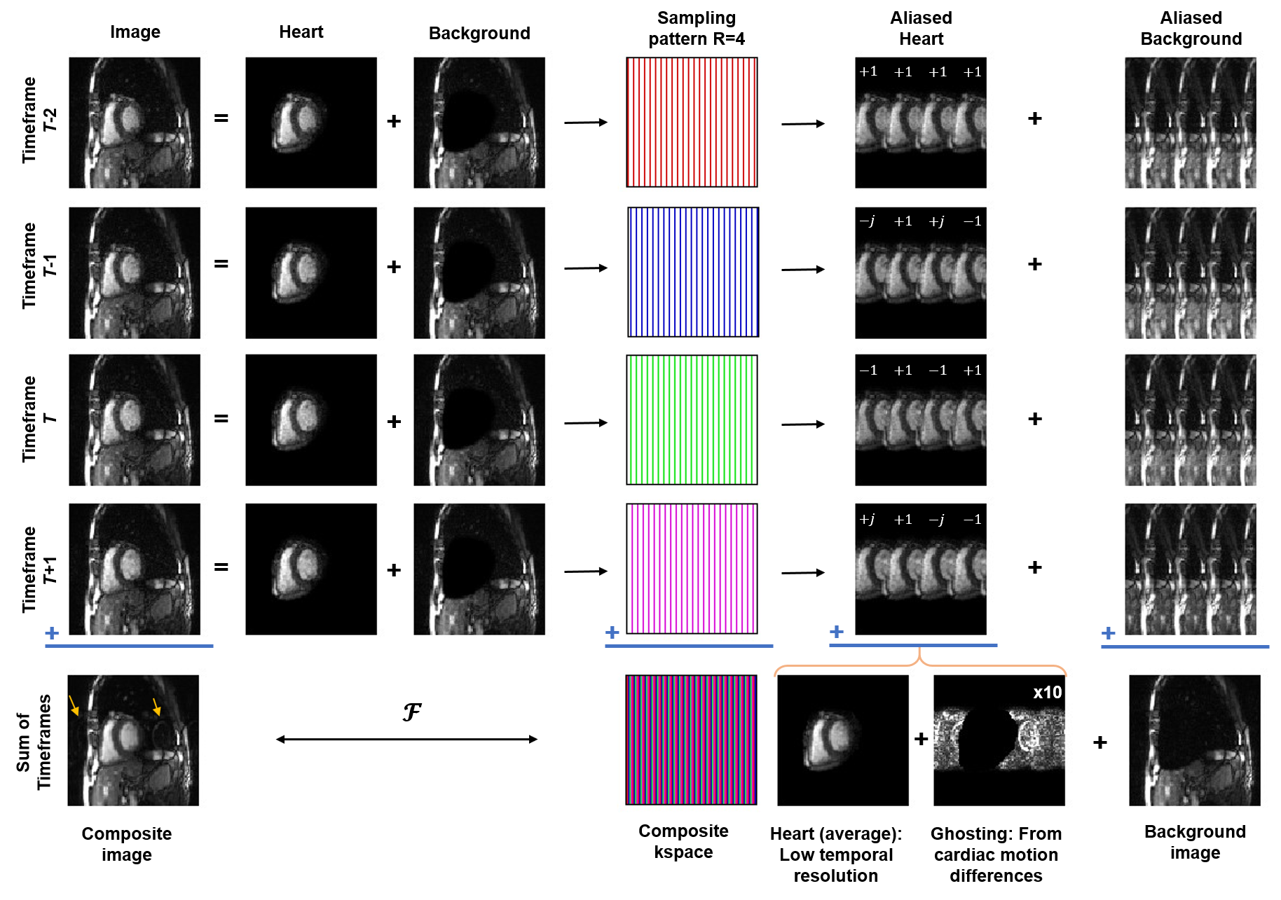
Consider a time-interleaved TGRAPPA/TSENSE2,3 uniform undersampling pattern at rate R. R-adjacent timeframes can be merged to form a fully-sampled composite k-space/image with low-temporal-resolution. Previous works used this image to estimate extracardiac volume14. However, we posit that this image is a sum of outer-volume, a low-resolution heart image, and ghosting artifacts due to cardiac motion. We first note that the true underlying image for each timeframe is truly a combination of outer-volume and heart. Therefore, each undersampled time-frame contributes an R-folded heart and background image to the composite image. Due to k-space shifting, each foldover has varying phase modulation for different timeframes (Fig. 1). The foldedover heart at the true heart location has no phase, resulting in a temporally-averaged heart. Conversely, the other foldovers of the heart cancel out perfectly if there is no cardiac motion, but produce a pseudo-periodic ghosting effect in the background if there is cardiac motion. Therefore, the composite image $$$x_{com}(t)$$$ at time $$$t$$$ can be written $$x_{com}(t)=\overline{x}_{heart}(t)+x_{ghost}(t)+x_{background}(t)$$ where $$$\overline{x}_{heart}(t)$$$ is the temporally-averaged heart, $$$x_{ghost}(t)$$$ is the aforementioned ghosting effect, and $$$x_{background}(t)$$$ is the true outer-volume image.Methods
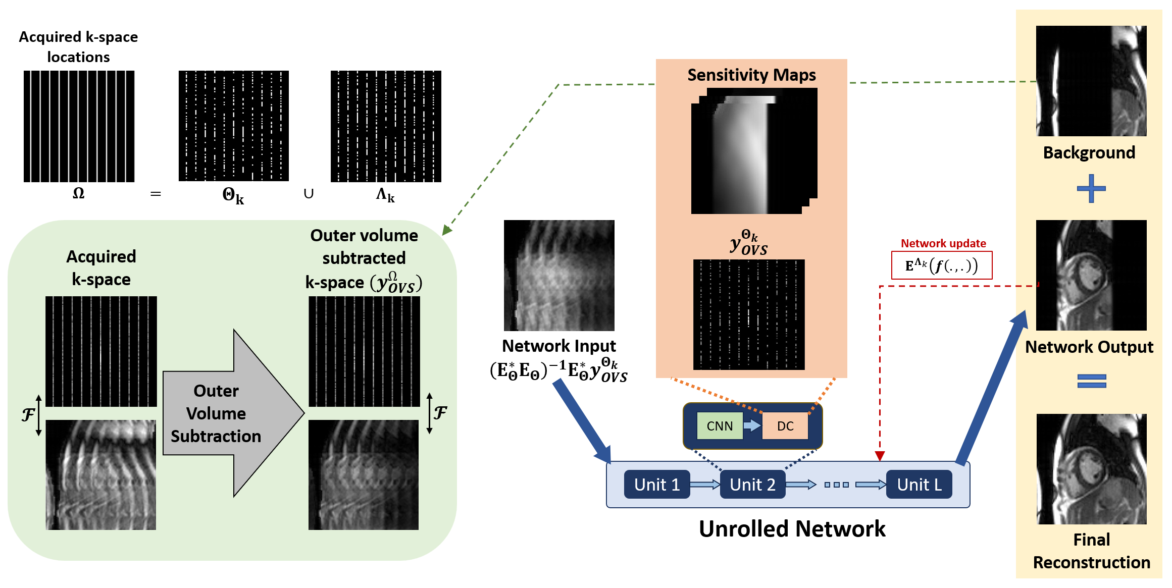
Deep learning for background estimation and outer volume subtraction: We use a neural network to estimate $$$x_{ghost}(t)$$$ from $$$x_{com}(t)$$$. The input to this neural network is the channel-wise concatenation of composite images for timeframes from $$$t_0-1$$$ to $$$t_0+2$$$, centered around the timeframe of interest t0 (denoted $$$x_{com}^{concat}(t_0)$$$), while the output is the ghosting component of the particular composite image, $$$x_{ghost}(t_0)$$$. To train the network, the reference ghosting image is generated by cropping the heart from the TGRAPPA R=4 images with a mask and folding with true modulation phases. The training was performed by minimizing $$$\mathcal{L}\left(x_{ghost}(t_0),f_{\theta}(x_{com}^{concat}(t_0))\cdot M_0\right)$$$ in expectation, with $$$f_{\theta}(x_{com}^{concat}(t_0))$$$ being network output with parameters $$$\theta$$$, and $$$M_0$$$ is a mask that is the complement to the heart location to ensure the averaged heart does not dominate the loss function due to its high intensity. Subsequently, this ghosting signal is subtracted from $$$x_{com}(t_0)$$$ to estimate the background signal.Physics-driven DL reconstruction implementation: The background signal was sub-sampled and subtracted from the corresponding timeframe’s undersampled data. This data, along with masked sensitivity maps were used for image reconstruction with an unrolled PD-DL network (Fig. 2). The network was trained with only undersampled data using multi-mask SSDU15 with K=3 masks.
Imaging experiments: Real-time cine CMR datasets were obtained from 13 subjects at 1.5T using time-interleaved Cartesian bSSFP acquisitions at R=4, resolution=2.25×2.93mm², FOV=360×270mm², temporal resolution=39ms, and slice thickness=8mm. Subsequently, the real-time data were retrospectively undersampled to nominal R=8 by retaining every 8th line while preserving the nearest ky line to central k-space.
Results
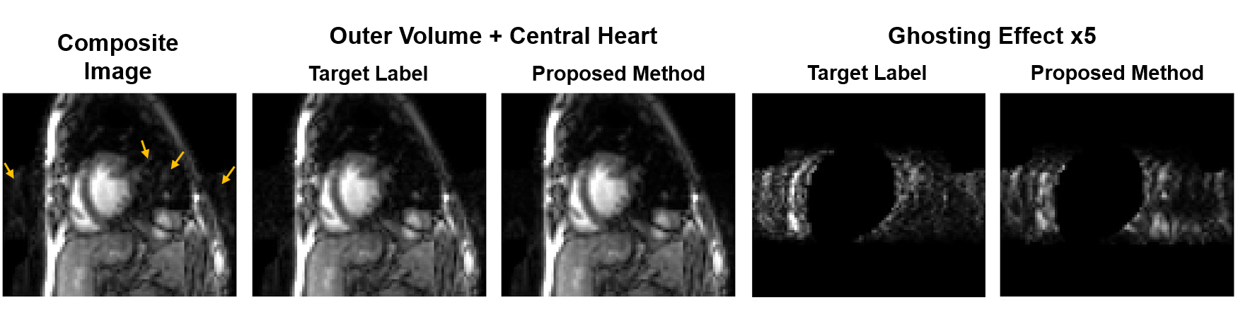
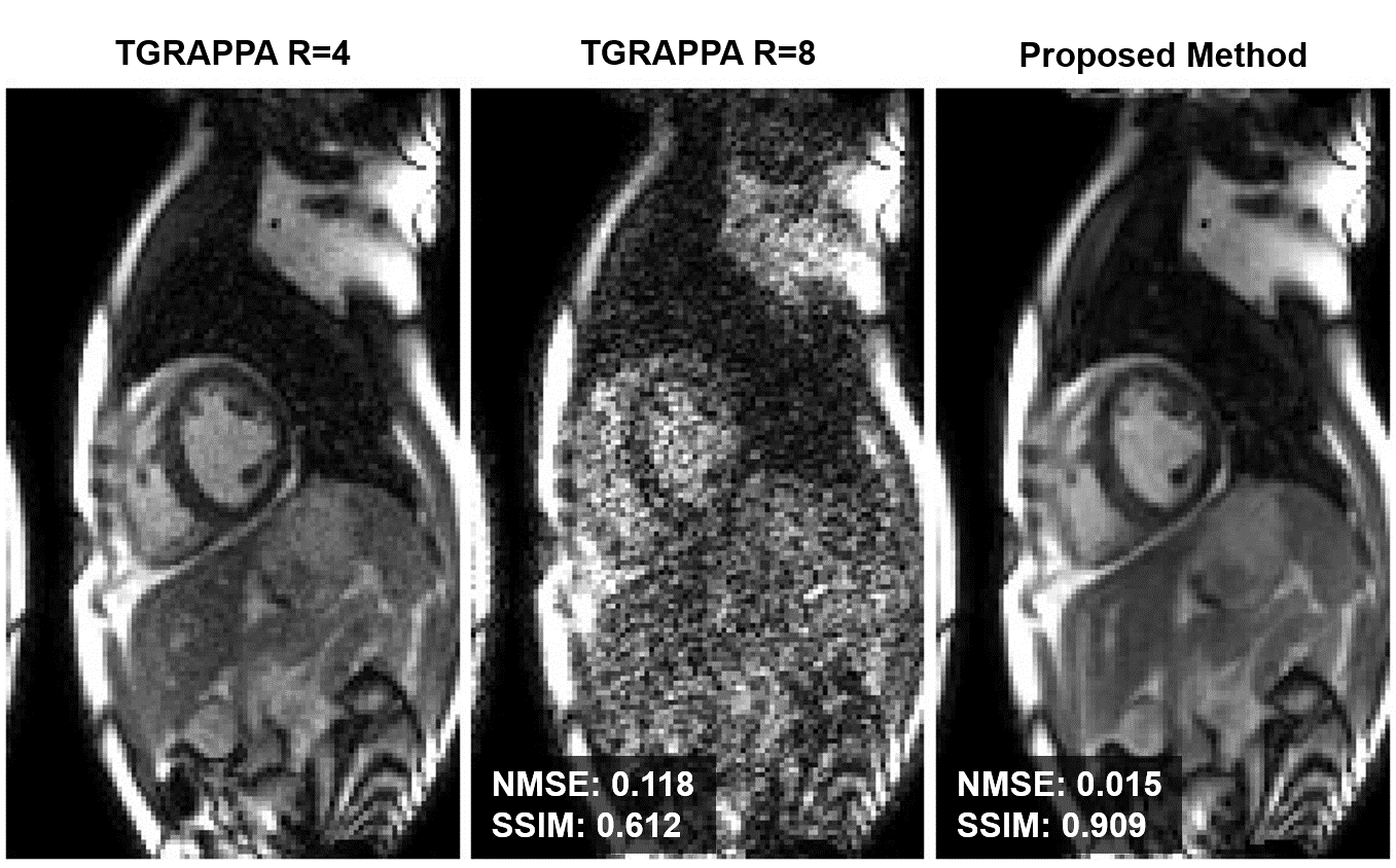
Fig. 3 shows that the proposed neural network approach accurately identifies pseudo-periodic ghosting effects, enabling subsequent estimation of ghosting-free background images.Fig. 4 depicts representative reconstruction results, including TGRAPPA at R=4, R=8, and the proposed method at R=8. Note all methods reconstruct individual timeframes without temporal regularization. The proposed pipeline enables clear depiction of myocardium-blood boundaries at this high acceleration rate, producing visibly similar images to TGRAPPA R=4.
Discussion
In this paper, we revisited outer-volume detection from time-averaged composite images using DL techniques to determine pseudo-periodic ghosting artifacts arising from cardiac motion. The detected outer-volume is subsequently subtracted from the corresponding undersampled timeframe, and reconstructed using self-supervised PD-DL. Our proposed reconstruction pipeline at nominal R=8 achieves comparable quality to baseline TGRAPPA R=4 real-time cine CMR.Acknowledgements
Grant support: NIH R01HL153146, NIH R01EB032830, NIH P41EB027061.References
[1] Beer, M., Stamm, H., Machann, W., Weng, A., Goltz, J. P., Breunig, F., ... & Köstler, H. (2010). Free breathing cardiac real-time cine MR without ECG triggering. International journal of cardiology, 145(2), 380-382.
[2] Kellman, P., Epstein, F. H., & McVeigh, E. R. (2001). Adaptive sensitivity encoding incorporating temporal filtering (TSENSE). Magnetic Resonance in Medicine: An Official Journal of the International Society for Magnetic Resonance in Medicine, 45(5), 846-852.
[3] Breuer, F. A., Kellman, P., Griswold, M. A., & Jakob, P. M. (2005). Dynamic autocalibrated parallel imaging using temporal GRAPPA (TGRAPPA). Magnetic Resonance in Medicine: An Official Journal of the International Society for Magnetic Resonance in Medicine, 53(4), 981-985.
[4] Demirel, Ö. B., Zhang, C., Yaman, B., Gulle, M., Shenoy, C., Leiner, T., ... & Akçakaya, M. (2023). High-fidelity Database-free Deep Learning Reconstruction for Real-time Cine Cardiac MRI. bioRxiv.
[5] Feng, L., Axel, L., Chandarana, H., Block, K. T., Sodickson, D. K., & Otazo, R. (2016). XD‐GRASP: golden‐angle radial MRI with reconstruction of extra motion‐state dimensions using compressed sensing. Magnetic resonance in medicine, 75(2), 775-788.
[6] Hauptmann, A., Arridge, S., Lucka, F., Muthurangu, V., & Steeden, J. A. (2019). Real‐time cardiovascular MR with spatio‐temporal artifact suppression using deep learning–proof of concept in congenital heart disease. Magnetic resonance in medicine, 81(2), 1143-1156.
[7] Kellman, P., Chefd'hotel, C., Lorenz, C. H., Mancini, C., Arai, A. E., & McVeigh, E. R. (2009). High spatial and temporal resolution cardiac cine MRI from retrospective reconstruction of data acquired in real time using motion correction and resorting. Magnetic Resonance in Medicine: An Official Journal of the International Society for Magnetic Resonance in Medicine, 62(6), 1557-1564.
[8] Hansen, M. S., Sørensen, T. S., Arai, A. E., & Kellman, P. (2012). Retrospective reconstruction of high temporal resolution cine images from real‐time MRI using iterative motion correction. Magnetic Resonance in Medicine, 68(3), 741-750.
[9] Feng, L., Srichai, M. B., Lim, R. P., Harrison, A., King, W., Adluru, G., ... & Kim, D. (2013). Highly accelerated real‐time cardiac cine MRI using k–t SPARSE‐SENSE. Magnetic resonance in medicine, 70(1), 64-74.
[10] Weingärtner, S., Moeller, S., Schmitter, S., & Akçakaya, M. (2018). Highly-Accelerated Simultaneous Multi-Slice CMR Using Outer Volume Suppression: Time-Efficient Characterization of Cardiac Function In A Single Breath-hold. Proceedings of the Society for Cardiovascular Magnetic Resonance.
[11] Yang, Y., Zhao, L., Chen, X., Chow, K., Shaw, P. W., Gonzalez, J. A., ... & Salerno, M. (2016). Reduced field-of-view stack-of-spirals enables high spatiotemporal resolution 3D perfusion imaging. Journal of Cardiovascular Magnetic Resonance, 18, 1-3.
[12] Menon, R. G., Miller, G. W., Jeudy, J., Rajagopalan, S., & Shin, T. (2017). Free breathing three‐dimensional late gadolinium enhancement cardiovascular magnetic resonance using outer volume suppressed projection navigators. Magnetic resonance in medicine, 77(4), 1533-1543.
[13] Gómez-Talavera, S., Fernandez-Jimenez, R., Fuster, V., Nothnagel, N. D., Kouwenhoven, M., Clemence, M., ... & Sánchez-González, J. (2021). Clinical validation of a 3-dimensional ultrafast cardiac magnetic resonance protocol including single breath-hold 3-dimensional sequences. Cardiovascular Imaging, 14(9), 1742-1754.
[14] Blaimer, M., Breuer, F. A., Jacob, P. M., Kellman, P., & Griswold, M. A. (2008). A sparse TSENSE approach for improved dynamic parallel MRI. In Proceedings of the 16th Annual Meeting of ISMRM (p. 1270).
[15] Yaman, B., Gu, H., Hosseini, S. A. H., Demirel, O. B., Moeller, S., Ellermann, J., ... & Akçakaya, M. (2022). Multi‐mask self‐supervised learning for physics‐guided neural networks in highly accelerated magnetic resonance imaging. NMR in Biomedicine, 35(12), e4798.
Figures