1370
A Phase-Injected Complex Forward-Distortion Approach for Deep Unsupervised Correction of Susceptibility Artifacts in EPI1Department of Electrical and Electronics Engineering, Bilkent University, Ankara, Turkey, 2National Magnetic Resonance Research Center (UMRAM), Bilkent University, Ankara, Turkey, 3Neuroscience Graduate Program, Bilkent University, Ankara, Turkey
Synopsis
Keywords: Artifacts, Artifacts, susceptibility artifacts, echo planar imaging, reversed phase-encoding, deep learning, unsupervised learning
Motivation: Classical susceptibility-artifact correction methods are impractical in clinical settings given their computational burden.
Goal(s): Fast and effective correction of susceptibility artifacts in EPI via physics-driven unsupervised deep learning by utilizing phase-injected complex-valued forward-distortion.
Approach: Previous methods apply distortion correction on magnitude images, potentially yielding suboptimal performance near regions of signal dropout/pileup. We propose a novel model, compFD-Net, employing phase-injected complex forward-distortion that leverages a predicted phase image, additionally to the magnitude image and displacement field estimates, for improved capture of signal dropout/pileup artifacts in EPI images.
Results: The proposed model boosts susceptibility-artifact correction performance, notably improving predicted image and field quality.
Impact: Robust emulation of signal-dropout/pileup via the complex forward-distortion formulation boosts reliability in unsupervised artifact correction. compFD-Net facilitates rapid and performant correction of susceptibility artifacts in EPI, with possible impact in time-sensitive applications in clinical settings.
Introduction
Echo planar imaging (EPI) is a rapid imaging sequence often preferred for diffusion MRI (dMRI) and functional MRI (fMRI). Nevertheless, susceptibility artifacts distort the anatomical accuracy of EPI images1, necessitating correction for subsequent quantitative assessments2. A common correction framework relies on reversed-phase-encode (reversed-PE) acquisitions to predict the susceptibility-induced displacement field from EPI data3. However, classical correction methods are computationally intensive and therefore impractical in clinical settings.To enable a leap in computational efficiency, we recently proposed Forward-Distortion Network (FD-Net) for unsupervised correction of susceptibility artifacts4. FD-Net takes as input the warped magnitude images acquired in reversed-PE directions, and predicts the displacement field and underlying anatomically-correct magnitude image. Unsupervised learning is achieved by explaining the input images as forward-distortion of the anatomically-correct image with the field. In this work, we propose a more generalized formulation, compFD-Net, that additionally predicts a phase image and applies complex-valued forward-distortion on the phase-injected anatomically-correct image. This generalization enables truer capture of signal dropout/pileup artifacts in EPI images. We demonstrate superiority of compFD-Net over the original FD-Net model and a supervised baseline, and validate it with respect to TOPUP.
Methods
Classical Correction ApproachIn reversed-PE image pairs (i.e., blip-up/-down EPI images), susceptibility-induced distortions primarily occur in opposite directions along the PE dimension5. As such, an inverse problem can be cast to solve for an anatomically-correct image that best accounts for the warped images given an estimated field6. Here we adopt TOPUP from FSL3 as a classical "benchmark" method following this strategy. While TOPUP is widely accepted as a gold standard for EPI distortion correction, it requires extensive computation times7.
Proposed Method
Previous deep-learning methods for susceptibility-artifact correction in EPI commonly employ a reverse-distortion framework to unwarp distorted images via an estimated field8,9. While enforcing similarity between unwarped images for reversed-PE directions permits unsupervised learning9, this framework can suffer from suboptimal performance due to neglecting fidelity to actual EPI measurements4.
Adopting a forward-distortion framework, compFD-Net instead estimates a complex-valued anatomically-correct image along with a displacement field (Fig. 1). Unsupervised learning is achieved by enforcing consistency between forward-distortions of the complex-valued anatomically-correct image and measured reversed-PE images. Note that compFD-Net still takes warped magnitude EPI images as input, but effectively performs phase injection onto the anatomically-correct image to better capture signal dropout/pileup artifacts during forward-distortion. As such, a complex K-Unit is developed as inspired by the magnitude K-Unit4 (Fig. 2). To address rigid-body motion across blip-up/-down acquisitions, an affine-transformation is applied for aligning one of the PE directions4,6. compFD-Net’s overall loss is:
$$L_{compFD-Net}=\sum_{m}{\omega_{m}[L_{MSE}^{(m)}+\lambda_{m}(L_{BE,F}^{(m)}+10^{3}L_{valley}^{(m)}+10^{-2}L_{BE,P}^{(m)})]}+{{\gamma}L_{rigid}},$$where $$$m$$$ indicates the multiresolution step4, superscript $$$(m)$$$ denotes the value of a loss function at step $$$m$$$; $$$\omega_m$$$/$$$\lambda_m$$$ are weighting and regularization parameters over the smoothness of the field/phase for step $$$m$$$, and $$$\gamma$$$ is the rigid loss weight. $$$L_{MSE}^{(m)}$$$ denotes mean-squared-error between measured and forward-distorted images averaged across PE directions, $$$L_{BE,F}^{(m)}$$$/$$$L_{BE,P}^{(m)}$$$ denote the bending energy regularizers over the field/phase, $$$L_{valley}^{(m)}$$$ is the valley loss for the field to curb early training loss explosions, and $$$L_{rigid}$$$ is the rigid loss for affine-transformation parameters.
Modeling procedures
Unprocessed dMRI data from 54 subjects randomly selected from HCP 1200 Subjects Data Release10 were analyzed. A (42, 4, 8)-subject split was used for training, validation and testing. For each subject, a single b0-volume comprising 111 slices with 168x144 image-matrix, acquired with single-short EPI readouts in right-to-left/left-to-right reversed-PE directions were utilized. compFD-Net was implemented with a multiblur strategy4, and loss parameters were tuned to minimize validation loss. compFD-Net was compared against TOPUP, FD-Net and a supervised variant trained to reproduce TOPUP results (i.e., supervised baseline). Models were trained via Adam optimizer for 200 epochs.
Results
Correction of a volume took $$$\sim\!{8}$$$ seconds for all network-based models, compared to $$$\sim\!{50}$$$ mins for TOPUP. Representative forward-distortion and correction results for compFD-Net are given in Fig. 3, demonstrating fidelity to measured blip-up/-down images and perceptual quality of the predicted images. Fig. 4 shows a comparison between FD-Net and compFD-Net. Error maps relative to TOPUP highlight the advantages of the phase-injected complex formulation. Quantitative evaluations in Fig. 5 show that compFD-Net boosts image quality by $$$0.29\text{dB}$$$ PSNR/$$$2.29\%$$$ SSIM over FD-Net and $$$1.68\text{dB}$$$ PSNR/$$$12.21\%$$$ SSIM over the supervised baseline. compFD-Net also boosts field quality by $$$1.05\text{dB}$$$ PSNR/$$$3.52\%$$$ SSIM over FD-Net, with a $$$0.13\text{dB}$$$ PSNR/$$$1.56\%$$$ SSIM cost against the supervised baseline.Conclusion
The proposed compFD-Net provides results comparable to TOPUP by predicting a self-consistent image and field, while improving upon the original FD-Net by injecting additional phase information. Our unsupervised deep-learning approach provides rapid correction of susceptibility artifacts in EPI, while maintaining high performance.Acknowledgements
This work was supported by the Scientific and Technological Council of Turkey (TUBITAK) via Grant 117E116. Data were provided by the Human Connectome Project, WU-Minn Consortium (Principal Investigators: David Van Essen and Kamil Ugurbil; 1U54MH091657) funded by the 16 NIH Institutes and Centers that support the NIH Blueprint for Neuroscience Research; and by the McDonnell Center for Systems Neuroscience at Washington University. Many thanks to Atakan Topcu for his fruitful discussions and valuable insights during the course of this work.
References
- P. Mansfield, "Multi-planar image formation using NMR spin echoes," J. phys., vol. 10, no. 3, pp. L55-L58, 1977.
- J.-D. Tournier, S. Mori, and A. Leemans, "Diffusion tensor imaging and beyond: Diffusion Tensor Imaging and Beyond," Magn. Reson. Med., vol. 65, no. 6, pp. 1532-1556, 2011.
- S. M. Smith et al., "Advances in functional and structural MR image analysis and implementation as FSL," Neuroimage, vol. 23 Suppl 1, pp. S208-19, 2004.
- A. Zaid Alkilani, T. Çukur, and E. U. Saritas, "FD-Net: An unsupervised deep forward-distortion model for susceptibility artifact correction in EPI," Magn. Reson. Med., pp. 1-17, 2023.
- D. Holland, J. M. Kuperman, and A. M. Dale, "Efficient correction of inhomogeneous static magnetic field-induced distortion in Echo Planar Imaging," Neuroimage, vol. 50, no. 1, pp. 175-183, 2010.
- M. S. Graham, I. Drobnjak, M. Jenkinson, and H. Zhang, "Quantitative assessment of the susceptibility artefact and its interaction with motion in diffusion MRI," PLoS One, vol. 12, no. 10, p. e0185647, 2017.
- J. L. R. Andersson, S. Skare, and J. Ashburner, "How to correct susceptibility distortions in spin-echo echo-planar images: application to diffusion tensor imaging," Neuroimage, vol. 20, no. 2, pp. 870-888, 2003.
- S. T. M. Duong, S. L. Phung, A. Bouzerdoum, and M. M. Schira, "An unsupervised deep learning technique for susceptibility artifact correction in reversed phase-encoding EPI images," Magn. Reson. Imaging, vol. 71, pp. 1-10, 2020.
- B. Zahneisen, K. Baeumler, G. Zaharchuk, D. Fleischmann, and M. Zeineh, "Deep flow-net for EPI distortion estimation," Neuroimage, vol. 217, no. 116886, p. 116886, 2020.
- D. C. Van Essen, S. M. Smith, D. M. Barch, T. E. J. Behrens, E. Yacoub, and K. Ugurbil, "The WU-Minn Human Connectome Project: An overview," Neuroimage, vol. 80, pp. 62-79, 2013.
Figures
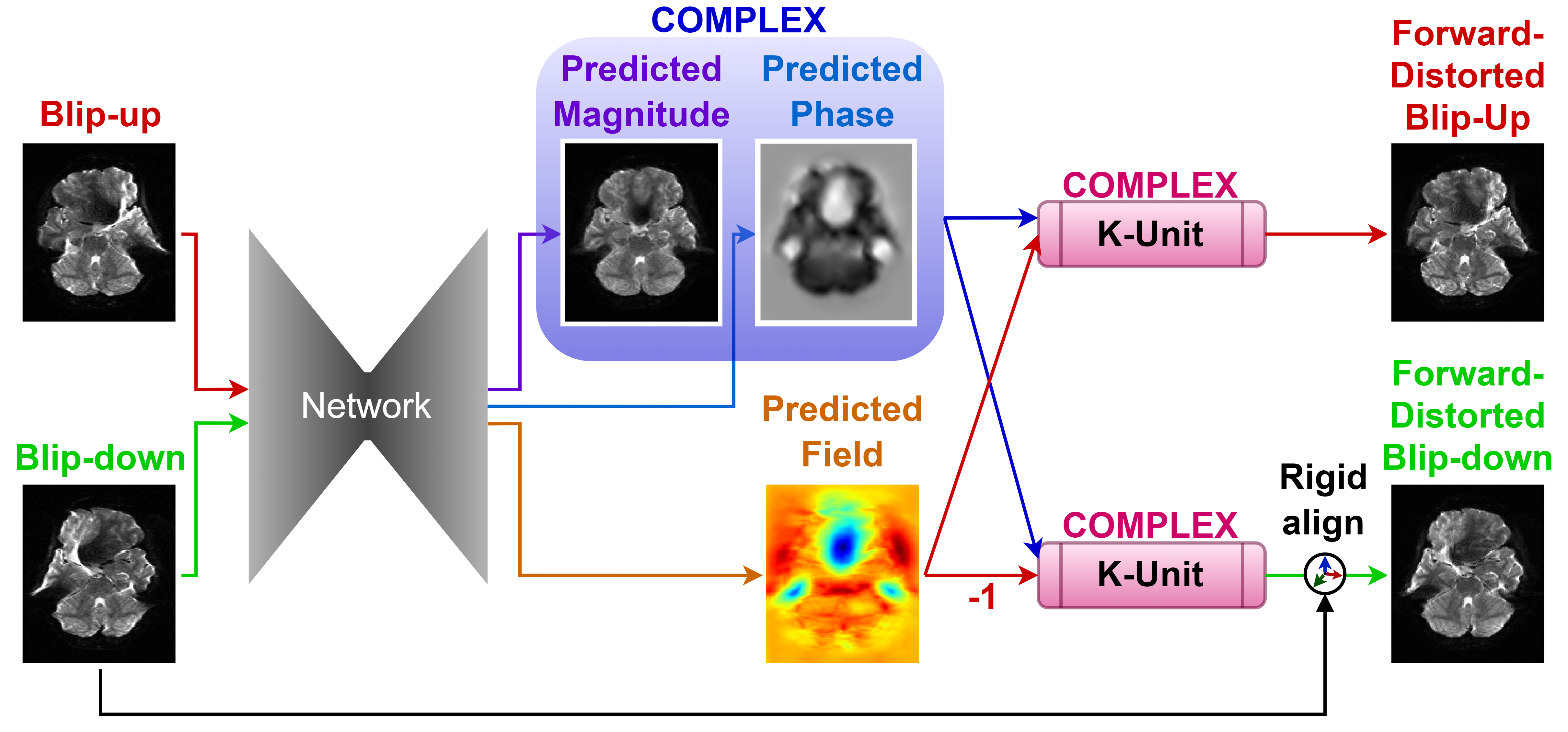
Figure 1. The proposed complex Forward-Distortion Network (compFD-Net). Starting from magnitude blip-up/-down EPI images, the main network predicts a displacement field and a complex-valued underlying image (i.e., both magnitude and phase). The complex K-Unit applies forward-distortion on the complex-valued anatomically-correct image to faithfully capture signal dropout/pileup artifacts. Affine transformation compensates for rigid-body motion between the forward-distorted and input blip-down images
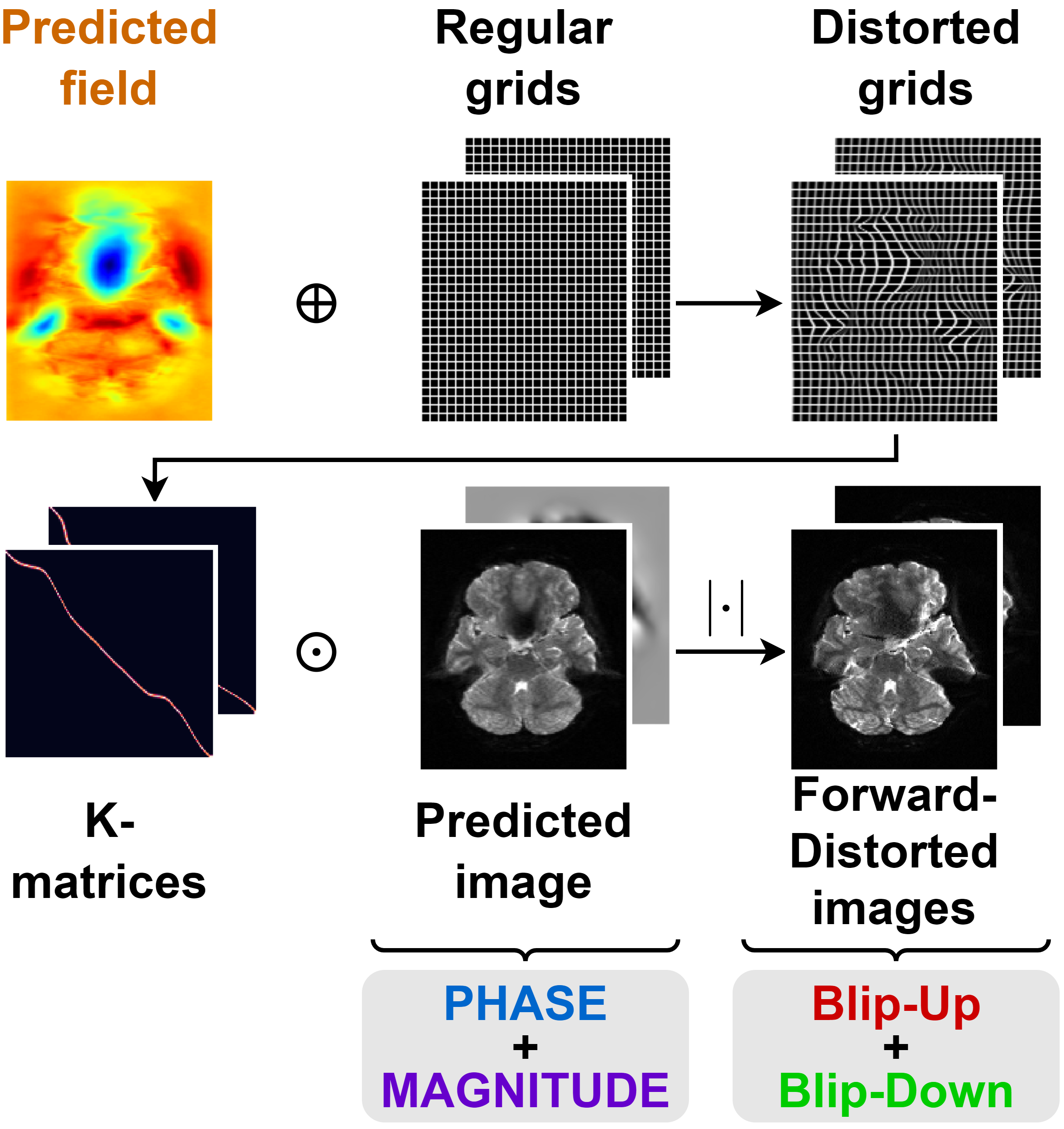
Figure 2. Detailed view of the complex K-Unit for forward-distortion. Regular grids are modulated by the predicted field to produce the distorted grids, which in turn are used to form the K-matrices. Multiplication of the complex-valued predicted image with the K-matrices produces the desired forward-distorted images. Here, $$$\lvert\cdot\rvert$$$ indicates magnitude operator.
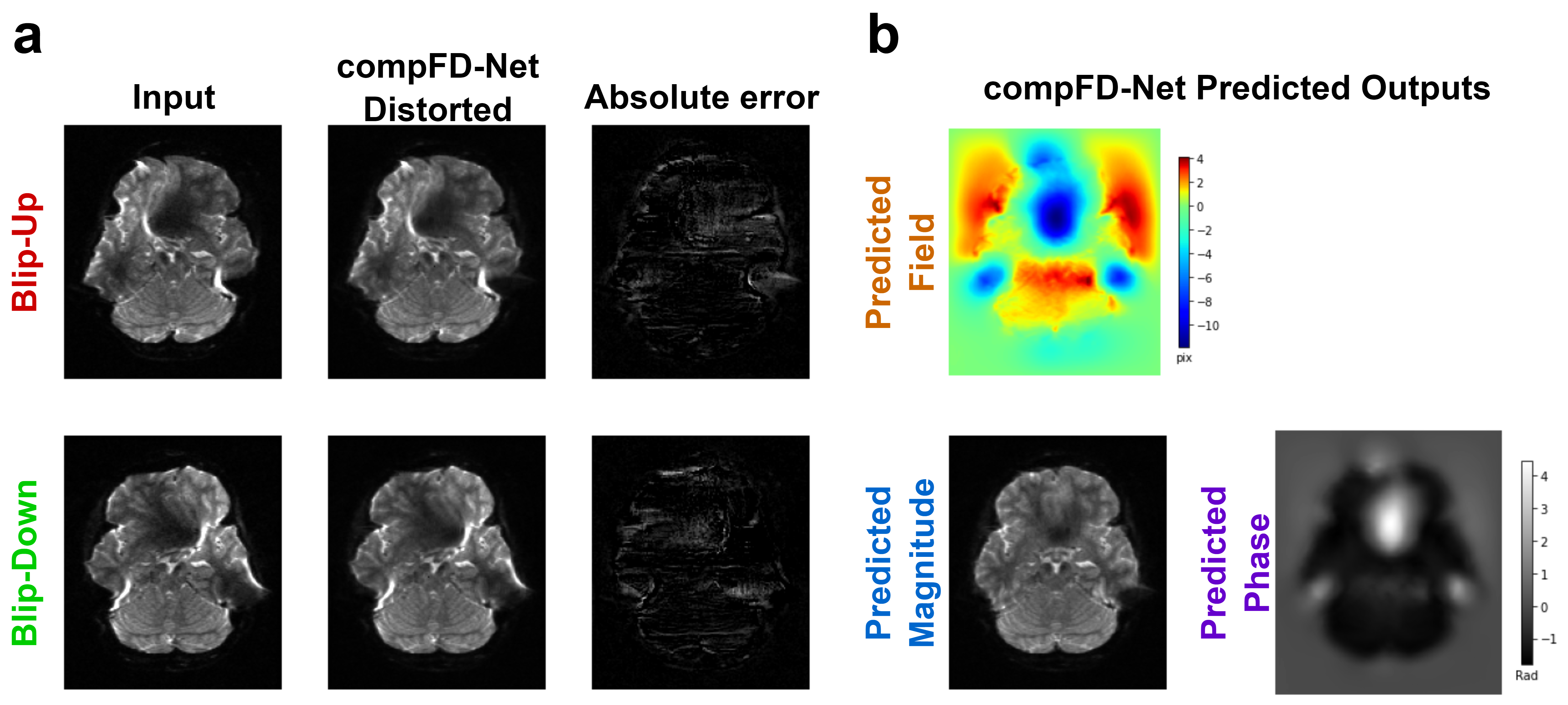
Figure 3. Representative result from compFD-Net. a) Comparisons of the input blip-up/-down EPI images and the forward-distorted images from compFD-Net show an excellent match. b) The outputs of FD-Net: the predicted field and the complex-valued anatomically-correct image (predicted as a magnitude image and a phase image). (Blip-up: Left-to-Right PE; blip-down: Right-to-Left PE).
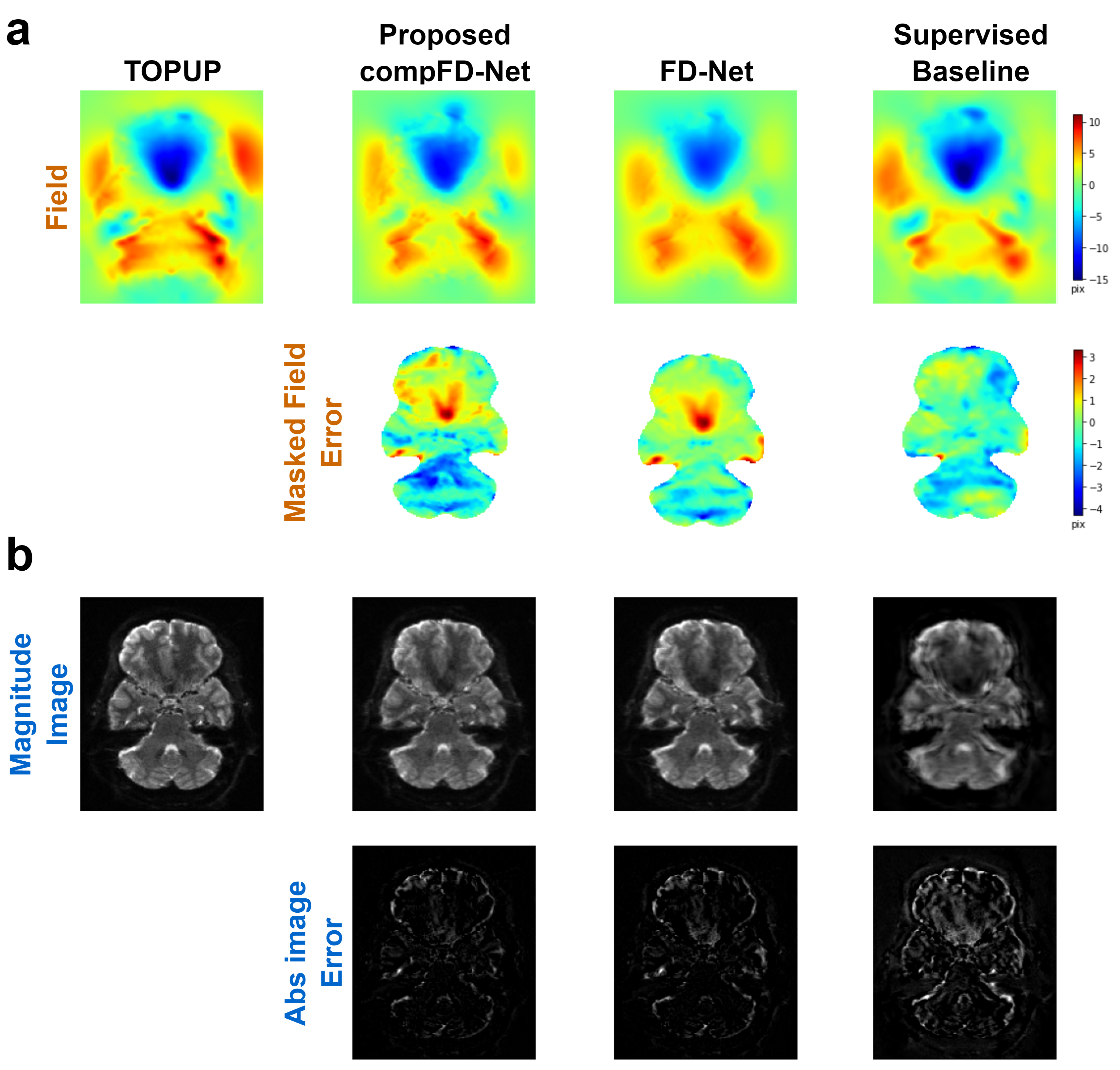
Figure 4. Representative results for compFD-Net against the original FD-Net and supervised baseline, with respect to the TOPUP benchmark. a) Predicted field and respective errors relative to TOPUP. Error maps for the field were masked via a median Otsu threshold over the TOPUP image to remove background regions. b) Corrected images and respective errors relative to TOPUP.
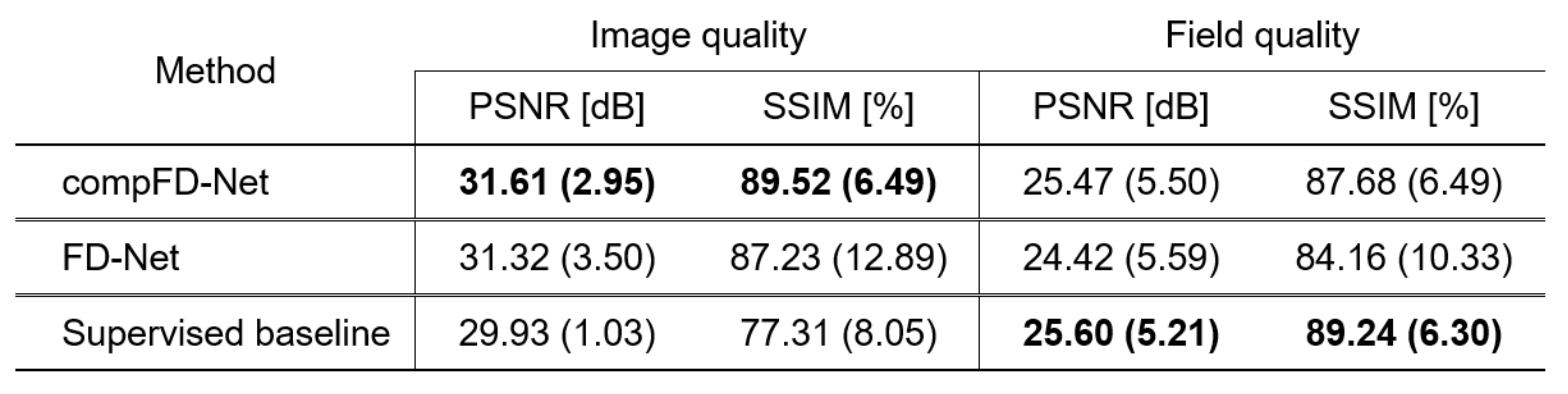
Figure 5. Table of performance metrics reported as mean and standard deviation (in brackets) across 8 test subjects for compFD-Net and competing methods. PSNR and SSIM were measured based on the image/field from benchmark TOPUP. The proposed compFD-Net outperforms the original FD-Net in all assessments. It also outperforms the supervised baseline in image quality, and performs competitively in terms of field quality.