1369
A Positive and Negative Learning based Image Decomposition Network for Phase Unwrapping and Background Removal1Department of Electronic Science, Xiamen University, Xiamen, China
Synopsis
Keywords: Quantitative Imaging, Quantitative Imaging, phase processing, background removal, deep learning, image decomposition, phase unwrapping
Motivation: Phase images contain important information useful in many fields. However, the phase data is often wrapped into a specific range, while background or noise signal in imaging scene may bring significant interference.
Goal(s): To obtain the exact information, phase images need an accurate processing that includes the unwrapping and the background removal.
Approach: In this paper, we propose a positive and negative learning based image decomposition network (PNnet) to accomplish the phase processing by a single network.
Results: Experimental results demonstrate that PNnet can achieve excellent performance and efficient generalization, even for complex wrapping and inhomogeneous background.
Impact: Except magnitude images, phase data in MRI also contain important information that is useful in many fields and scenarios. This work proposed a SOTA method for phase processing with high accuracy and excellent performance.
INTRODUCTION
For complex data, magnitude images are always applied for the object recognition, classification and segmentation, but phase images also contain important information that is useful in many fields and scenarios. However, extracting the local field from its measured phase in MRI is nontrivial because phase data are uniquely folded in the principal value range of (−𝜋, 𝜋]. Meanwhile, field shifts suffer from the overwhelming background field generated from sources outside the volume of interest (VOI), and those background or noise signal in imaging scene may bring significant interference. Therefore, phase images need an accurate processing that includes the unwrapping and background removal, which has high requirements on quantitative accuracy and reliability. However, traditional phase unwrapping approaches [1-3] may yield large error in presence of noise and phase discontinuities, while conventional background removal methods[4-7] are not capable to deal with regions of large susceptibility variations. At present, there are only a few deep leaning based phase processing attempts[8-10], nonetheless phase unwrapping and background removal are mostly conducted separately or combined with suceptibility reconstruction[11-13], which is not convenient and also suffers from error propagation.METHODS
With respect to the echo time TE, the measured MRI phase can be formulated into$$φ_{un}=-γTE{\cdot}B_{tol}=-γTE{\cdot}(B_{loc}+B_{bkg})$$
In object VOI, total field $$$B_{tol}$$$ can be decomposed into local field $$$B_{loc}$$$ and background field $$$B_{bkg}$$$. Meanwhile, the relationship between unwrapped phase $$$φ_{un}$$$ and wrapped phase $$$φ_{w}$$$ can be represented by
$$K=(φ_{un}-φ_{w})⁄2π$$
where K is the map of wrap count and 2πK is called as wrapping map. Therefore, we could derive $$$B_{loc}$$$ from $$$φ_{w}$$$ with
$$B_{loc}=(φ_w+2πK)⁄(-γTE)-B_{bkg}$$
Normally, the end-to-end network could obtain a calculation of $$$B_{loc}$$$, and the mapping of $$$φ_w→B_{loc}$$$ belongs to positive learning. However, this unidirectional framework lacks of necessary feedback and corrective mechanisms, meanwhile without any concern about intermediate variables K and $$$B_{bkg}$$$. In this view, we propose to explore them in the form of negative learning.
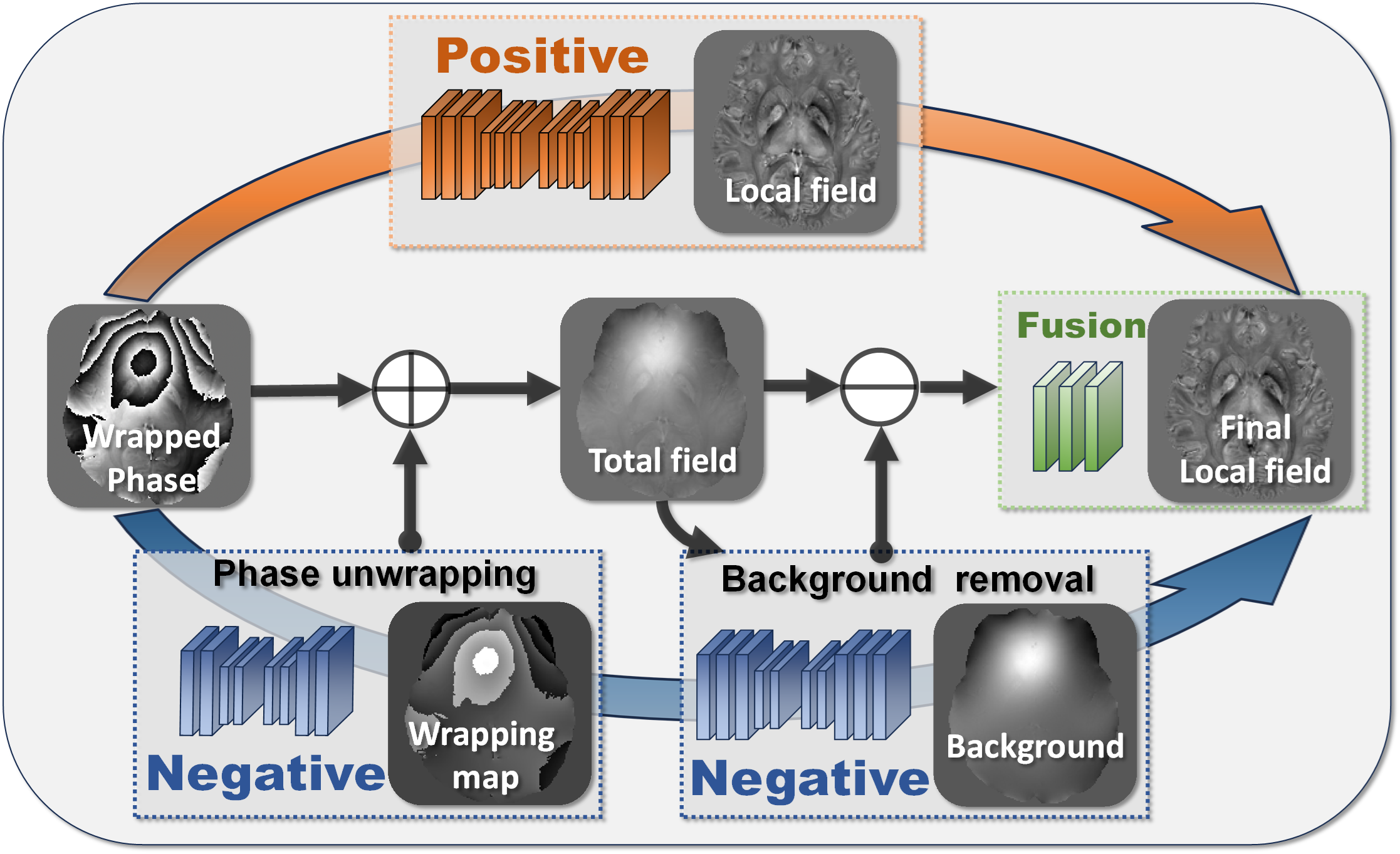
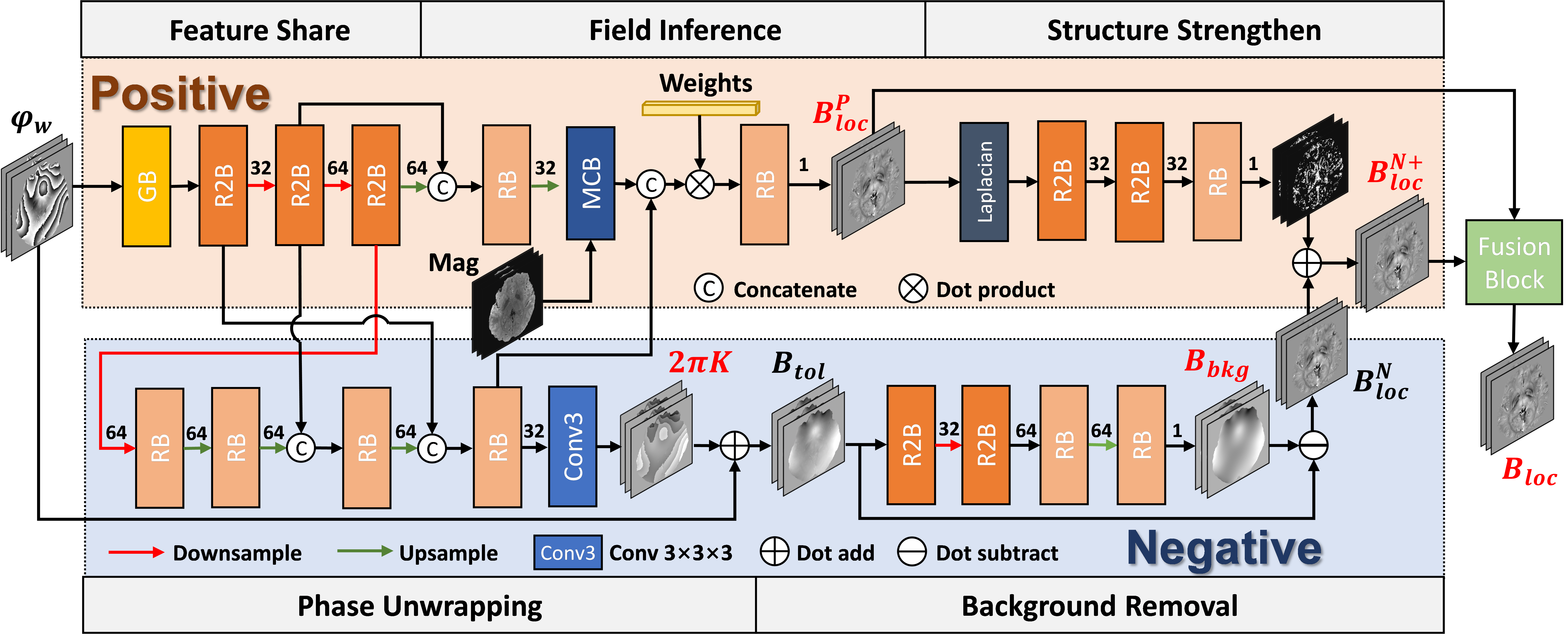
As shown in Fig. 1, the framework is constituted by a positive branch, a negative branch and an adaptively weighted fusion block, while the wrapped phase can be decomposed into wrapping map, background field and local field. For positive learning, it is directly trained by local field to establish a quantitative relationship between phase images and field shifts. Reversely, our negative learning is trained by the wrapping map and the background field, which intends to extract the interference information from actual phase signals. In this work, a positive and negative learning based image decomposition network PNnet is proposed to accomplish two tasks in phase processing with a single framework, as illustrated in Fig. 2, which includes five subnets of different roles.
RESULTS
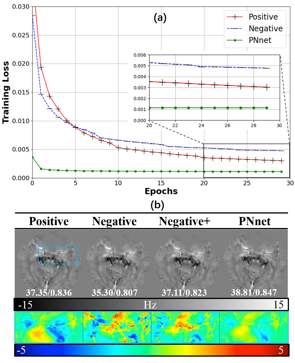
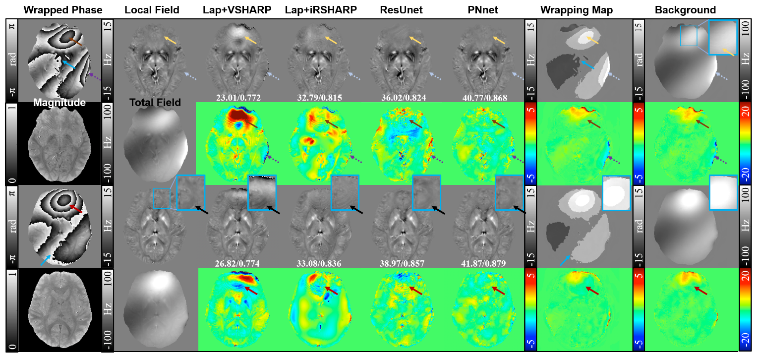
Fig. 3a compares curves of training loss between PNnet and its ablated versions, in which only positive learning or negative learning is employed separately. The training loss of PNnet is significantly better than those of Positive and Negative, demonstrating the outperformance of bidirectional learning. Their phase processing results are analyzed in Fig.3b. We can see that the discrepancies between models are remarkable. Neither Positive model nor Negative model is able to carry out an accurate processing. Adding with structure strengthen, errors in Negative+ model (i.e. $$$B_{loc}^{N+}$$$ in Fig.2) are less than Negative model but still quite obvious. In contrast, PNnet obtains the best result with the least residual errors and the highest PSNR/MSSIM scores.Fig. 4 shows local field results on synthetic dataset obtained by different methods. To investigate the efficiency of negative learning, we exhibit the wrapping map and background field output by PNnet and their difference maps. PNnet has the best performance over the sinus area with no obvious remaining turbulence and no noticeable structural errors, as well as the skull region marked by dash arrows.
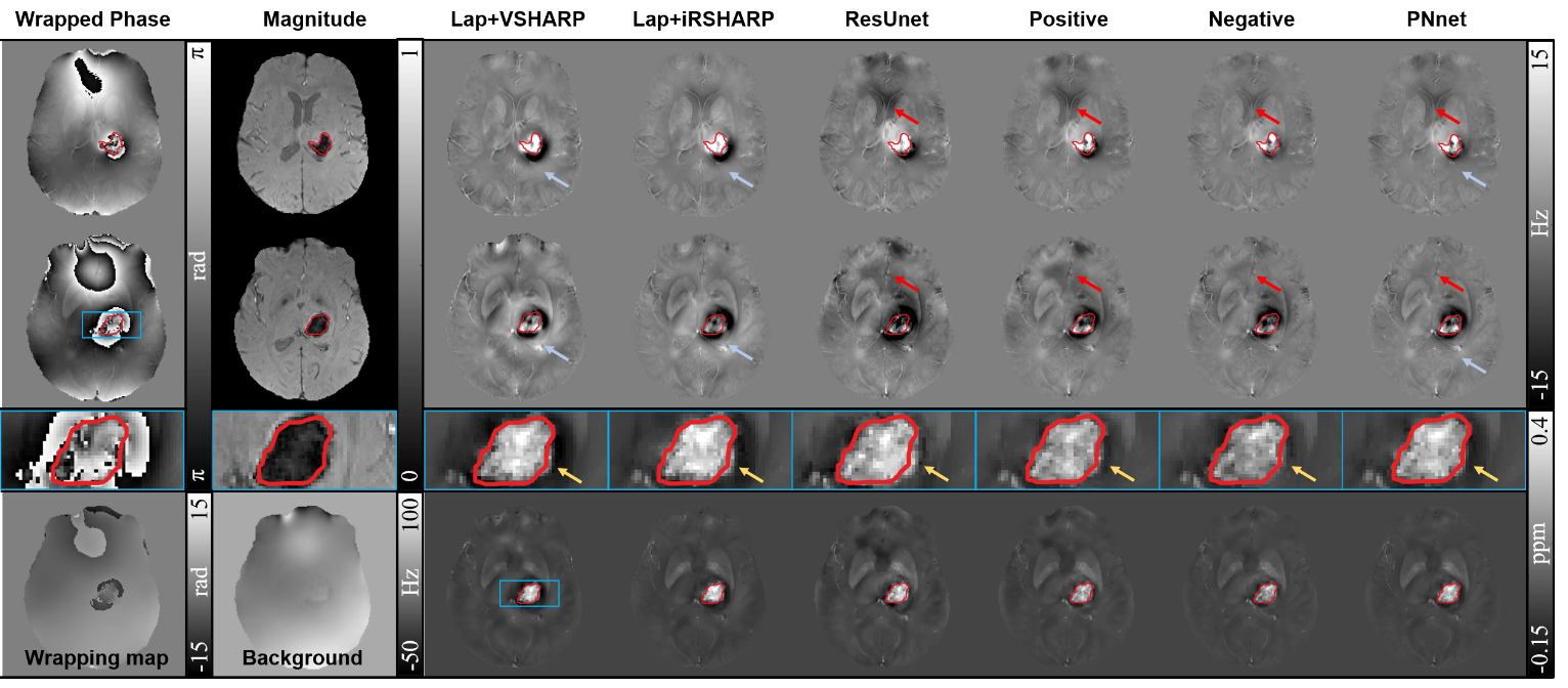
Fig. 5 evaluates the network generalization on cerebral hemorrhage data without any fine-tuning. The susceptibility artifacts are so overwhelming that is unable to recognize the lesion boundary, even partly occurring at the upper side indicated with red arrows. Compared to the other methods, hematoma sections are better recovered in PNnet with more reliable information and less artifacts.
DISCUSSION
It is common that a unidirection framework is applied following the positive learning scheme, i.e., the data in solution to be network input while the final goal as label data. However, a comprehensive understanding of both positive and negative information can benefit network to realize more solid determination and accurate analysis. Hence, a bidirectional network is proposed by assembling positive and negative learning. The wrapping map and the background field are regarded as "negative information", while the local field is "positive information".Acknowledgements
This work was supported in part by the National Natural Science Foundation of China under Grant 62071405.References
[1] Schofield M. A., Zhu Y., Fast phase unwrapping algorithm for interferometric applications, Opt Lett., 2003; 28(14):1194-1196.
[2] Jenkinson M., Fast, automated, N-dimensional phase-unwrapping algorithm, Magn. Reson. Med., 2003; 49(1):193-197.
[3] Karsa A., Shmueli K., SEGUE: A speedy region-growing algorithm for unwrapping estimated phase, IEEE Trans. Med. Imaging, 2019; 38(6):1347-1357.
[4] Liu T. et al., A novel background field removal method for MRI using projection onto dipole fields (PDF), NMR Biomed., 2011; 24(9):1129-1136.
[5] Schweser F. et al., Quantitative imaging of intrinsic magnetic tissue properties using MRI signal phase: an approach to in vivo brain iron metabolism, Neuroimage, 2011; 54(4):2789-2807.
[6] Ozbay P. S. et al., A comprehensive numerical analysis of background phase correction with V-SHARP, NMR Biomed., 2017; 30(4): e3550.
[7] Fang J. S. et al., Background field removal for susceptibility mapping of human brain with large susceptibility variations, Magn. Reson. Med., 2019; 81(3): 2025-2037.
[8] G. Spoorthi, R. K. S. S. Gorthi, and S. Gorthi, PhaseNet 2.0: Phase unwrapping of noisy data based on deep learning approach, IEEE Trans. Image Process., 2020; 29: 4862-4872.
[9] Bollmann S. et al., SHARQnet - Sophisticated harmonic artifact reduction in quantitative susceptibility mapping using a deep convolutional neural network, Z. Med. Phys., 2019; 29(2):139-149.
[10] Liu J., Koch K. M., Deep gated convolutional neural network for QSM background field removal, Med Image Comput Comput Assist Interv., 2019; 83-91.
[11] Cognolato F. et al., NeXtQSM-A complete deep learning pipeline for data-consistent Quantitative Susceptibility Mapping trained with hybrid data, Med Image Anal., 2023; 84:102700.
[12] Li Z. et al., Reconstruction of quantitative susceptibility mapping from total field maps with local field maps guided UU-Net, IEEE J. Biomed. Health Inform., 2023; 27(4): 2047-2058.
[13] Gao Y. et al., Instant tissue field and magnetic susceptibility mapping from MRI raw phase using Laplacian enhanced deep neural networks, Neuroimage, 2022; 259:119410.
Figures