1367
Image space formalism of k-space interpolation networks for analytical expression of noise characteristics1Experimental Physics 5, University of Würzburg, Würzburg, Germany, 2Magnetic Resonance and X-ray Imaging Department, Fraunhofer Institute for Integrated Circuits IIS, Division Development Center X-Ray Technology, Würzburg, Germany, 3Molecular and Cellular Imaging, Comprehensive Heart Failure Center, University Hospital Würzburg, Würzburg, Germany
Synopsis
Keywords: Machine Learning/Artificial Intelligence, Parallel Imaging, complex-valued convolutional neural networks, RAKI, GRAPPA, ReLU
Motivation: Robust Artificial Neural Networks for k-space Interpolation (RAKI) exhibit superior image reconstructions compared to traditional Parallel Imaging. It is crucial to thoroughly characterize RAKI to gain insights into its functionality and stimulate further enhancements.
Goal(s): Exploring how k-space interpolation with convolutional neural networks can be transformed into image domain to obtain an analytical description of noise characteristics.
Approach: The nonlinear activation in k-space is expressed as elementwise multiplication. This can be transformed into convolution in image space.
Results: The proposed image space formalism yields image reconstructions quasi-equivalent to k-space interpolation. The analytical expression of noise characteristics is in correspondence with Monte Carlo simulations.
Impact: We propose an image space formalism for k-space interpolation with convolutional neural networks. This enables an analytical expression of the noise characteristics, analogous to g-factor maps in traditional parallel imaging methods.
Introduction
GRAPPA [1] is a Parallel Imaging (PI) technique that interpolates missing k-space signals by convolution in k-space. Recently, RAKI [2] was introduced which generalizes GRAPPA with additional convolution layers, on which nonlinear activation functions are applied. Analogous to GRAPPA, convolution kernels in RAKI are calibrated using scan-specific training samples from auto-calibration signals. RAKI has proved to be more noise resilient than GRAPPA. However, a quantitative description of noise performance in RAKI, analogous to the geometry (g-)factor for PI [3,4], has yet not been presented. We introduce an analytical term for noise enhancement in RAKI. To this end, an image space formalism of RAKI is presented.Methods
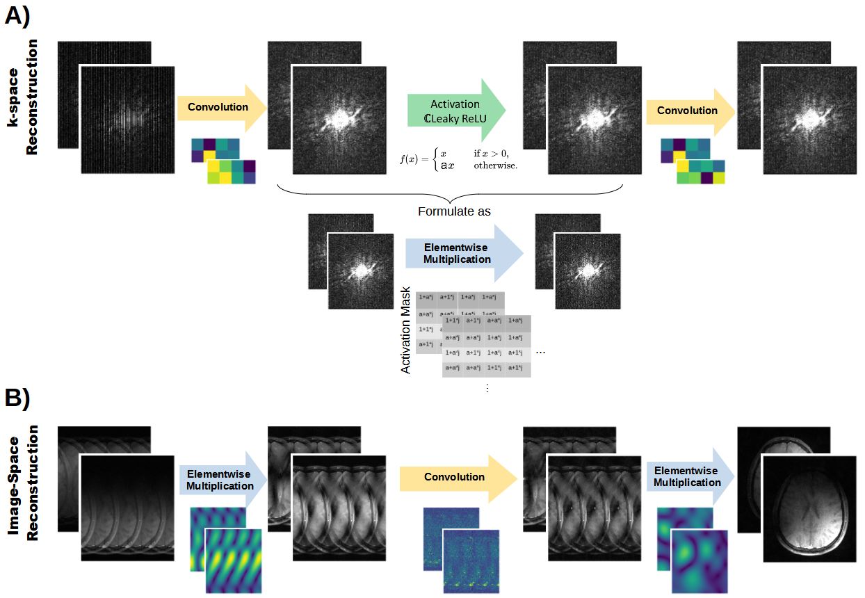
Image space formalism It is well known how to translate trained convolution kernels from k-space to image space [4]. However, a proper transformation of nonlinear activations of hidden layers is required to formulate an image domain reconstruction. The nonlinear activations are typically obtained via the (complex-valued) Rectifier Linear Unit[5] (i.e. identity operator for inputs>0, otherwise, input scaled with pre-defined constant a). Thus, the activation of a hidden layer Shid with Nchhidd channels and k-space size NxxNy can be formulated as elementwise multiplication with an activation mask Mact of same shape:$$S_{hid}^{act} = M_{act} \odot S_{hid} \;\;\; (1)$$
Mact is derived as follows:
- Define binary masks Mreal, Mimag:
Mreal/imag=1, where Shid,real/imag>0, otherwise Mreal/imag =a - Mact‘ = Mreal * Shid,real + i Mimag* Shid,imag
- Mact = (Mact‘ * ShidH) / |Shid|²
$$ IFFT(S^{act}_{hid}) = IFFT(M_{act}) \otimes IFFT(S_{hid}) \;\;\; (2)$$
Eq. (2) enables to formulate the image reconstruction (i.e. inference) entirely in image domain. Assuming the simplest network with one hidden layer (Figure 1A), the image space reconstruction is formulated with three image-space weight-matrices W (1,2,3) (Figure 1B). W(1) is attributed to the first convolution in k-space (shape [N, Nchhidd,Nc], where Nc is number of coils and N is pixel-number). W(2) is attributed to the activation of the hidden layer (Eq. (2)) and can be re-written to a Toelpitz-matrix to re-formulate the convolution into matrix multiplication (shape [Nchhidd, N, N]). W(3) is attributed to the second, final convolution (shape [N, Nc, Nchhidd]). The reconstructed h-th pixel in the unfolded c-th coil image Ih,c can be expressed as:
$$I_{h,c} = \sum_{l=1}^{N_{hidd}} \sum_{k=1}^{N} \sum_{c^{'}=1}^{N_{c}} ((( I_{k,c^{'}}^{'} \cdot W^{1}[k,l,c^{'}] ) \cdot W^2[l,k,h] ) \cdot W^3[h,c,l])\;\;\; (3)$$
where I’k,c‘ is the k-th pixel of the folded c‘-th coil image.
Analytical g-factor maps Eq. (3) enables the formulation of the Jacobian J for each reconstructed pixel of each coil image w.r.t. each pixel in the folded coil images:
$$J_{h,c;k,c^{'}} = \frac{\partial I_{h,c} } {\partial I^{'}_{k,c^{'}} } = \sum_{l=0}^{N_{hidd}} W^{1}[k,l,c^{'}] \cdot W^2[l,k,h] \cdot W^3[h,c,l] \;\;\; (4)$$
Note that J is of shape [N, Nc, N, Nc]. The analytical RAKI g-factor of the h-th pixel in the coil combined image can then be calculated by
$$g_h=\frac { \sqrt{(p^T \cdot J_h) \Sigma^2 (p^T \cdot J_h)^H} } {\sqrt{(p^T \cdot 1) \Sigma^2 (p^T \cdot 1)^H } } \;\;\; (5)$$
where vector p denotes the coil-combination weights for the h-th pixel, and Σ is the covariance of the receiver coils.
Experiments: 2D T1w TSE brain imaging was performed on a volunteer on a 3T Magnetom Skyra (Siemens Healthineers, Erlangen, Germany) with TR/TE=500/10ms, FOV=220x193mm2 (ROxPE), matrixsize: 256x224 (16 receiver coils, 4-fold retrospectively undersampled). The RAKI network was assigned one hidden layer (64 channels), a=0.5, kernelsizes = [5,2] and [3,1] in (ROxPE) for first and second convolution layers, respectively, and the reconstructed coil images were combined via sum-of-squares. The analytical g-factor maps were validated using Monte Carlo simulations [6] and Jacobians obtained by Pytorch-autodifferentiation framework [7].
Results
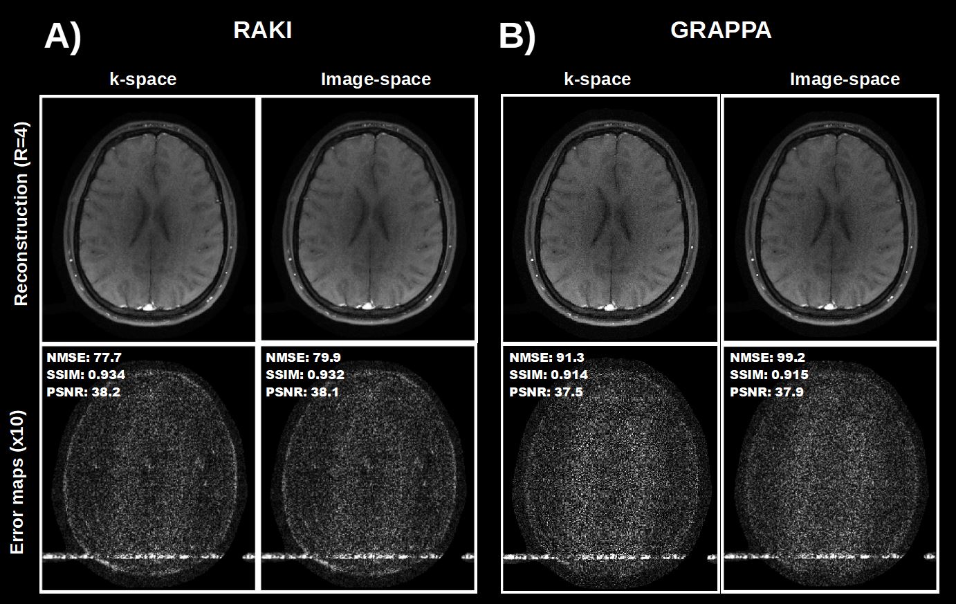
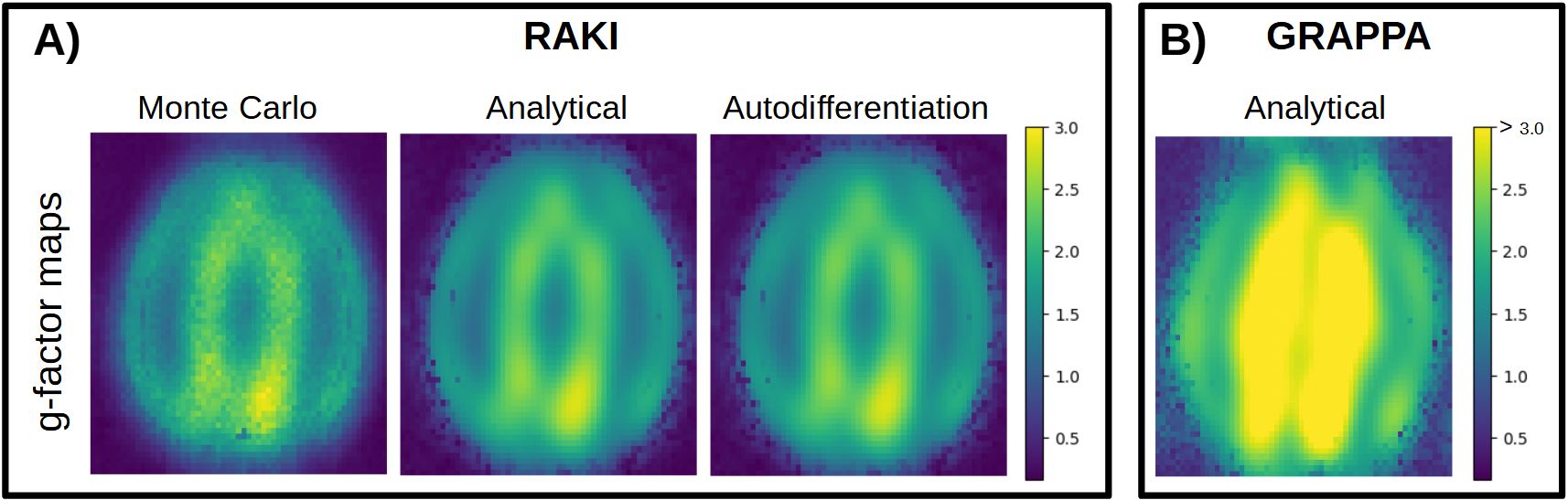
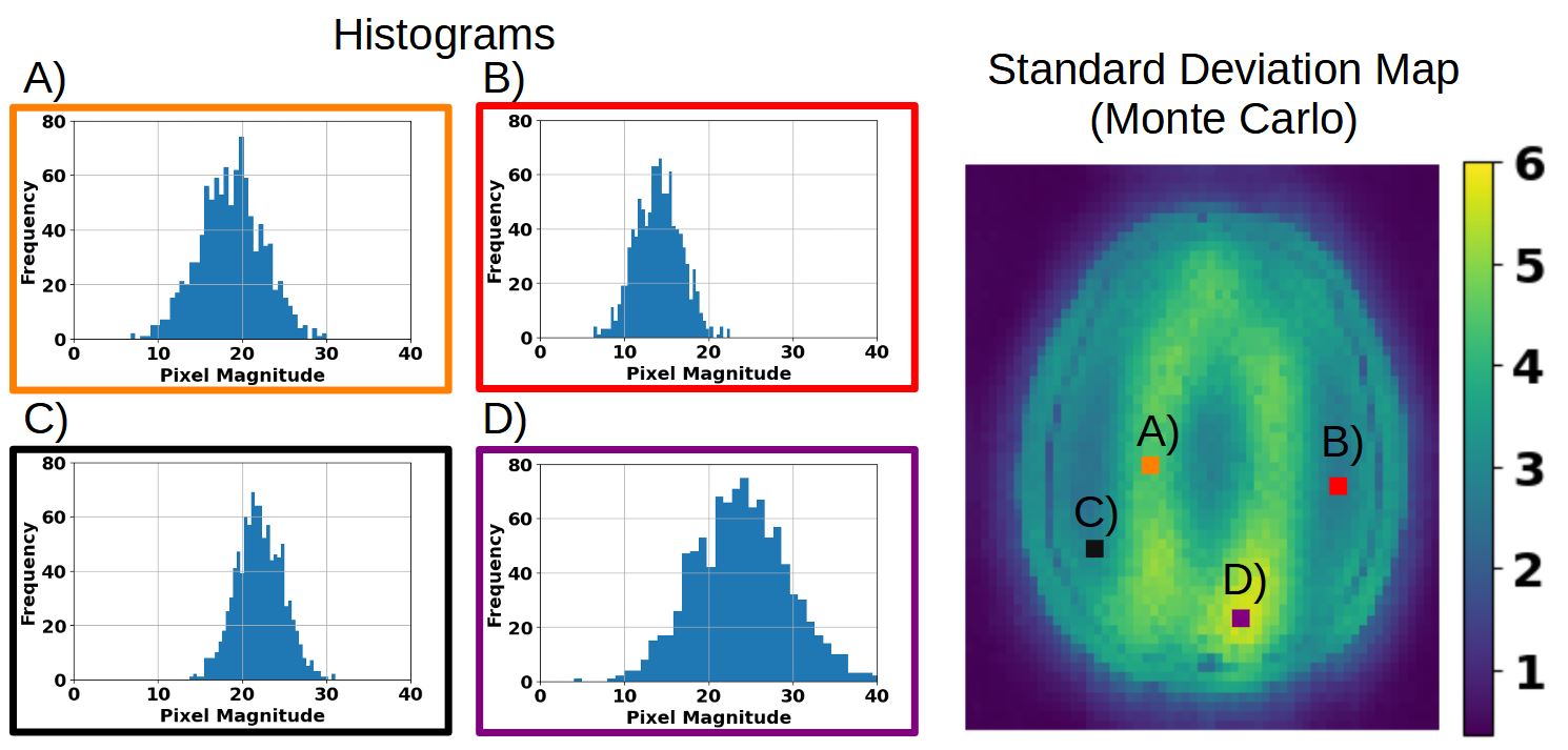
The RAKI reconstruction conducted in image space is quasi-identical to the k-space reconstruction, underlined by corresponding quantitative metrics (Figure 2A). Minor deviations may be attributed to convolution kernel padding. G-factor maps of RAKI obtained from the analytical expression correspond with those obtained via Monte Carlo simulations, as well as via autodifferentiation (Figure 3A), but are computed faster. The noise resilience is well characterized in comparison to GRAPPA (Figure 2B, Figure 3B). Monte Carlo simulations demonstrate Gaussian distributions of pixel magnitudes in the standard deviation maps, as evidenced by histograms (Figure 4).Discussion
The suppression of noise enhancement in RAKI can be attributed to nonlinear activations of hidden layers. This work introduces an image space formalism of RAKI and analytical expression for the g-factor. While common quantitative metrics (NMSE, SSIM) demand fully sampled references, the analytical g-factor may be used as a metric when no fully sampled references are available. The formalism can be extended to architectures with more hidden layers, or varying kernel sizes.Acknowledgements
Funded by the Bavarian Ministry of Economic Affairs, Infrastructure, Transport and Technology.References
[1] Akçakaya M, Moeller S, Weingärtner S, Uğurbil K. Scan-specific robust artificial-neural-networks for k-space interpolation (RAKI) reconstruction: Database-free deep learning for fast imaging. Magnetic Resonance in Medicine 2019;81(1):439–453.[2] Griswold MA, Jakob PM, Heidemann RM, Nittka M, Jellus V, Wang J, et al. Generalized autocalibrating partially parallel acquisitions (GRAPPA). Magnetic Resonance in Medicine 2002;47(6):1202–1210.
[3] Pruessmann, KP, Weiger, M, Scheidegger, M, Boesiger P. SENSE: Sensitivity encoding for fast MRI. Magnetic Resonance in Medicine 1999;42(5):952-62.
[4] Breuer, FA, Kannengiesser S, Blaimer A, Seiberlich N, Jakob PM, Griswold, M. General formulation for quantitative G-factor calculation in GRAPPA reconstructions. Magnetic Resonance in Medicine 2009;62(3):739-46.
[5] Cole E, Cheng J, Pauly J, Vasanawala S. Analysis of deep complex-valued convolutional neural networks for MRI reconstruction and phase-focused applications. Magnetic Resonance in Medicine 2021;86(2): 1093-1109.
[6] Robson P, Grant A, Madhuranthakam A, Lattanzi R, Sodickson D, McKenzie C. Comprehensive quantification of signal-to-noise ratio and g-factor for image-based and k-space-based parallel imaging reconstructions. Magnetic Resonance in Medicine 2008;60(4):895-907.
[7] Wang X, Ludwig D, Rawson M, Balan R, Ernst T. Estimating Noise Propagation of Neural Network Based Image Reconstruction Using Automated Differentiation. Proceedings of the int. Soc. Magn Reson Med. 2022, London. Abstract Number 0500.
Figures