1353
Investigating the relationship between multi-scale perfusion and myelin content in MS1Barrow Neurological Institute, Phoenix, AZ, United States
Synopsis
Keywords: Multiple Sclerosis, Multiple Sclerosis
Motivation: This study investigates the complex pathology of multiple sclerosis (MS), focusing on the relationship between cerebral perfusion and myelin integrity.
Goal(s): Utilizing selective inversion recovery (SIR) to determine myelin content through the macromolecular (PSR), and a combined spin- and gradient-echo (SAGE) sequence to assess micro- and macrovascular perfusion,
Approach: This study aims to uncover the interplay of hemodynamic impairments and myelin repair in MS.
Results: In a cohort of individuals with RRMS and healthy controls, we found significant correlations between PSR and microvascular perfusion within lesions, suggesting a possible link between myelin integrity and cerebral blood flow.
Impact: Uncovering specific pathological changes in MS lesions may enhance diagnostic accuracy and disease monitoring. Such insights could also drive the creation of new therapies focused on remyelination and neuroprotection, potentially altering the treatment landscape for MS.
Introduction
Multiple sclerosis (MS) is an immune-mediated disease with complex pathological features (e.g., de/remyelination, axonal loss, edema, inflammation) that often present concurrently. Imaging biomarkers have improved our understanding of the role of these pathologies in disease progression1,2, including for the evaluation of myelin loss and repair and the potential role hemodynamic impairments play in this process3,4. In particular, selective inversion recovery (SIR) has shown promise in evaluating myelin content changes (via the macromolecular pool-size-ratio, PSR), which relate to MS severity and progression5,6,7. On the other hand, perfusion MRI has been used to investigate cerebral blood flow (CBF) and volume (CBV) changes in MS patients, which have been associated with both MS phenotypes and disability levels8,9 and may reflect widespread brain dysfunction in MS10. Previous work has demonstrated that non-specific measures of myelination (from ratios of T1- and T2-weighted images) relate to the perfusion status of lesions11. The objective of this study is to systematically explore this relationship by obtaining i) myelin-specific PSR values and ii) measures of micro- and macro-vascular perfusion in a cohort of persons with relapsing-remitting MS (RRMS). The long-term goal is to better understand the role cerebral perfusion plays in myelin repair, potentially contributing to a deeper understanding of MS pathology and paving the way for novel prognostic biomarkers of remyelination.Method
The study encompassed 19 participants (9 RRMS patients, 40-53 years old, 8 F and 10 healthy controls, 23-67 years old, 4 F). SIR images were collected using an inversion recovery prepared 3D turbo spin-echo (TSE) sequence, with field of view (FOV) 210 × 210 × 90 mm3, reconstructed matrix size of 224 × 224 × 40, and echo time (TE) of 65 ms. From SIR data, PSR maps were calculated6. A combined spin- and gradient-echo (SAGE) dynamic susceptibility contrast (DSC) sequence, with five echoes (TEs = 6.6, 24.7, 54, 72.1 and 90 ms, TR = 1.8s), was acquired to estimate both micro- and macro-vascular perfusion (FOV 240 × 240 mm2, resolution 2.5 × 2.5 × 5 mm3, 15 slices). For each participant, baseline scans were collected for 30 seconds, at which time a gadolinium-based contrast agent (0.1 mmol/kg) was injected, with the total acquisition lasting 4.5 minutes. SAGE-based CBV and CBF maps were generated from the DSC data, denoted as SE for spin-echo and GRE for gradient-echo measures. Of note, SE measures are more specific to microvascular perfusion, while GRE measures reflect total perfusion. FreeSurfer and the Lesion Segmentation Toolbox (LST) were utilized to compute normal-appearing white matter (NAWM) and lesion (LES) masks, respectively. Spearman’s correlation was used to assess the relationship between PSR and perfusion in NAWM and lesion regions of interest (ROIs).Results
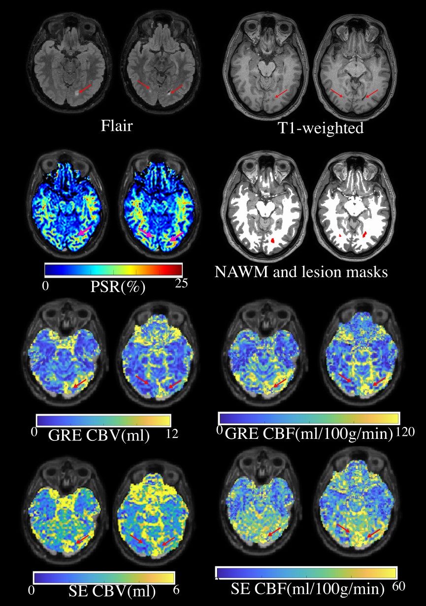
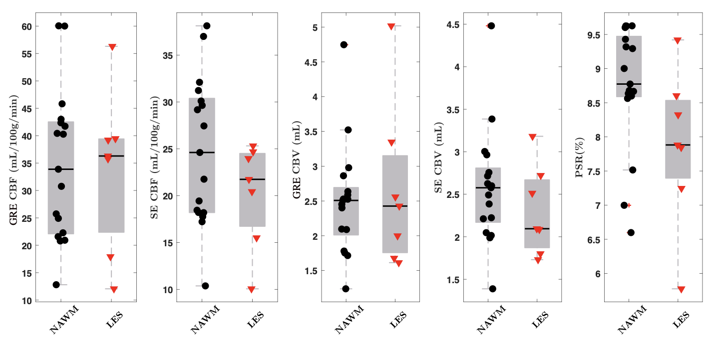
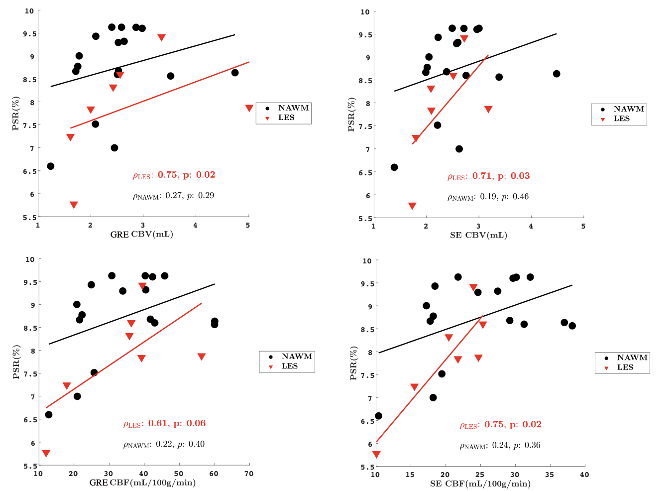
Representative images and maps, with corresponding ROIs, in a person with RRMS are shown in Figure 1. Box plots demonstrate the distribution of perfusion (SE- and GRE- CBV and CBF) and PSR for both lesion and NAWM ROIs (Figure 2). Lesions were characterized by lower PSR and microvascular perfusion compared to NAWM, while total perfusion was similar between lesions and NAWM. Figure 3 shows the correlation between PSR and perfusion metrics. The correlation between PSR and DSC parameters for NAWM was not significant (ρ=0.19-0.27), while significant correlations were observed in lesion ROIs (ρ=0.61-0.75). Notably, the correlations between lesion PSR and microvascular perfusion (ρ=0.71-0.75 p<0.02) demonstrate that microvascular perfusion may relate to myelin integrity.Discussion and Conclusions:
This study assessed the relationship between PSR obtained from SIR and perfusion metrics from SAGE-DSC, with comparisons between lesion in individuals with MS and white matter in both MS patients and healthy control subjects. In lesion ROIs, a notable correlation between PSR and perfusion metrics was observed, signifying a potential link between microstructural and hemodynamic alterations. Conversely, no significant correlations were observed in NAWM, which may indicate stability of myelin in these regions. These findings bolster our previous findings using nonspecific measures of white matter integrity11. The disparity in correlations between lesion and NAWM regions underscores the nuanced pathological dynamics in MS, highlighted by the joint analysis of myelin and perfusion metrics. Work is ongoing to characterize repeatability of perfusion and myelin in a larger cohort and to understand differences in myelin and perfusion between MS patients and healthy controls. Long-term, we aim to understand whether perfusion is indicative of myelin changes, which may have important implications for novel remyelinating and/or neuroprotective therapies.Acknowledgements
We thank NIH R21 NS125535 and National MS Society RG-2111-3872 for support. We would also like to thank our MRI Tech Sharmeen Maze.References
1. Lapointe, E., Li, D.K.B., Traboulsee, A.L. and Rauscher, A., 2018. What have we learned from perfusion MRI in multiple sclerosis? American Journal of Neuroradiology, 39(6), pp.994-1000.
2. Laganà, M.M., Pelizzari, L. and Baglio, F., 2020. Relationship between MRI perfusion and clinical severity in multiple sclerosis. Neural regeneration research, 15(4), p.646.
3. Ge, Y., Law, M., Johnson, G., Herbert, J., Babb, J.S., Mannon, L.J. and Grossman, R.I., 2005. Dynamic susceptibility contrast perfusion MR imaging of multiple sclerosis lesions: characterizing hemodynamic impairment and inflammatory activity. American journal of neuroradiology, 26(6), pp.1539-1547.
4. de la Peña, M.J., Peña, I.C., García, P.G.P., Gavilán, M.L., Malpica, N., Rubio, M., González, R.A. and de Vega, V.M., 2019. Early perfusion changes in multiple sclerosis patients as assessed by MRI using arterial spin labeling. Acta Radiologica Open, 8(12), p.2058460119894214.
5. Bagnato, F., Franco, G., Ye, F., Fan, R., Commiskey, P., Smith, S.A., Xu, J. and Dortch, R., 2020. Selective inversion recovery quantitative magnetization transfer imaging: Toward a 3 T clinical application in multiple sclerosis. Multiple Sclerosis Journal, 26(4), pp.457-467.
6. Wang, P., Sisco, N., Yoo, W., Borazanci, A., Karis, J. and Dortch, R., 2023. Rapid whole‐brain myelin imaging with selective inversion recovery and compressed SENSE. Magnetic resonance in medicine, 89(3), pp.1041-1054.
7. Bagnato, F., Hametner, S., Franco, G., Pawate, S., Sriram, S., Lassmann, H., Gore, J., Smith, S.E. and Dortch, R., 2018. Selective inversion recovery quantitative magnetization transfer brain MRI at 7T: clinical and postmortem validation in multiple sclerosis. Journal of Neuroimaging, 28(4), pp.380-388.
8. Koudriavtseva, T., Sbardella, E. and Mainero, C., 2015. Brain perfusion and vasoreactivity in multiple sclerosis. American Journal of Neuroradiology, 36(4), pp. E27-E28.
9. Marshall, O., Lu, H., Brisset, J.C., Xu, F., Liu, P., Herbert, J., Grossman, R.I. and Ge, Y., 2014. Impaired cerebrovascular reactivity in multiple sclerosis. JAMA neurology, 71(10), pp.1275-1281.
10. Bakshi, R., Thompson, A.J., Rocca, M.A., Pelletier, D., Dousset, V., Barkhof, F., Inglese, M., Guttmann, C.R., Horsfield, M.A. and Filippi, M., 2008. MRI in multiple sclerosis: current status and future prospects. The Lancet Neurology, 7(7), pp.615-625.
11. Sisco, N.J., Borazanci, A., Dortch, R. and Stokes, A.M., 2021. Investigating the relationship between multi-scale perfusion and white matter microstructural integrity in patients with relapsing-remitting MS. Multiple Sclerosis Journal–Experimental, Translational and Clinical, 7(3), p.20552173211037002.
Figures