1349
Identification of constitutive parameters of carotid atherosclerotic plaques by CINE MR imaging1Center for Biomedical Imaging Research, Department of Biomedical Engineering, School of Medicine, Tsinghua University, Beijing, China, 2Department of Radiology, Peking University Third Hospital, Beijing, China, 3Department of Neurosurgery, Peking University Third Hospital, Beijing, China, 4Department of Vascular Surgery, Beijing Hospital, National Center of Gerontology, Institute of Geriatric Medicine, Chinese Academy of Medical Sciences, Beijing, China
Synopsis
Keywords: Vessel Wall, Atherosclerosis, mechanical constitutive property, inverse problem
Motivation: Beyond structural features of carotid plaque and hemodynamic parameters, the hemodynamic and structural metrics were determined by image-based computer simulations and the constitutive characteristics of carotid plaques were ignored.
Goal(s): We aimed to propose a method for identification of carotid plaque constitutive parameters using CINE MR images.
Approach: A novel MRI-based method to identify constitutive parameters of carotid plaque compositions was proposed and the calculated values from the proposed method were compared with the reference and ex-vivo validation.
Results: We found that calculated values were consistent with the reference and experimental results. The constant D2 could identify plaque compositions with different stiffness.
Impact: Our study proposed a novel MRI-based method for identifying constitutive parameters of carotid plaque compositions. The constant D2 could indicate different components of carotid plaques.
Introduction
Carotid atherosclerotic disease is known as one of the major etiologies for ischemic stroke1, 2. A number of studies demonstrated that the key features of carotid vulnerable plaques are associated with cerebrovascular events3. Beyond structural features of carotid plaque and hemodynamic parameters4-6, the structural mechanical characteristics are also essential for indicating the vulnerability of carotid plaques7. Nevertheless, the hemodynamic and structural metrics were determined by image-based computer simulations5-7 and the constitutive characteristics of carotid plaques were ignored8, 9. In this study, we proposed a method for identification of carotid plaque constitutive parameters to determine different compositions of carotid plaques using CINE MR images.Methods
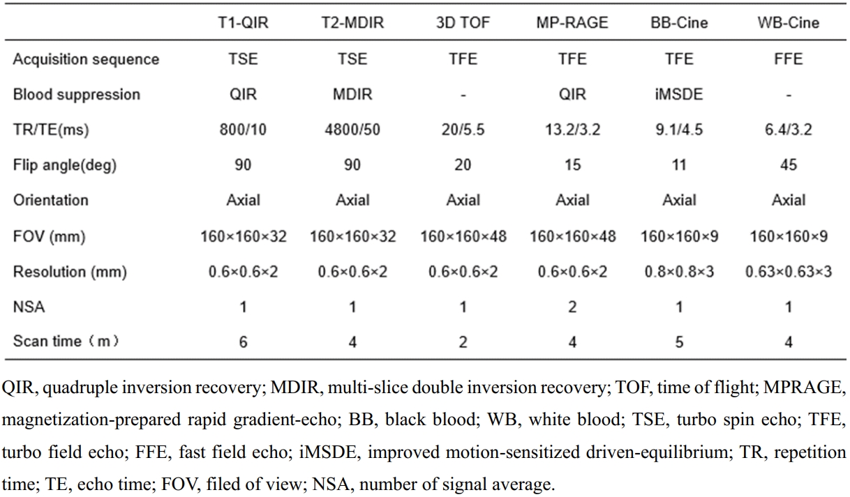
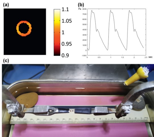
Study population: Thirty-six patients with moderate-to-severe carotid atherosclerotic stenosis who were scheduled for carotid endarterectomy (CEA) were recruited. The study protocol was approved by institutional review board and written consent form was obtained from all subjects. MR imaging protocol: Multi-contrast carotid MRI and CINE MRI were conducted on a whole-body 3.0T MR scanner (Ingenia TX, Philips Healthcare) with 8-channel carotid coil within one week before CEA. The detailed imaging parameters are listed in Table 1. MR image analysis: The lumen and outer wall boundaries of carotid arteries were outlined on MR images which were matched to the CINE MR images. The carotid plaque compositions were identified according to multi-contrast MR images by two experienced radiologists. Image post-processing and property identification: The contours of lumen, outer wall and different plaque compositions were mapped onto different frames of CINE MR images. To calculate the displacement field of carotid plaque contour, different frames of CINE MR images were registered by using an open-source toolkit Elastix. The blood pressure utilized in the 2D finite element (FE) model was presented in Figure 1(b). In the FE procedure, the modified Mooney-Rivlin strain energy density function (as Eq 1) was utilized to describe the stress-stretch relationship and the arterial wall and plaque components were assumed as hyper-elastic, isotropic and incompressible. With the specific strain and pressure, the zero-pressure algorithm by Huang et al was implemented10. The constitutive parameters of carotid plaques were determined by the FE analysis. Ex-vivo validation analysis: The ex-vivo carotid plaques samples of 6 patients from CEA surgery within 24 hours were segmented into stripes for uniaxial tensile test (Figure 1(d)). The stress-stretch curve of each stripe was fitted by using ordinary least square fitting method to obtain constitutive parameters11.$$$W=c_1 (I ̅_1-2)+D_1(exp(D_2 (I ̅_1-3))-1)+κ (J-1) $$$ Eq 1
Results
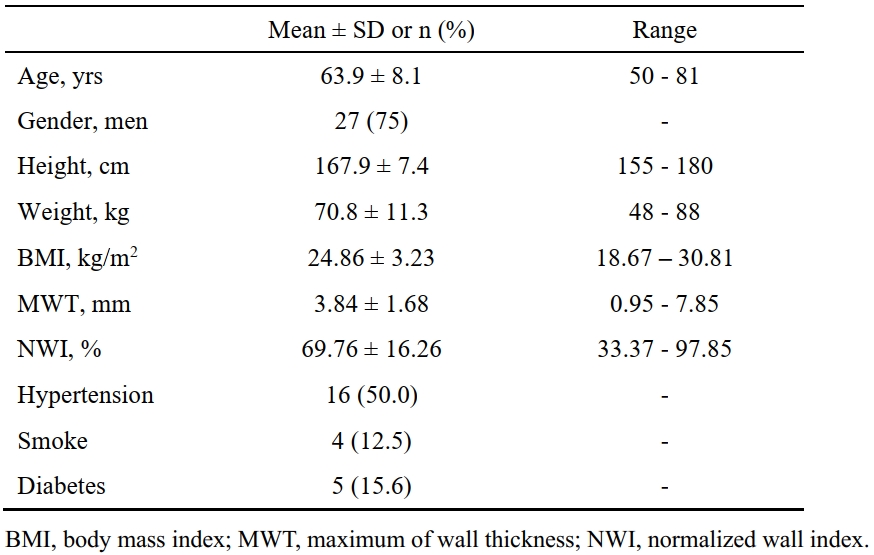
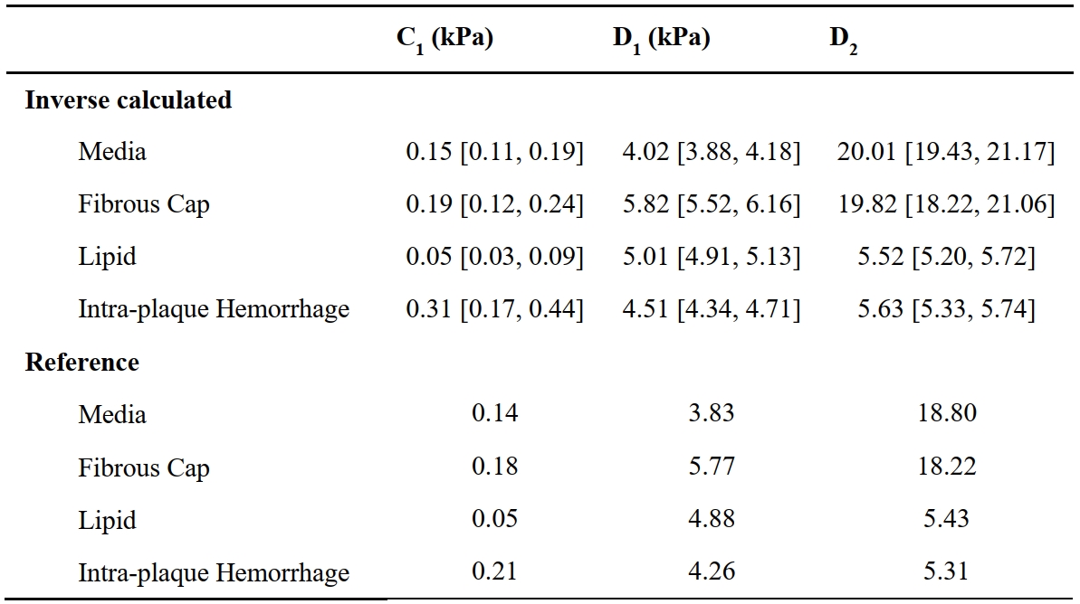
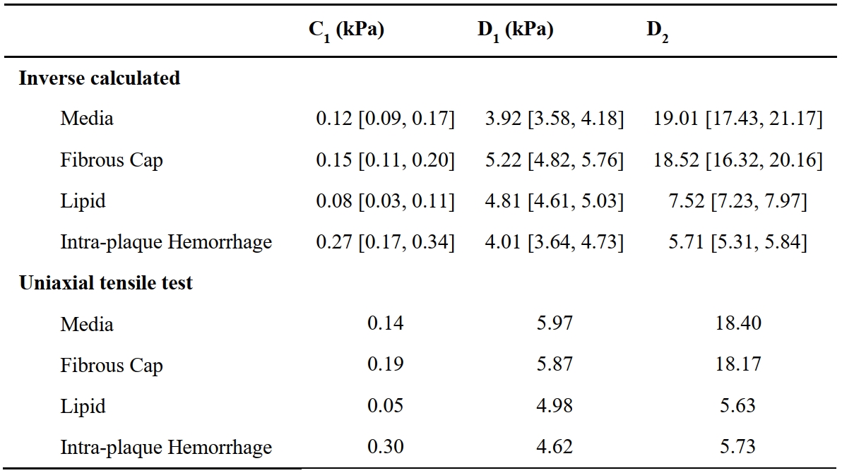
Of 36 patients (mean age: 63.9±8.1 years; 27 males) enrolled in this study, 52 slices with CINE MR images were matched with the multi-contrast MR images. The clinical characteristics of the study population are summarized in Table 2. The constitutive parameters of each tissue type were: media, c1 = 0.15 [0.11, 0.19] kPa, D1 = 4.02 [3.88, 4.18] kPa and D2 = 20.01 [19.43, 21.17]; fibrous cap, c1 = 0.19 [0.12, 0.24] kPa, D1 = 5.82 [5.52, 6.16] kPa and D2 = 19.82 [18.22, 21.06]; lipid, c1 = 0.05 [0.03, 0.09] kPa, D1 = 5.01 [4.91, 5.13] kPa and D2 = 5.52 [5.20, 5.72]; and intraplaque hemorrhage (IPH), c1 = 0.31 [0.17, 0.44] kPa, D1 = 4.51 [4.34, 4.71] kPa and D2 = 5.63 [5.33, 5.74]. Of all the parameters, the D2 of media and fibrous cap is much larger than those of the lipid and intraplaque hemorrhage. In the validation experiment, the fresh carotid plaque samples of 6 patients were segmented into 11 media stripes, 10 fibrous cap stripes, 8 lipid stripes and 6 IPH stripes. The ex-vivo validation demonstrated that the identified parameters of different compositions are consistent with the averaged values from the uniaxial tensile test.Discussion and Conclusion
In this study, we proposed a new MRI-based method to identify carotid plaque constitutive property and found that the constitutive parameters could identify carotid plaque compositions with different stiffness. All the calculated values of different compositions of all slices were consistent with both experimental results in the validation and a previous study12. In addition, the value of D2 indicates that the media and fibrous cap are stiffer than the lipid and IPH. This finding suggests that the D2 constant is an effective indicator for composition identification. However, there are still some limitations: (1) some tissue stripes may contain more than one tissue types; (2) the 2D FE model only determined the material property in the circumferential direction. In conclusion, a novel MRI-based method for carotid plaque constitutive parameters identification was proposed and validated with ex-vivo uniaxial tensile test and the constant D2 could identify compositions of different stiffness in carotid atherosclerotic plaques.Acknowledgements
NoneReferences
1. Benjamin, E. J., Muntner, P., Alonso, A., et al. Heart disease and stroke statistics-2019 update: a report from the American Heart Association. Circulation. 2019, 139, e56–528.
2. Markus, H. Stroke: causes and clinical features. Medicine. 2016, 44, 515–520.
3. Michel JB, Virmani R, Arbustini E, Pasterkamp G. Intra-plaque hemorrhages as the trigger of plaque vulnerability. European Heart Journal. 2011, 32:1977–1985.
4. Zhao X , Miller Z E , Yuan C . Atherosclerotic plaque imaging by carotid MRI. Current Cardiology Reports. 2009, 11(1):70-77.
5. Wang J., Paritala P. K., Mendieta J. B., et al. Carotid Bifurcation with tandem stenosis—a patient-specific case study combined in vivo imaging, in vitro Histology and in silico Simulation. Frontiers in Bioengineering and Biotechnology. 2019, 7(349):1-11.
6. Shen R., Tong X., Li D., et al. Slice-based and time-specific hemodynamic measurements discriminate carotid artery vulnerable atherosclerotic plaques. Comput Methods Programs Biomed. 2022, Oct;225:107050.
7. Huang Y, Teng Z, Sadat U, et al. The influence of computational strategy on prediction of mechanical stress in carotid atherosclerotic plaques: comparison of 2D structure-only, 3D structure-only, one-way and fully coupled fluid-structure interaction analyses. J Biomech. 2014 Apr 11;47(6):1465-71.
8. Teng Z, Yuan J, Feng J, et al. The influence of constitutive law choice used to characterise atherosclerotic tissue material properties on computing stress values in human carotid plaques. J Biomech. 2015 Nov 5;48(14):3912-21.
9. Yuan J, Teng Z, Feng J, et al. Influence of material property variability on the mechanical behaviour of carotid atherosclerotic plaques: a 3D fluid-structure interaction analysis. Int J Numer Method Biomed Eng. 2015 Aug;31(8):e02722.
10. Huang Y, Wang S, Luo T, et al. Estimation of the zero-pressure computational start shape of atherosclerotic plaques: Improving the backward displacement method with deformation gradient tensor. J Biomech. 2022 Jan;131:110910.
11. Wang S, Tokgoz A, Huang Y, et al. Bayesian Inference-Based Estimation of Normal Aortic, Aneurysmal and Atherosclerotic Tissue Mechanical Properties: From Material Testing, Modeling and Histology. IEEE Trans Biomed Eng. 2019 Aug;66(8):2269-2278.
12. Teng Z, Zhang Y, Huang Y, et al. Material properties of components in human carotid atherosclerotic plaques: a uniaxial extension study. Acta Biomater. 2014 Dec;10(12):5055-5063
Figures