1344
Hippocampal amide proton transfer value is associated with the imaging marker and total burden of cerebral small vessel disease1Department of Radiology, Nanxishan Hospital of Guangxi Zhuang Autonomous Region, Guilin, China, 2Philips Healthcare, Guangzhou, China, 3Philips Healthcare, Beijing, China
Synopsis
Keywords: Blood Vessels, CEST & MT
Motivation: Exploring the CSVD imaging markers can provide insights into the pathophysiology of CSVD, which is crucial for understanding its etiology, development, and clinical interventions.
Goal(s): To investigate potential variations in hippocampal APT values among individuals with CSVD imaging markers and varying degrees of CSVD total burden.
Approach: All the participants conducted the cognitive assessment and the MRI scans. And statistical analysis was used for evaluation.
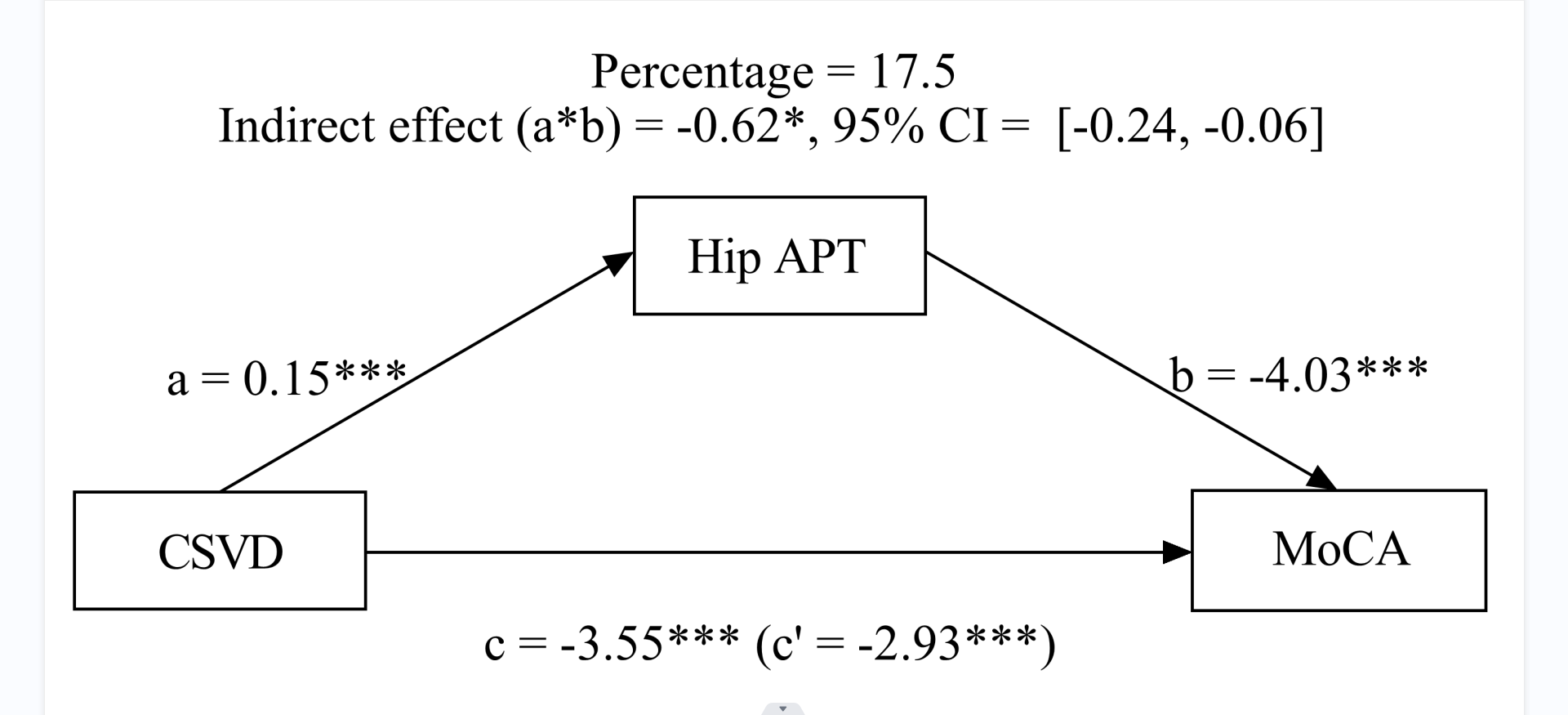
Results: The hippocampal APT values among different CSVD total load groups were significantly different (p<0.001). The mediation models demonstrated that the APT values of the hippocampus partially mediated the association between CSVD total load and MoCA score.
Impact: Hippocampal APT values may serve as a biomarker for the early detection of neurodegeneration in CSVD patients.
Introduction
Cerebral small vessel disease (CSVD) has been considered to be a disorder that primarily affects cerebral microvessels, making it the most prevalent form of vascular cognitive impairment and a significant contributor to mixed dementia [1,2]. Exploring the CSVD imaging markers can provide insights into the pathophysiology of CSVD, which is crucial for understanding its etiology, development, and clinical interventions. Amide proton transfer (APT) imaging is an MRI technique that reflect the protein and polypeptide content in tissues [3]. In this study, we aimed to explore potential differences in hippocampal APT values between CSVD imaging markers presence and absence groups and between groups with different CSVD total burden. The correlations between APT values and CSVD imaging markers were analyzed. The mediating effect of the hippocampal APT values in the association between CSVD total loads and Montreal Cognitive Assessment (MoCA) score was also assessed.Methods
A total of 674 stroke-free subjects were recruited from the local community. Forty-eight age- and education matched healthy subjects were selected as normal control (Figure 1). All the participants conducted the Beijing version of the Montreal Cognitive Assessment (MoCA) for the cognitive assessment. And the MRI scans were performed using a 3.0-T MR system (Ingenia CX; Philips Healthcare, Best, The Netherlands) with 32-channel head coils. A radiologist, following the definitions established in previous studies [4], identified the lacunar infarction (LI), white matter hyperintensity (WMH), enlarged perivascular space (EPVS), and cerebral microbleed (CMB). The CSVD total burden was assessed using an ordinal scale from 1 to 4. The differences of hippocampal APT values between CSVD imaging makers presence or absence groups and different CSVD total burden groups were compared using an independent t-test and one-way analysis of variance (ANOVA), and the Bonferroni correction was used for post-hoc tests. Pearson correlation analysis was used to study correlations between APT values, CSVD imaging markers, and brain volume. A mediation analysis model was used to investigate the mediating effect of the mean hippocampus APT value in the association between CSVD total loads and MoCA score.Results
The hippocampal APT values among different CSVD total load groups were significantly different (p<0.001) (Table 1, Figure 2). The hippocampal APT values were significantly different between the imaging markers presence and absence groups. The p-values for the LI, WMH, EPVS and CMB presence or absence groups were <0.001, <0.001, 0.034, and 0.002, respectively(Table 2, Figure 3). The hippocampal APT values were significantly correlated with CMB(r = 0.290, P < 0.001), LI(r = 0.346, P < 0.001) and WMH (r = 0.375, P < 0.001). The mediation models demonstrated that the APT values of the hippocampus partially mediated the association between CSVD total load and MoCA score, the proportion of mediation attributable was calculated as 17.50% (Table 3, Figure 4).Discussion
The first result of this study showed a higher hippocampal APT value in the imaging markers presence groups compared to the imaging markers absence groups. These findings suggest a potential association between the presence of CSVD imaging markers and neurodegeneration. The relationship between CSVD imaging markers and neurodegeneration is likely a bidirectional pathophysiological process. Firstly, neurodegeneration may accelerate the development of certain CSVD imaging markers. For example, elevated b-secretase levels, as a key enzyme in amyloidogenesis of amyloid precursor proteins, can result in white matter contraction and damage, thereby disrupting the connection of projection fibers in this area [5,6]. Tau protein, playing a crucial role in AD pathology, ultimately disrupts neuronal microstructure through the formation of neurofibrillary tangles, leading to gray and white matter damage and secondary brain atrophy [7,8]. Secondly, we observed a gradual increase in hippocampal APT values with an increasing total CSVD load. A higher CSVD total load may further accelerate the occurrence of secondary neurodegenerative diseases [9]. An increased CSVD total load induces secondary gray and white matter loss in affected brain regions, resulting in brain atrophy and neurodegeneration. An additional finding of this study is that the mediation models revealed the partial mediating effect of the hippocampal APT value on the relationship between the CSVD total load and the MoCA score. It is worth noting that mixed neurodegeneration and CSVD pathology are frequently observed in the same group of patients, and this pattern becomes more common with increasing age [10].Conclusion
Hippocampal APT values were associated with cerebral small vessel disease imaging markers and total burden. Hippocampal APT values may serve as a biomarker for the early detection of neurodegeneration in CSVD patients.Acknowledgements
The authors express their gratitude to the dedicated staff of The Neurology Subject Group at Nanxishan Hospital of Guangxi Zhuang Autonomous Region.References
[1] Wardlaw JM, William M. Feinberg Award for Excellence in Clinical Stroke: Small Vessel Disease; a Big Problem, But Fixable. Stroke. 2018;49:1770-1775.
[2] Pantoni L. Cerebral small vessel disease: from pathogenesis and clinical characteristics to therapeutic challenges. Lancet Neurol. 2010;9:689–701.
[3] Zhou J, Heo HY, Knutsson L, van Zijl PCM, Jiang S. APT-weighted MRI: Techniques, current neuro applications, and challenging issues. J Magn Reson Imaging. 2019 Aug;50(2):347-364.
[4] Wardlaw JM, Smith EE, Biessels GJ, et al. Neuroimaging standards for research into small vessel disease and its contribution to ageing and neurodegeneration. lancet Neurol. 2013;12:822-838.
[5] Fukumoto H, Cheung BS, Hyman BT, Irizarry MC. Beta-secretase protein and activity are increased in the neocortex in Alzheimer disease. Arch Neurol. 2002;59(9):1381-1389.
[6] Chalmers K, Wilcock G, Love S. Contributors to white matter damage in the frontal lobe in Alzheimer's disease. Neuropathol Appl Neurobiol. 2005;31(6):623-631.
[7] Sanford AM. Mild Cognitive Impairment. Clin Geriatr Med. 2017 Aug;33(3):325-337.
[8] Cummings JL, Cole G. Alzheimer disease. JAMA. 2002;287(18):2335-2338.
[9] Wardlaw JM, Smith C, Dichgans M. Small vessel disease: mechanisms and clinical implications. Lancet Neurol. 2019 Jul;18(7):684-696.
[10] Attems J, Jellinger KA. The overlap between vascular disease and Alzheimer’s disease—Lessons from pathology. BMC Med. 2014;12(1):1–12.
Figures
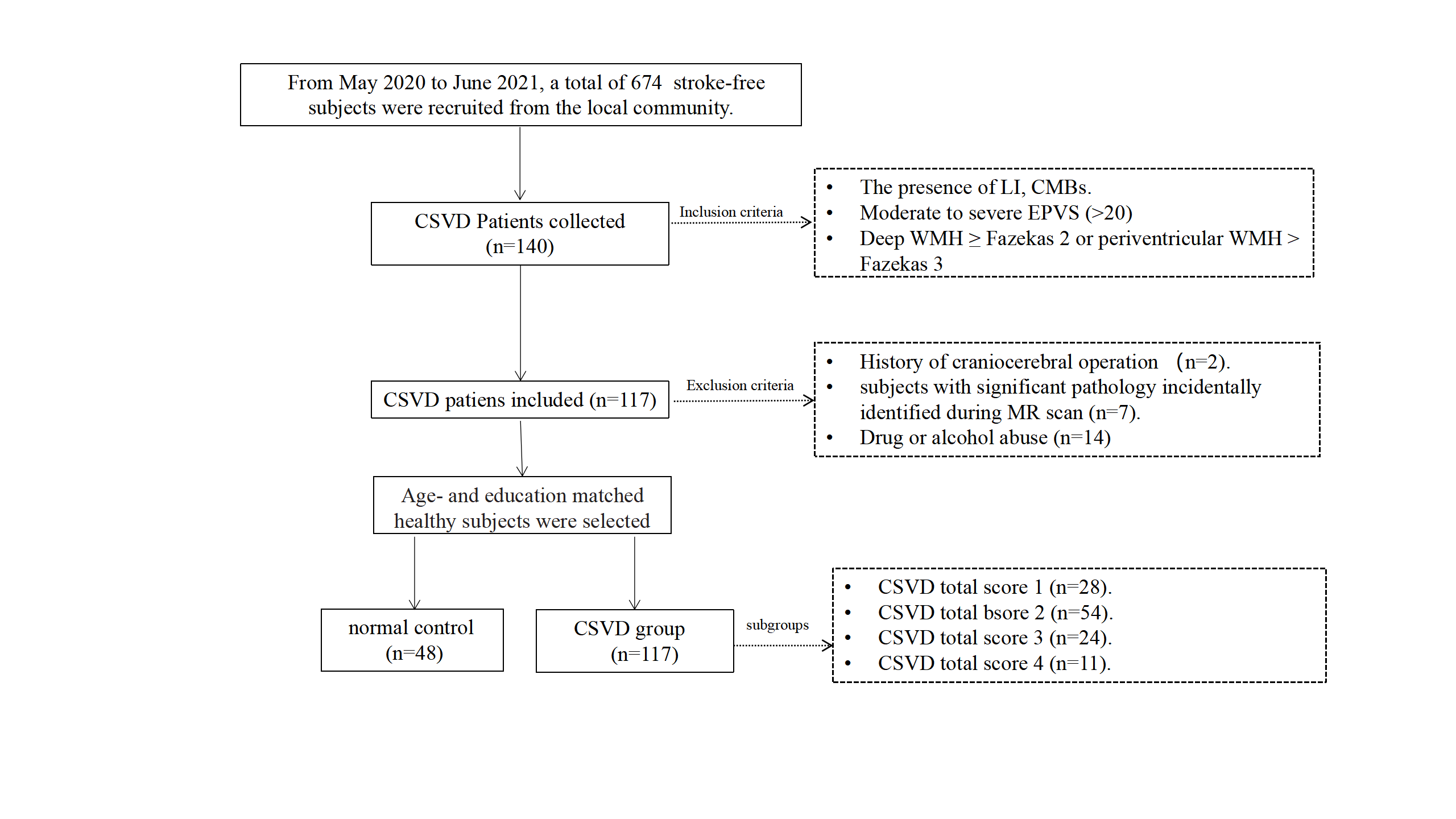
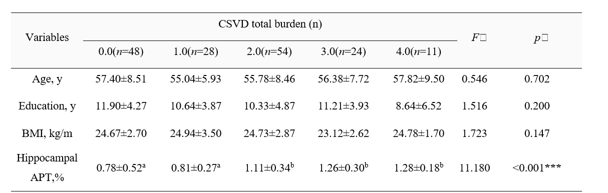
Table 1. The deference of demographic characteristics, hippocampus APT value and brain volume between the groups of CSVD total burden.
y, year; CSVD, cerebral small vessel disease; APT, amide proton transfer; a&b, no difference between the same letters; ***, P<0.001.
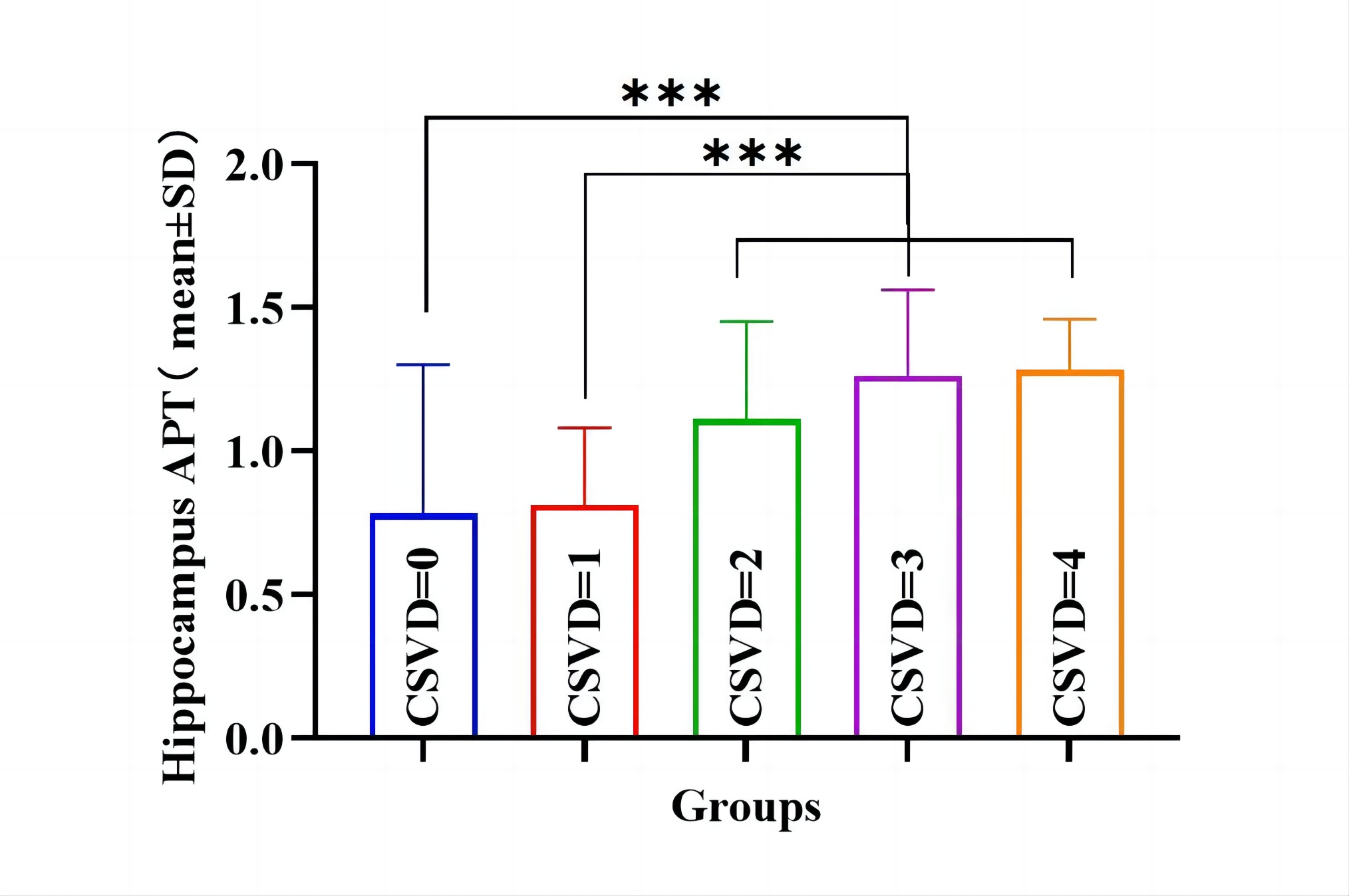
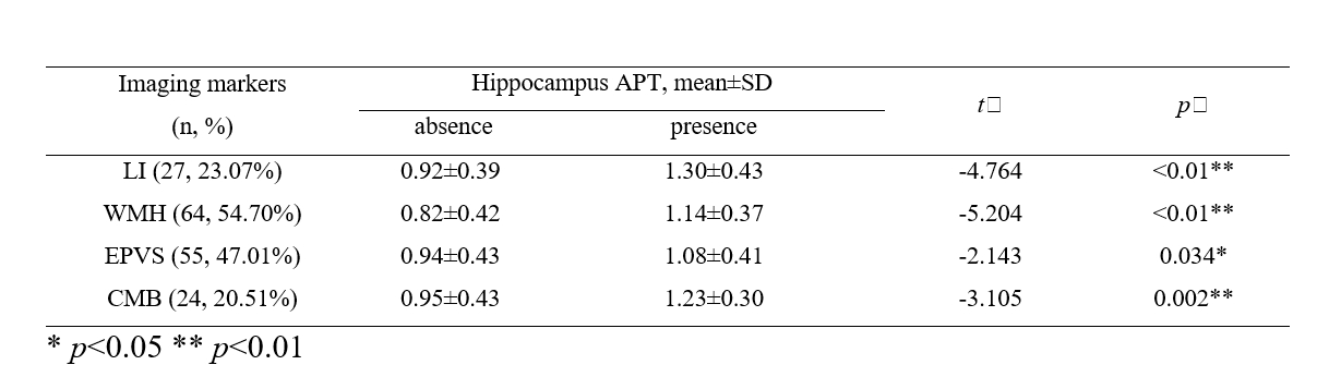
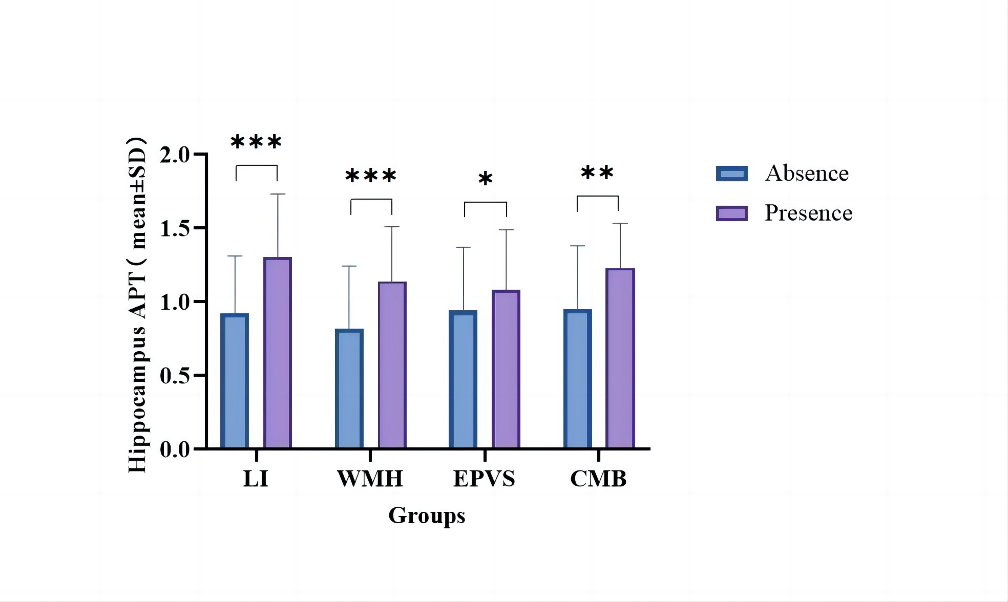
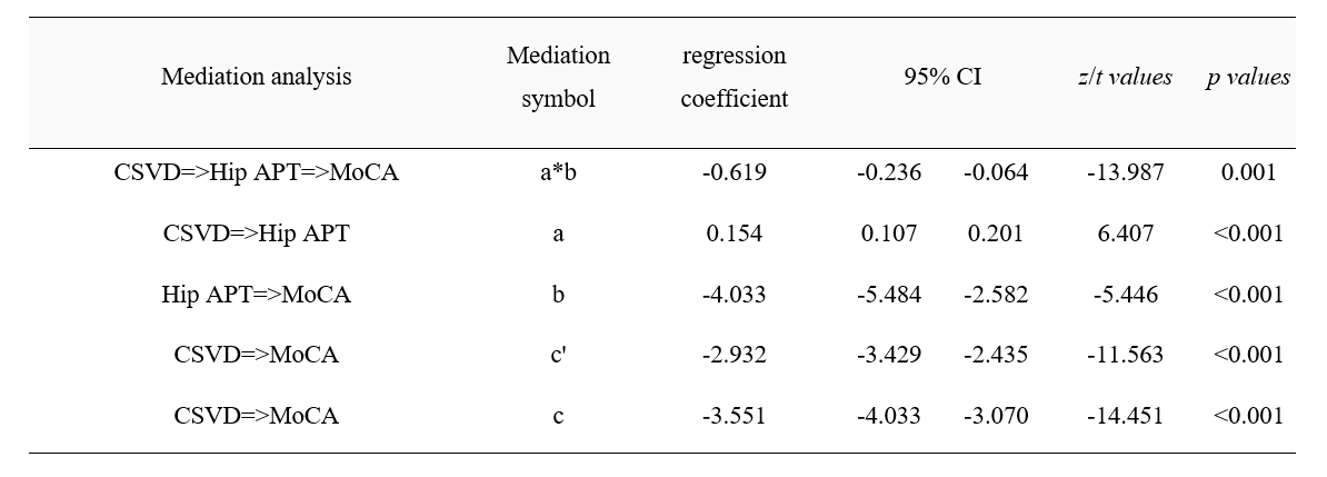
Table 3. Mediation effect of hippocampal APT on cognitive impairment caused by CSVD total load.
CSVD, cerebral small vessel disease; APT, amide proton transfer; MoCA, Montreal Cognitive Scale; CI, confidence interval. Hip, hippocampus.