1342
Changes in vessel density in genetic cerebral small vessel disease (CADASIL) revealed by high-resolution black-blood MRI: A pilot study1Department of Biomedical Engineering, University of Southern California, Los Angeles, CA, United States, 2Department of Radiology, Beijing Chaoyang Hospital, Beijing, China, 3Department of Neurology, National Center for Neurological Disorders, Shanghai, China, 4Department of Neurology, University of Southern California, Los Angeles, CA, United States
Synopsis
Keywords: Blood Vessels, Dementia, Black Blood, CADASIL, small vessel disease
Motivation: Cerebral small vessel disease (cSVD) is a leading cause of vascular dementia in the elderly worldwide. No existing MRI methods can directly visualize cerebral small vessels.
Goal(s): Evaluation of a novel pipeline for mapping small blood vessels using high-resolution black-blood MRI in genetic cSVD (CADASIL) patients.
Approach: Small blood vessels were segmented and quantified in 10 CADASIL patients and 10 matched healthy controls.
Results: Significantly lower vessel density has been found in the hippocampus of the patients, whereas the vessel density is significantly higher in cortical white matter of patients compared to the control.
Impact: Visualization and quantification methods of small cerebral blood vessels from high-resolution black blood MRI which would facilitate the study of cSVD mechanisms.
Introduction
Cerebral Autosomal Dominant Arteriopathy with Subcortical Infarcts and Leukoencephalopthy (CADASIL) is the most prevalent form of genetic cerebral small vessel disease (cSVD). The prevalence of CSVD is estimated to be 8% to 30% (Toledo et al., 2013), and it is considered a major cause of cognitive impairment and dementia in the elderly. Despite various reports on the associations of cSVD with changes in cognitive abilities (Li et al., 2018) and a significantly increased risk of stroke incidents, the underlying mechanisms are largely vailed. Previous study has demonstrated the feasibility of high resolution black-blood MRI using T1-weighted turbo spin-echo with variable flip-angles sequence (T1w TSE-VFA) to delineate finer blood vessels on healthy subjects (Ma et al., 2019) at 3T. Here we pilot a novel 3D analysis pipeline (Sarabi et al., 2023) based on black-blood MRI to characterize small blood vessels on 10 CADASIL patients compared with 10 matched controls.Methods
Ten genetically confirmed CADASIL patients (46±14.9yrs, 5 males) and 10 healthy controls (44.7±10.7 yrs, 4 males) free of general neurological and psychiatric disorders underwent MRI on a Siemens 3T Prisma scanner with a 64-channel head coil. The imaging protocol included 3D MPRAGE and black-blood MRI. The “black blood” images were acquired using a T1w TSE-VFA sequence (Ma et al., 2019) with the following parameters: TR/TE=900/15ms, turbo factor=52, matrix size =384×288, FOV=196x147mm2, iso-0.5mm spatial resolution, 240 sagittal slices with 7% oversampling, GRAPPA factor=2, and a total acquisition time of 7:23min. All data processing steps were implemented using custom MATLAB2023b scripts. The MPRAGE and black-blood images were skull-stripped and bias-corrected using Statistical Parametric Mapping (SPM) toolbox. Block-wise non-local means (NLM) filters were applied to denoise the black-blood image (Coupe et al., 2008) with a window size of 3×3×3 in a block of size 32×32×32. The Jerman vesselness filter (Jerman et al., 2015) with a voxel size of iso-0.5mm consisting of vessel size scales of [0.10:0.05:0.60] was used for vesselness segmentation. Responses were normalized from 0.0 to 1.0 with a threshold 0.048 based on ROC analysis. Superficial veins, dural sinuses, and the middle cerebral artery (MCA) were removed using the ASEG map from the Freesurfer software to result in the final segmentation of small vessels (Sarabi et al., 2023). The segmented vessels were visually checked and vessel density (defined as vessel volume divided by ROI volume) was calculated in the whole brain, cortical white matter, cerebral gray matter, and hippocampus given their relevance with cSVD. Group difference was calculated using two-sample t-test in each of the four regions, a p value <0.05 is considered significant.Results
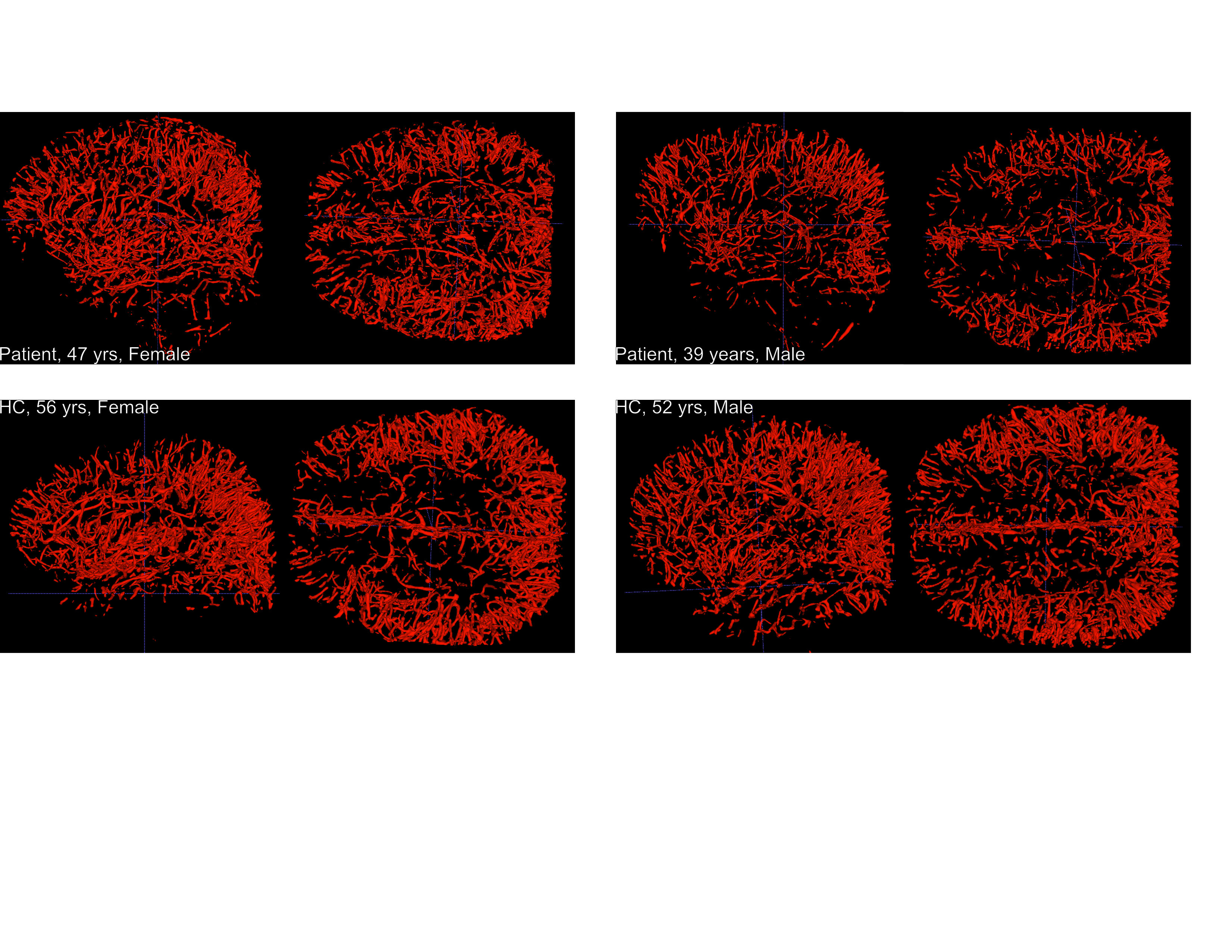
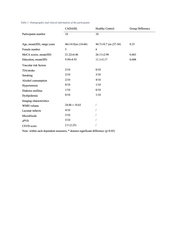
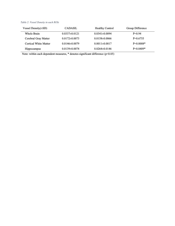
Table 1 shows the demographic and clinical information of the subjects. No significant difference was found between two groups in age, sex or years of education. MoCA scores showed a trend of reduction (p=0.063) in CADASIL patients. Figure 1 shows 3D rendering of segmented small cerebral blood vessels in 2 representative CADASIL patients and 2 healthy controls of similar demographics. In general, similar distribution of small vessels can be seen in both patients (upper) and controls (lower). Paired sample t-test revealed significantly higher vessel density in the hippocampus (p=0.0409) in control subjects compared to CADASIL patients. In the cortical white matter, however, significantly lower vessel density (p=0.0008) was identified in healthy controls (Table 2). Vessel density of the cerebral gray matter or the entire brain do not show significant difference.Discussion
We presented a pilot study of the application of high-resolution black-blood MRI in the study of genetic cSVD. Significantly lower vessel density in the hippocampus was identified in the CADASIL patients compared to the controls. The hippocampus is an area involved in learning, memory, and emotion, and it is implicated in various neurocognitive diseases including genetic cSVD (Yamamoto et al., 2021). Increased vessel density in white matter of CADASIL patients may indicate vascular dilation which requires further investigation. The promising results suggest that the high-resolution T1w TSE-VFA sequence, alongside with our analysis pipeline, may be able to characterize small vascular changes associated with cSVD, and be beneficial in the screening and management of genetic and sporadic cSVD. Further study with more diverse patient and control groups may be necessary in fully demonstrate the potential of current techniques used.Conclusion
We provide the first demonstration of changes in small blood vessel density in genetic cSVD (CADASIL) using a novel 3D analysis pipeline and high-resolution black-blood MRI with near whole-brain coverage at 3T. This technique may be applied to both genetic and sporadic cSVD.Acknowledgements
No acknowledgement found.References
Coupe, P., Yger, P., Prima, S., Hellier, P., Kervrann, C., & Barillot, C. (2008). An Optimized Blockwise Nonlocal Means Denoising Filter for 3-D Magnetic Resonance Images. IEEE Transactions on Medical Imaging, 27(4), 425–441. https://doi.org/10.1109/TMI.2007.906087
Jerman, T., Pernuš, F., Likar, B., & Špiclin, Ž. (2015, March). Beyond Frangi: an improved multiscale vesselness filter. In Medical imaging 2015: Image processing (Vol. 9413, pp. 623-633). SPIE.
Li, Q., Yang, Y., Reis, C., Tao, T., Li, W., Li, X., & Zhang, J. H. (2018). Cerebral Small Vessel Disease. Cell Transplantation, 27(12), 1711–1722. https://doi.org/10.1177/0963689718795148
Ma, S. J., Sarabi, M. S., Yan, L., Shao, X., Chen, Y., Yang, Q., Jann, K., Toga, A. W., Shi, Y., & Wang, D. J. J. (2019). Characterization of lenticulostriate arteries with high resolution black-blood T1-weighted turbo spin echo with variable flip angles at 3 and 7 Tesla. NeuroImage, 199, 184–193. https://doi.org/10.1016/j.neuroimage.2019.05.065
Sarabi, M. S., Ma, S. J., Jann, K., Ringman, J. M., Wang, D. J. J., & Shi, Y. (2023). Vessel Density Mapping of Cerebral Small Vessels on 3D High Resolution Black Blood MRI [Preprint]. Neuroscience. https://doi.org/10.1101/2023.03.18.533300
Toledo, J. B., Arnold, S. E., Raible, K., Brettschneider, J., Xie, S. X., Grossman, M., Monsell, S. E., Kukull, W. A., & Trojanowski, J. Q. (2013). Contribution of cerebrovascular disease in autopsy confirmed neurodegenerative disease cases in the National Alzheimer’s Coordinating Centre. Brain, 136(9), 2697–2706. https://doi.org/10.1093/brain/awt188
Yamamoto, Y., Hase, Y., Ihara, M., Khundakar, A., Roeber, Sigrun., Duering, M., & Kalaria, R. N. (2021). Neuronal densities and vascular pathology in the hippocampal formation in CADASIL. Neurobiology of Aging, 97, 33–40. https://doi.org/10.1016/j.neurobiolaging.2020.09.016
Figures