1340
Reproducibility of intracranial vascular pulsatility on 3D cine black-blood MRI1Department of Bioengineering, University of Washington, Seattle, WA, United States, 2Department of Radiology, University of Washington, Seattle, WA, United States, 3Department of Radiology and Imaging Sciences, University of Utah, Salt Lake City, UT, United States, 4Electrical and Computer Engineering, University of Washington, Seattle, WA, United States, 5Department of Surgery, University of Washington, Seattle, WA, United States
Synopsis
Keywords: Vessel Wall, Vessels, Cine Vessel Wall Imaging
Motivation: This research investigates the lesser-explored domain of lumen area changes in intracranial vascular pulsatility, providing insight into cerebral vasculature's mechanical behavior.
Goal(s): To employ cine black-blood MRI for delineating and quantifying intracranial artery pulsatility by monitoring cardiac cycle-induced lumen variations.
Approach: We utilized a sophisticated vessel analysis system to discern pulsatility within the lumen and adjacent tissues, validating our findings with reproducibility scans.
Results: Cine black-blood imaging successfully visualized intracranial vascular pulsatility. The method displayed robust reproducibility in detecting lumen area changes, proving comparable to velocity pulsatility measurements.
Impact: This study illuminates the often-overlooked aspect of lumen area pulsatility, with implications for a holistic assessment of vascular health and disease.
Introduction
Understanding the pulsatility of intracranial vessels is vital for diagnosing and managing diseases like aneurysms and dementia1,2. While the significance of velocity pulsatility has been well-documented3, the pulsatility related to vessel wall compliance and diameter changes remains understudied. Recently 3D cine black-blood MRI has been suggested as a means to measure vascular pulsatility and compliance4 but reproducibilty of this method has not been assessed. This study evaluates the reproducibility of measurements using a 3D isotropic black-blood cine imaging technique, and a new post-processing technique for this purpose.Method
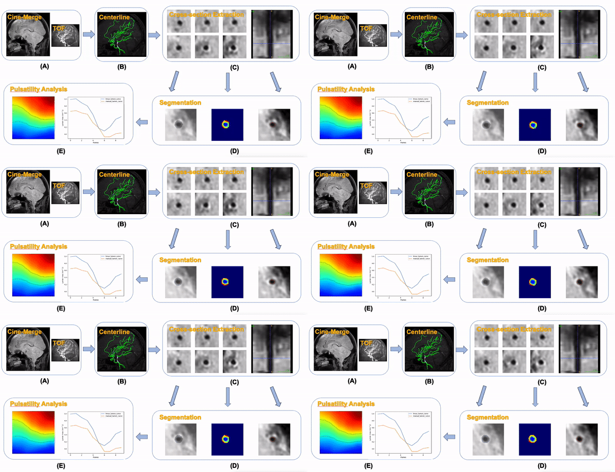
MRI protocol: We conducted two reproducibility scans utilizing a Philips Ingenia 3T whole-body scanner with a 32-channel head coil. Pulse sequence design details of the Cine-Merge sequence are available in reference4. Peripheral pulse gating (PPG) was employed for cardiac triggering, supplemented by ECG monitoring. Each heartbeat captured ten segments, correlating to ten distinct cardiac phases. Imaging resolution was set at 1x1x1mm, interpolated to 0.5x0.5x0.5mm, covering an AP/FH/RL field of view of 20x20x16cm, utilizing a GRE TR/TE of 10/4ms and a flip angle of 6 degrees, scan time 4.5 minutes. Additionally, Time-of-Flight (ToF) imaging was performed with a 0.25mm in-plane resolution and a 0.5mm slice thickness. The cine-Merge scan was repeated in the same scan session after a time interval of 5 minutes after the first cine-Merge scan. Reproducibility was assessed by vessel measurements on the two cine-Merge scans.Post-processing: The whole pipeline was shown in Figure 1. Initially, ToF images were registered with Cine Merge. Vessel centerlines of Circle-of-Willis arteries were traced on ToF using iCafe software5. The centerlines were then transferred onto Cine Merge. Utilizing a previously described method for reformatting images along the vessel centerline6, cross-sectional images of the basilar artery (BA), posterior cerebral artery (P1), middle cerebral artery (M1), and vertebral artery (VA) were derived from the centerlines. Lumen and wall were segmented using a transformer-based deep learning model7. In order to visualize the lumen and wall changes with vessel pulsation, a new post-processing method that takes into account sub-pixel signal changes with time was implemented. Lumen area quantification involved pixel counting within the identified lumen. These pixels, along with those delineating the wall area, were color-coded, restructured into a singular column, and concatenated with pixels across all cardiac phases. Contours were derived based on threshold values of lumen and wall boundaries. The pulsatility index for the lumen area was calculated using the formula (max-min)/mean across the cardiac cycle.
Result
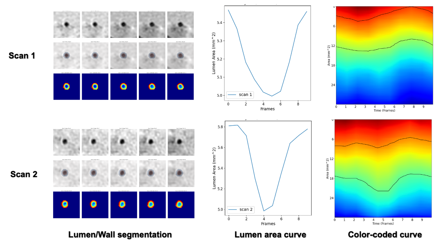
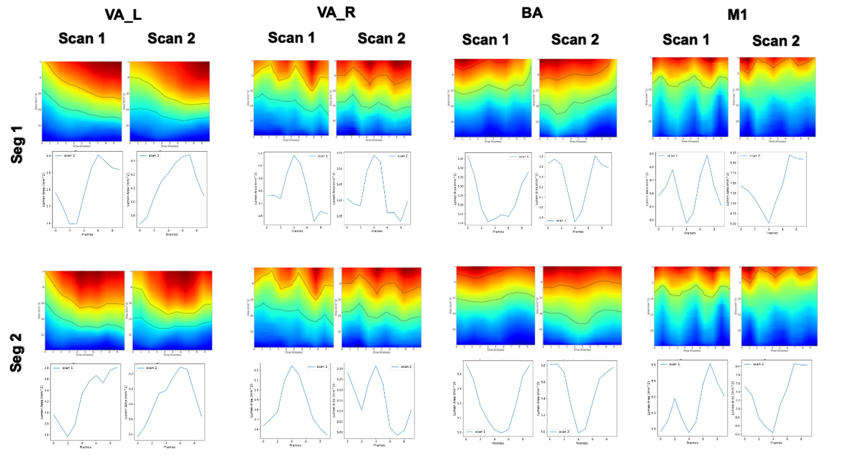
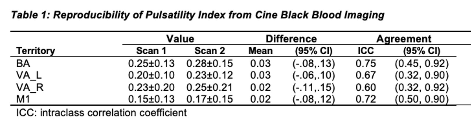
We achieved consistent and reproducible lumen area change curves, exemplified in Figure 2, focusing on the proximal segment of the basilar artery near the P1 segment, an area less influenced by artifacts. Figure 3 delineates the variability in lumen change patterns across different arterial segments, underscoring the nuanced nature of vascular pulsatility. Each artery was divided into two segments and the pattern agreed with each other. Table 1 shows the reproducibility result of pulsatility index values of different territories.Conclusion
The cine black-blood imaging sequence has proven effective in capturing the pulsatility of intracranial vessels, with reproducible results that align closely with velocity pulsatility metrics. This study reinforces the potential of lumen area pulsatility as a significant factor in cerebral vascular assessment, suggesting its integration with velocity measurements for a comprehensive evaluation. Our findings pave the way for a deeper understanding of vascular dynamics, promising advancements in the diagnosis and treatment of cerebrovascular diseases.Acknowledgements
No acknowledgement found.References
- Gottwald LM, Töger J, Bloch KM, Peper ES, Coolen BF, Strijkers GJ, Van Ooij P, Nederveen AJ. High spatiotemporal resolution 4D flow MRI of intracranial aneurysms at 7T in 10 minutes. American Journal of Neuroradiology. 2020 Jul 1;41(7):1201-8.
- Pahlavian SH, Wang X, Ma S, Zheng H, Casey M, D’Orazio LM, Shao X, Ringman JM, Chui H, Wang DJ, Yan L. Cerebroarterial pulsatility and resistivity indices are associated with cognitive impairment and white matter hyperintensity in elderly subjects: a phase-contrast MRI study. Journal of Cerebral Blood Flow & Metabolism. 2021 Mar;41(3):670-83.
- Wåhlin A, Ambarki K, Birgander R, Wieben O, Johnson KM, Malm J, Eklund A. Measuring pulsatile flow in cerebral arteries using 4D phase-contrast MR imaging. American Journal of Neuroradiology. 2013 Sep 1;34(9):1740-5.
- Balu N, Zhang K, Hatsakami T, Yuan C. 3D Isotropic black-blood cine MRI of intracranial arteries. ISMRM 2023
- Chen L, Mossa‐Basha M, Balu N, Canton G, Sun J, Pimentel K, Hatsukami TS, Hwang JN, Yuan C. Development of a quantitative intracranial vascular features extraction tool on 3 D MRA using semiautomated open‐curve active contour vessel tracing. Magnetic resonance in medicine. 2018 Jun;79(6):3229-38.
- Guo Y, Canton G, Chen L, Sun J, Geleri DB, Balu N, Xu D, Mossa‐Basha M, Hatsukami TS, Yuan C. Multi‐Planar, Multi‐Contrast and Multi‐Time Point Analysis Tool (MOCHA) for Intracranial Vessel Wall Characterization. Journal of Magnetic Resonance Imaging. 2022 Sep;56(3):944-55.
- Yu Q, Wang H, Qiao S, Collins M, Zhu Y, Adam H, Yuille A, Chen LC. k-means Mask Transformer. InEuropean Conference on Computer Vision 2022 Oct 22 (pp. 288-307). Cham: Springer Nature Switzerland.
Figures