1336
Simultaneous assessment of arterial pulsatility at different segments of cerebral arteries using multiband dual-VENC Phase-Contrast MRI1Department of Radiology, Northwestern University, Chicago, IL, United States, 2Department of Biomedical Engineering, Northwestern University, Chicago, IL, United States, 3Siemens Medical Solutions USA, Chicago, IL, United States
Synopsis
Keywords: Blood Vessels, Blood vessels
Motivation: Arterial pulsatility is used for assessing cerebral vascular dysfunction. However, 2D PC-MRI and 4D flow have limitations including multiple separate scans, prolonged scan time, and flow saturation effects.
Goal(s): Our study aims to develop a Multi-band Dual-Venc PC-MRI (MB-DV) senquence to measure pulsatility for larger and distal small vessels within short scan time.
Approach: MB-DV scans were performed on 8 subjects to image velocity waveforms from ICA, M2, M3, LSA, and M4 simultaneously. For comparison, multiple standard single-slice PC-MRI were performed.
Results: MB-DV PC-MRI successfully measured flow waveforms and provided reliable PI measurements with no significant difference from PI measured by standard PC-MRI.
Impact: MD-DV PC-MRI enables simultaneously assessing arterial pulsatility at multiple vessel segments in the brain within a short scan time, which could be a potentially useful imaging tool to study pulsatility in the cerebral vascular tree for cerebrovascular and neurodegenerative diseases.
Introduction
Phase-contrast MRI (PC-MRI) has become a useful imaging tool for assessing arterial pulsatility in the brain, including 2D PC-MRI and 4D flow1,2. 2D PC-MRI typically measures blood flow velocities in a single slice that is perpendicular to the target vessel. However, multiple acquisitions are generally needed to measure different segments of cerebral arteries, such as the internal carotid artery (ICA), middle cerebral arteries (MCA), posterior cerebral artery (PCA), and anterior cerebral artery (ACA). As an alternative, 4D flow enables accessing cerebral arterial pulsatility in all major branches of brain vessels simultaneously given its volumetric acquisition. However, 4D flow suffers from long acquisition time, inadequate temporal resolution due to the three flow encoding directions, and sophisticated postprocessing procedures. Furthermore, it’s challenging to measure deep perforating arteries, such as lenticulostriate arteries (LSAs), using 4D flow due to flow saturation effects. To overcome the limitations of both conventional 2D PC-MRI and 4D flow, in this study we propose and evaluate a multi-band dual-Venc PC-MRI (MB-DV PC-MRI) sequence to measure arterial pulsatility at multiple cerebral arterial segments simultaneously within a short scan time while preserving good hemodynamic delineation of both larger and deep distal small vessels.Methods
MB-DV PC-MRI Sequence Design, Acquisition, and Image reconstructionMultiband SINC RF pulses were used for simultaneous excitation in this study, represented as $$$RF_{MB}(t) = sinc(t)\times\sum_Ne^{i\Delta\omega_nt+\varphi_n}$$$ where N represents the number of slices simultaneously excited or multiband factor, Δω is the frequency off-resonant of each slice, φ is the phase. Different from conventional multi-band RF implementation, Δω varied across slices depending on slice position. Figure 1 illustrates the positions of 4 simultaneously excited slices by MB-DV PC-MRI using TOF as a reference to cover ICA(slice 1), M2 and LSA(slice 2), M2/3(slice 3), and M4(slice 4) with average gaps of 20+/-2.4, 11.5+/-2.4, and 67.3+/-9.1mm between two adjacent slices. The CAPIRINA was applied by adding a phase to the multiband RF pulses to shift individual slices by FOV/N3. A reference scan of each slice was acquired using a standard PC-MRI sequence without cardiac-gating was incorporated into the MB-DV PC-MRI sequence in the end and used for the MB-DV PC-MRI image reconstruction with SENSE4.
MRI experiments
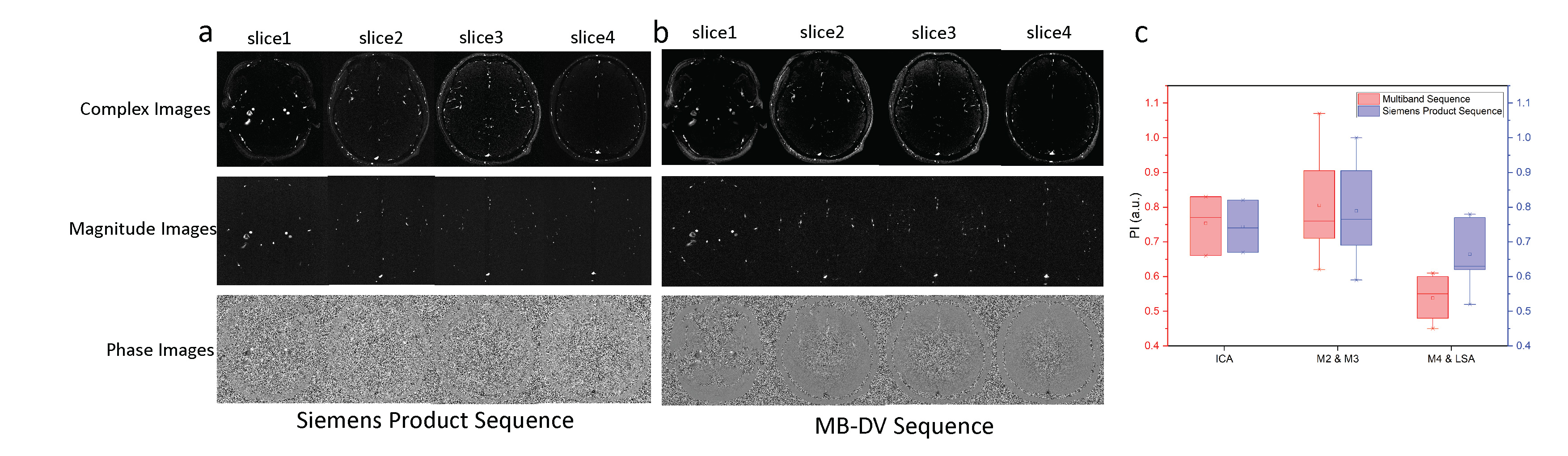
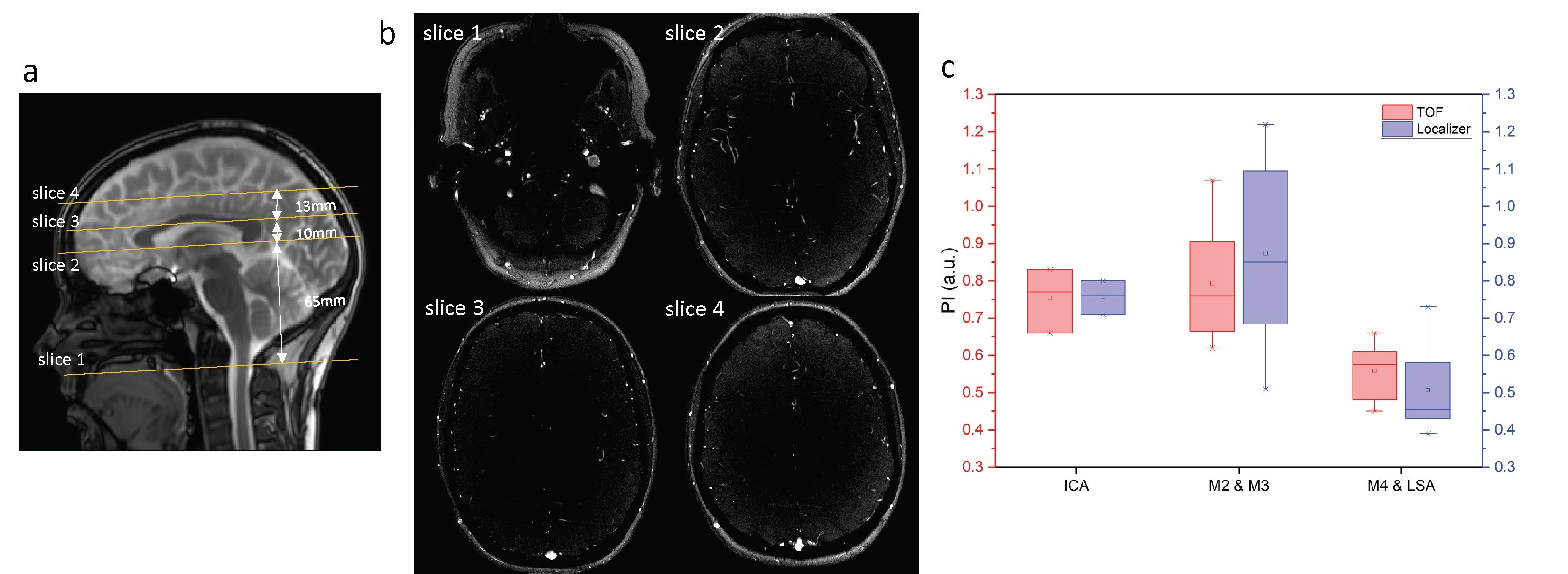
ECG retrospectively-gated MB-DV PC-MRI sequence was performed on 8 participants (age 31.4 +/ 11.2 years, 7 female) on a SIEMENS Prisma 3T MRI scanner using a 64ch head-neck coil with the following parameters: TR/TE = 88.26/9.54ms, flip angle = 60o, FOV = 200 x 200 mm2, spatial resolution= 480mm, voxel size = 0.4 x 0.4 x 2.2 mm3, dual VENC = 40 and 80 cm/s, bandwidth = 248 Hz/pixel, 20 cardiac phases, multiband factor = 4, scan time = 3-4min depending on heart rate. A time-of-flight scan was performed before PC-MRI scans as a reference for MB-DV PC-MRI slice planning. To evaluate the performance of MB DV PC-MRI, a standard 2D dual-VENC PC-MRI sequence with the same imaging coverage and closely matched parameters was performed on 3 participants, and repeated scans were performed on two participants. The feasibility of positioning MB-DV PC-MRI slices based on anatomical landmarks (e.g. localizer) was also evaluated through a comparison with the TOF method on 3 participants.
Results
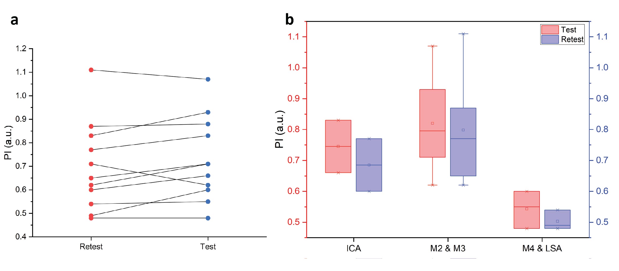
MB-DV PC-MRI scans were successfully performed on all participants. Figure 1 shows an example of the inline composited MB-DV PC-MRI image and reconstructed images of individual slices. One can appreciate a good depiction of blood vessels at different vessel segments. Blood velocity waveforms extracted from MB-DV PC-MRI images are shown in Figure 2 from a representative case. The average velocities from ICA to downstream MCA segments gradually decrease. Figure 3 shows preliminary test-retest results, indicating MB-DV PC-MRI provides overall reliable PI measurements at different vessel segments. The comparison between the MB-DV PC-MRI and standard PC-MRI sequences is shown in Figure 4. No significant difference in the PI measurements was found between MB-DV PC-MRI and standard PC-MRI (p = 0.56). Figure 5 illustrates MB-DV PC-MRI slice positioning on anatomical images. The PI measurements using the two positioning strategies did not reveal any significant differences (p = 0.93).Discussion and Conclusion
This proof-of-concept study demonstrates the feasibility of using multi-band dual-VENC PC-MRI to simultaneously assess arterial pulsatility at multiple vessel segments in the brain within a short scan time. MB-DV PC-MRI could be a potentially useful imaging tool to study arterial pulsatility across different segments in the cerebral vascular tree in cerebrovascular and neurodegenerative diseases. Further work will be carried out to further optimize the imaging protocol of MB-DV PC-MRI and evaluate arterial pulsatility measurements in all branches of the circle of Willis.Acknowledgements
This work was partly supported by National Institute of Health (NIH) grants R01NS118019, RF1AG072490, and BrightFocus Foundation A20201411S.References
1. Srichai, Monvadi B., et al. "Cardiovascular applications of phase-contrast MRI." American Journal of Roentgenology 192.3 (2009): 662-675.
2. Vikner, Tomas, et al. "Characterizing pulsatility in distal cerebral arteries using 4D flow MRI." Journal of Cerebral Blood Flow & Metabolism 40.12 (2020): 2429-2440.
3. Breuer, Felix A., et al. "Controlled aliasing in parallel imaging results in higher acceleration (CAIPIRINHA) for multi‐slice imaging." Magnetic Resonance in Medicine: An Official Journal of the International Society for Magnetic Resonance in Medicine 53.3 (2005): 684-691.
4. MLAPruessmann, Klaas P., et al. "SENSE: sensitivity encoding for fast MRI." Magnetic Resonance in Medicine: An Official Journal of the International Society for Magnetic Resonance in Medicine 42.5 (1999): 952-962.
Figures
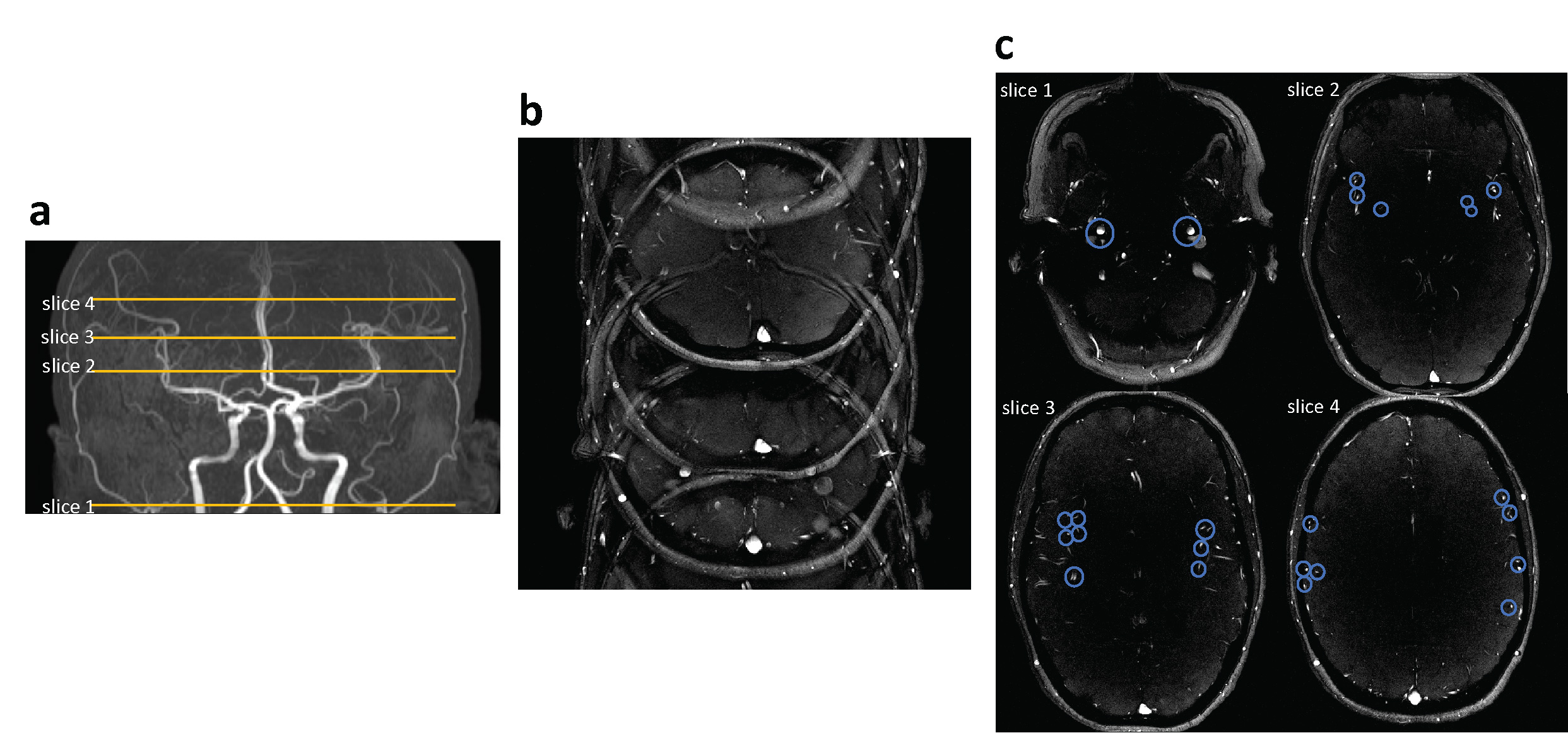
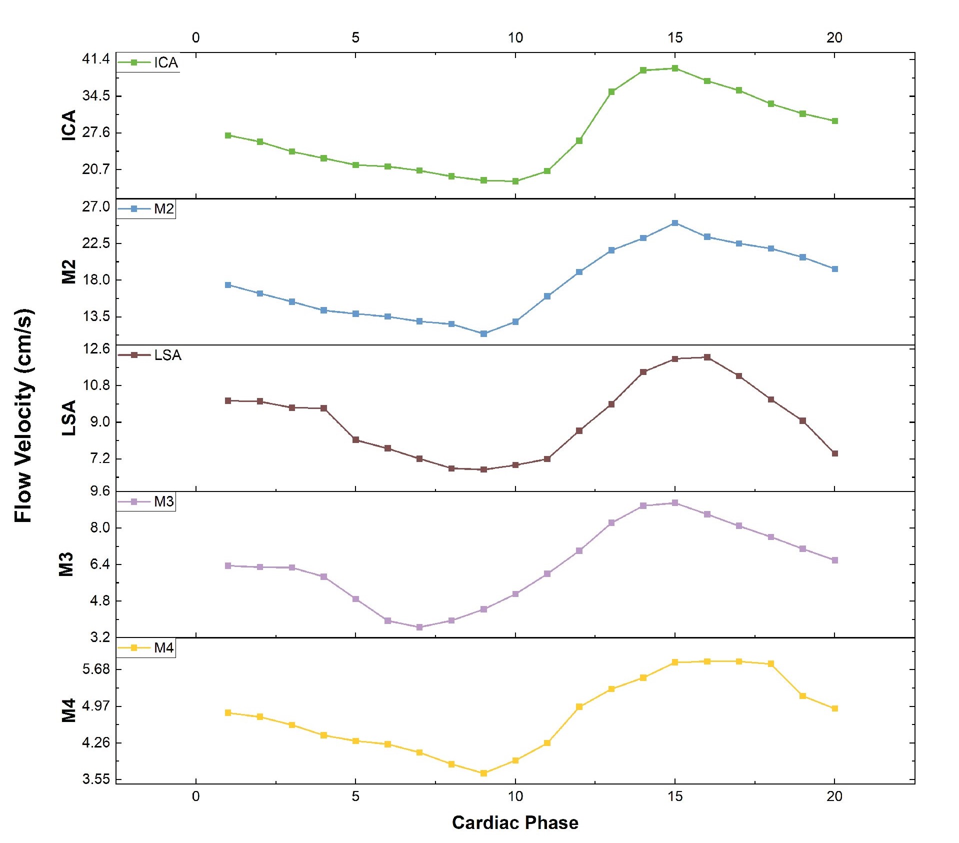
Figure 2. Mean flow velocity waveforms of ICA, M2, LSA, M3, and M4 from a representative participant.