1334
Magnetic Particle Imaging goes clinical routine? – first step to human-sized MPI-guided intervention in realtime1Experimental Physics 5, University of Würzburg, Würzburg, Germany, 2Pure Devices GmbH, Rimpar, Germany, 3Diagnostic and Interventional Neuroradiology, University Hospital Würzburg, Würzburg, Germany, 4Diagnostic and Interventional Radiology, University Hospital Würzburg, Würzburg, Germany, 5Radiologie Augsburg Friedberg, Augsburg, Germany
Synopsis
Keywords: Hybrid & Novel Systems Technology, Hybrid & Novel Systems Technology, Magnetic Particle Imaging, MPI
Motivation: The gold-standard for guiding minimally invasive cardiovascular interventions is X-ray (digital subtraction angiography - DSA). Can we reduce the radiation exposure for clinical staff and patients?
Goal(s): The use of alternative radiation-free imaging methods providing all necessary features can reduce the radiation exposure dramatically.
Approach: The imaging modality Magnetic Particle Imaging (MPI) uses iron-oxide-based nanoparticles as tracer for realtime visualization of dynamic processes. In a first step, this technique could be used to support clinical DSA treatment.
Results: In a first study, a lightweight and portable human-sized MPI scanner has been built and successfully tested under realistic conditions with vascular phantoms within a catheter-lab.
Impact: First simultaneous MPI/DSA hybrid imaging in human-sized phantoms demonstrates the feasibility of scaling-up the MPI technology. With a clinical approved tracer, MPI could be ready for clinical routine.
Introduction and Motivation
Patients with cardiovascular disease (CVD), such as peripheral artery disease or stroke, are increasingly receiving therapy with less invasive endovascular procedures 1. For that, guidewires and balloon catheters are used in interventional procedures to dissolve blood clots and reopen blocked veins. For these operations, digital subtraction angiography (DSA) and X-ray fluoroscopy are the gold-standard imaging modalities. Nevertheless, patients and medical personnel may be exposed to radiation while using X-ray-based techniques and in addition, the use of contrast agents containing iodine raises the risk of acute renal injury 2.Since Magnetic Particle Imaging (MPI) was introduced in 2005 3, this tracer-based imaging technique became a promising candidate for radiation-free functional and molecular realtime imaging 4. MPI uses dynamic magnetic fields to determine the spatial distribution of tracer agents composed of magnetic nanoparticles (MNPs) by utilizing the nonlinear magnetization response of those MNPs 3. MNP-based intravascular tracers can visualize the vasculature background-free as in DSA and have been used as MRI contrast agents in humans 5. MPI features fast and sensitive imaging with a high signal-to-noise ratio (SNR) and has no depth attenuation in human tissues. Due to technical reasons, MPI scanners were essentially large and stationary small animal systems with small fields-of-view (FOVs) of only a few centimeters in each dimension 4. Applications in the field of CVD have been limited to initial pre-clinical phantom studies so far 6,7.
The up-scaling of MPI scanners to human size remains challenging despite advances in hardware approaches 4. Recently published images from dedicated head scanners shows promising results for human-sized systems handling SAR and PNS limitations as well as the technical issues 9,10.
However, realtime visualization, which means high sensitivity detection of MNPs, high spatial resolution for fast encoding and (in-vivo) imaging in 2D and 3D as well as rapid data reconstruction and visualization with latency times below 100ms, is crucial for cardiovascular intervention and has only been shown in pre-clinical MPI scanners so far 12-16.
The major point to the translation from pre-clinical to clinical state is the lack of a scanner with sufficient bore size and field-of-view (FOV) operating with real-time visualization.
Methods
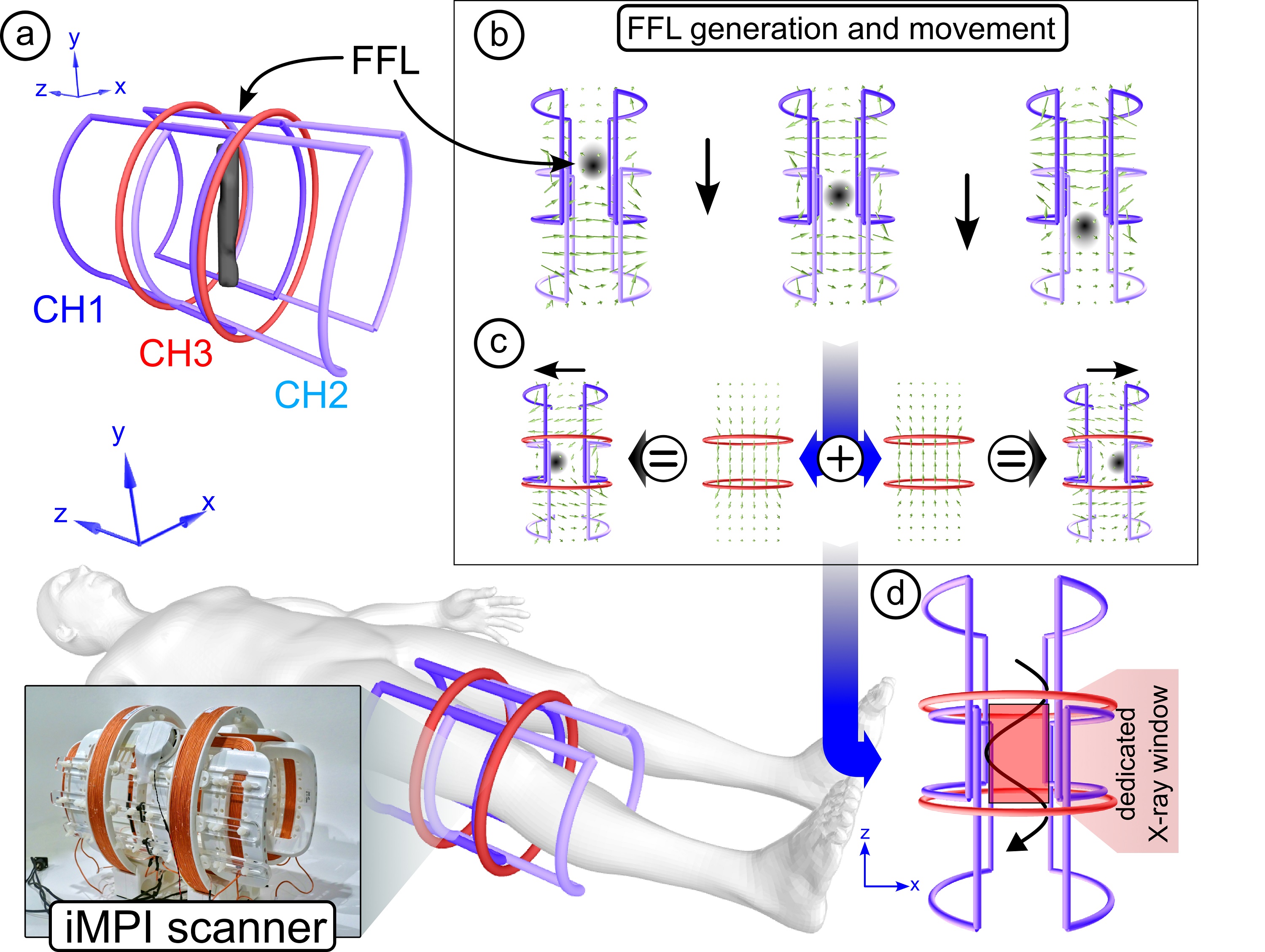
In MPI there are two basic encoding concepts available: using a point-by-point scanning method using a field-free-point (FFP) 3 or a projection-based methods using a field-free-line (FFL) 17. The latter concept provides a more sensitive and faster imaging process but only in 2D and with an enormous hardware effort 18.We found a novel approach for generating the required magnetic fields dynamically and fully electrically, which allows rapid scanning using the sensitive FFL encoding method 19,20. Furthermore, this new approach provides a more compact scanner design resulting in a lightweight and portable scanner design (Fig.1).
For initial experiments, a dedicated MPI scanner for intervention at human extremities has been designed and built. For evaluation, an open design with a dedicated ‘X-ray window’ has been chosen to enable simultaneous MPI/DSA imaging within a catheter laboratory 21.
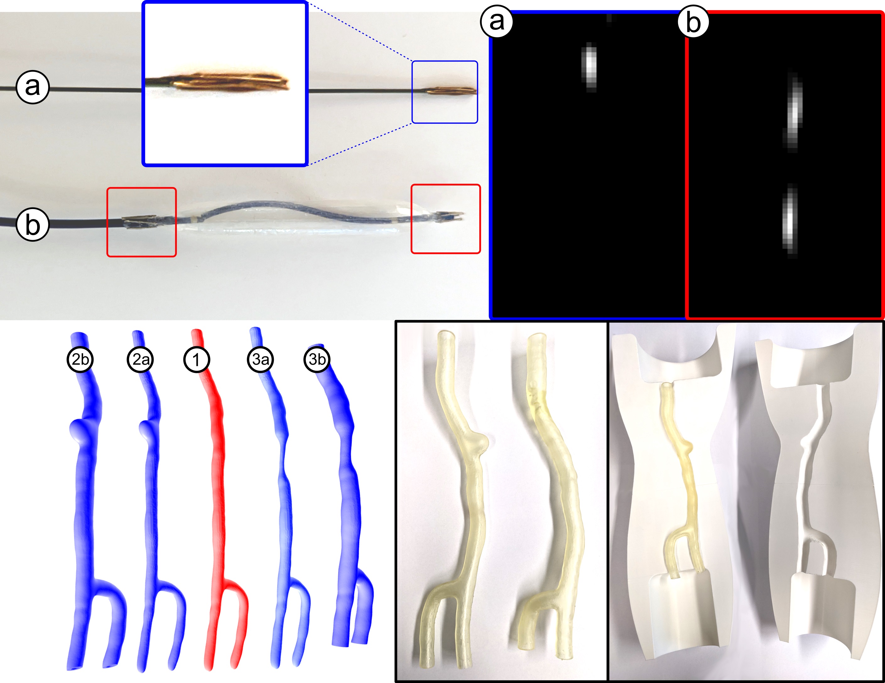
For tracking endovascular devices, such as guide-wires and balloon catheters, which are required for realtime PTA (percutaneous transluminal angioplasty), specific markers have been added 6,7. Furthermore, realistic human-sized phantoms with different indications (stenoses or aneurysms) have been prepared for imaging under realistic flow conditions 22 (fig.2).
Results
The results in fig.3 top shows the visualization of an entire MPI-guided PTA in realtime at 4 frames per second (single data acquisition 50ms). The covered FOV here is 11x12cm² within the scanners bore size of 20 cm. The peak pulsatile flow velocity in the experimental vessel system was 50cm/s. For angiography, a 1ml bolus of tracer (Perimag, Micromod GmbH, Germany) with iron concentration of 8.5mg/ml has been injected.The results in fig.3 bottom show the first simultaneous DSA/MPI hybrid images of a bolus of Perimag and iodinated contrast media mixture (ratio 1:1) tracked through an artificial vascular stenosis.
Conclusion
The presented concept for real-time image-guided vascular interventions using MPI demonstrates the feasibility of such a system from initial scanner characterizations to experimental interventions in dynamic vascular models. The possibility of simultaneous hybrid use in combination with gold standard X-ray technology could accelerate translation to clinical use in vascular interventions to decrease ionizing radiation levels for patients and clinical staff. iMPI holds particular promise for endovascular interventions without ionizing radiation, which will enable broader use of these treatment tools without extensive protective measures in the field of (cardio-)vascular diseases.Acknowledgements
The work was supported by the German Research Council (DFG), grant numbers: VO-2288/1-1, VO-2288/3-1, and BE 5293/1-2.References
- Roth, G. A. et al. Global burden of cardiovascular diseases and risk factors, 1990–2019: Update from the GBD 2019 Study. J. Am. Coll. Cardiol. 76(25), 2982–3021 (2020).
- Bartorelli, A. L. & Marenzi, G. Contrast-induced nephropathy. J. Interv. Cardiol. 21, 74–85 (2008).
- Gleich, B. & Weizenecker, J. Tomographic imaging using the nonlinear response of magnetic particles. Nature 435(7046), 1217–1217 (2005).
- Neumann, A. et al. Recent developments in magnetic particle imaging. JMMM 500, 169037 (2022).
- Wang, Y. X. et al. Superparamagnetic iron oxide contrast agents: Physicochemical characteristics and applications in MR imaging. Eur. Radiol. 11(11), 2319–2331 (2001).
- Herz, S. et al. Magnetic particle imaging-guided stenting. J. Endovasc. Ther. 26(4), 512–519 (2019).
- Herz, S. et al. Magnetic particle imaging guided real-time percutaneous transluminal angioplasty in a phantom model. Cardiovasc. Interv. Radiol. 41(7), 1100–1105 (2018).
- Mason, E. E. et al. Design analysis of an MPI human functional brain scanner. Int. J. Magn. Part. Imaging 3(1), 1703008 (2017).
- Graeser, M. et al. Human-sized magnetic particle imaging for brain applications. Nat. Commun. 10, 1936 (2019).
- www.mitsubishielectric.com/news/2023/pdf/0907-a.pdf
- Saritas, E. U. et al. Magnetostimulation limits in magnetic particle imaging. IEEE Trans. Med. Imaging 32(9), 1600–1610 (2013).
- Graeser, M. et al. Towards picogram detection of superparamagnetic iron-oxide particles using a gradiometric receive coil. Sci. Rep. 7, 6872 (2017).
- Vogel, P. et al. Micro-traveling wave magnetic particle imaging: Sub-millimeter resolution with optimized tracer LS-008. IEEE TMAG 55(10), 5300207 (2019).
- Vogel, P. et al. First in-vivo traveling wave magnetic imaging of a beating mouse heart. Phys. Med. Biol. 61(18), 6620–6634 (2016).
- Vogel, P. et al. Superspeed bolus visualization for vascular magnetic particle imaging. IEEE TMI 39(6), 2133–2139 (2020).
- Vogel, P. et al. Low latency real-time reconstruction for MPI systems. Int. J. MPI 3(2), 1707002 (2017).
- Weizenecker, J. et al. Magnetic particle imaging using a field free line. J. Phys. D: Appl. Phys. 41(10), 105009 (2008).
- Top, C. B. & Güngör, A. Tomographic field free line magnetic particle imaging with an open-sided scanner configuration. IEEE Trans. Med. Imaging 39(12), 4164–4173 (2020).
- Greiner, C. et al. Traveling Wave MPI utilizing a Field-Free Line. Int. J. Magn. Part. Imaging 8(1), 2203027 (2022).
- Vogel, P. et al. iMPI: portable human-sized magnetic particle imaging scanner for real-time endovascular interventions. Sci Rep 13, 10472 (2023).
- Vogel, P. et al. Magnetic particle imaging meets computed tomography: First simultaneous imaging. Sci. Rep. 9, 12672 (2019).
- Reichl, T. et al. Highly flexible human aneurysm models for realistic flow experiments with MPI and MRI. Int. J. Magn. Part Imaging 8(1), 2203035 (2022).
Figures
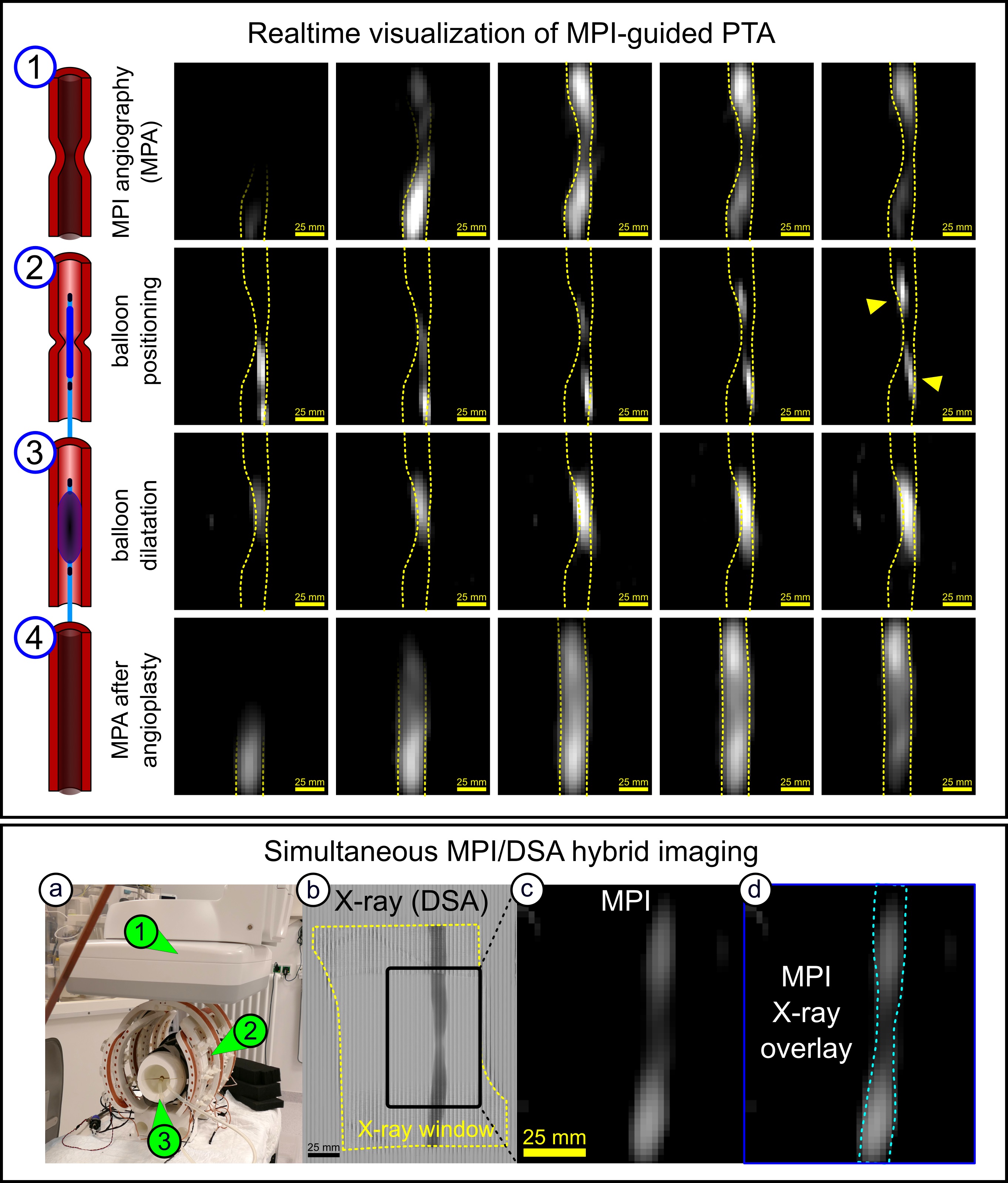
Top: Real-time visualization of an MPI-guided PTA. (1) MPI angiography (MPA) for stenosis location by injecting a 1 ml Perimag bolus. (2) Balloon positioning. (3) Balloon dilation by inflating the balloon catheter with tracer. (4) Second MPA visualized the successful treatment of the stenosis.
Bottom: First simultaneous MPI/DSA bolus tracking. (a) Image of the iMPI scanner (2) within the X-ray system (1). Simultaneous visualization of the bolus through an artificial vascular stenosis in a human-size knee model (3): (b) X-ray and (c) iMPI. (d) shows an overlay of both modalities.