1332
PET Recon with MR Priors: Application in Ambiguously Rated Amyloid PET Scans1Stanford University, Stanford, CA, United States
Synopsis
Keywords: PET/MR, PET/MR, MR Priors, Motion Correction
Motivation: Uptake in the supratentorial cortex defines clinical amyloid positivity. Because of PET’s low spatial resolution, there is a spill-in effect from adjacent white-matter to gray-matter in amyloid PET scans, which may cause inconsistent rating between trained readers.
Goal(s): Reduce the spill-in effect by using MR priors in PET recon to improve the consistency of reading amyloid PET scans.
Approach: A recently developed PET recon with MR-priors and motion correction is applied to a set of 18F-florbetaben (amyloid) PET/MRI scans and is compared with regular TOF-OSEM PET recon using SUVR.
Results: PET recon with MR priors improves reading of negative amyloid PET scans.
Impact: PET-recon with MR-priors can make identifying negative cases more consistent between readers. Given how common inconsistent ratings are for amyloid-PET, methods that improve the ability to distinguish intermediate amyloid levels may be valuable for the widespread use of this modality.
Objectives
Although amyloid PET in the clinical setting is typically interpreted as either positive or negative, a portion of amyloid PET scans fall into an intermediate range and will be rated inconsistently across trained raters. Because of PET’s low spatial resolution, there is a spill-in effect from adjacent white-matter to gray-matter that may cause inconsistent categorization between trained raters. PET recon with MR priors provides higher spatial resolution and minimizes the spill-in effect, which can improve consistency. We sought to determine the impact of advanced PET reconstructions that incorporate MRI priors and motion correction on quantitative standardized uptake value ratios (SUVRs) from these ambiguous scans.Methods
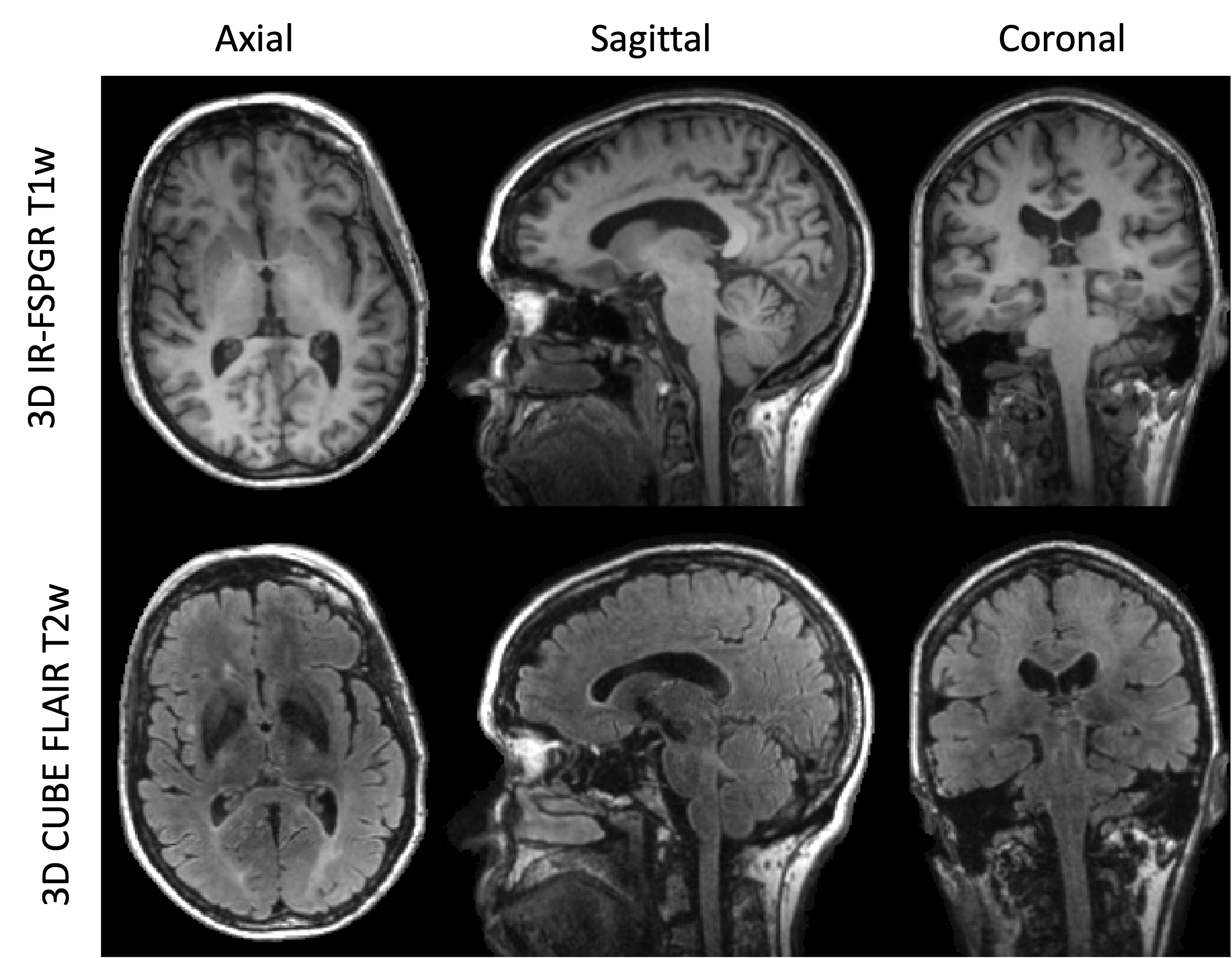
Subjects with a range of cognitive impairment were injected with 300 MBq of florbetaben and underwent a 20-minute brain scan 90 min after injection on a SIGNA 3T PET/MR (GE Healthcare, Waukesha, WI). 3D T1 IR FSPGR and T2 FLAIR CUBE images were acquired simultaneously with PET. The study was approved by Stanford's Institutional Review Board and all subjects provided written consent. PET images were first reconstructed using TOF-OSEM with 28 subsets and 4 iterations using 3 mm FWHM spatial filter and standard Z-filter. They were also reconstructed with MR-guided TOF-BSREM (MRgTOF-BSREM)1,2 with b =15 and bm=100, using short-frame motion correction1,3 and 3D T1 and T2 weighted MR images as priors. SUVRs were calculated using a global cortical composite ROI divided by the whole cerebellum for both TOF-OSEM and MRgTOF-BSREM recons.Results
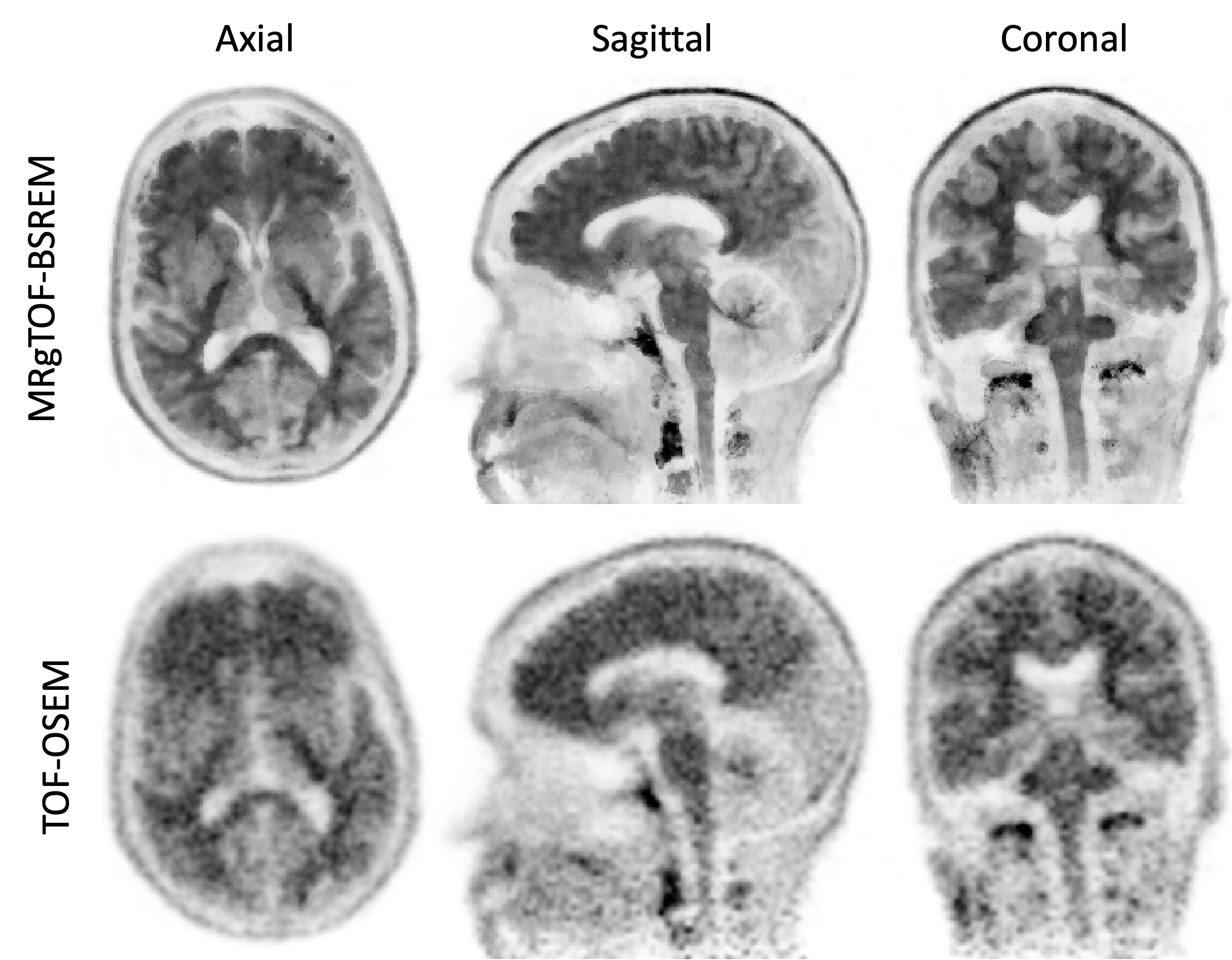
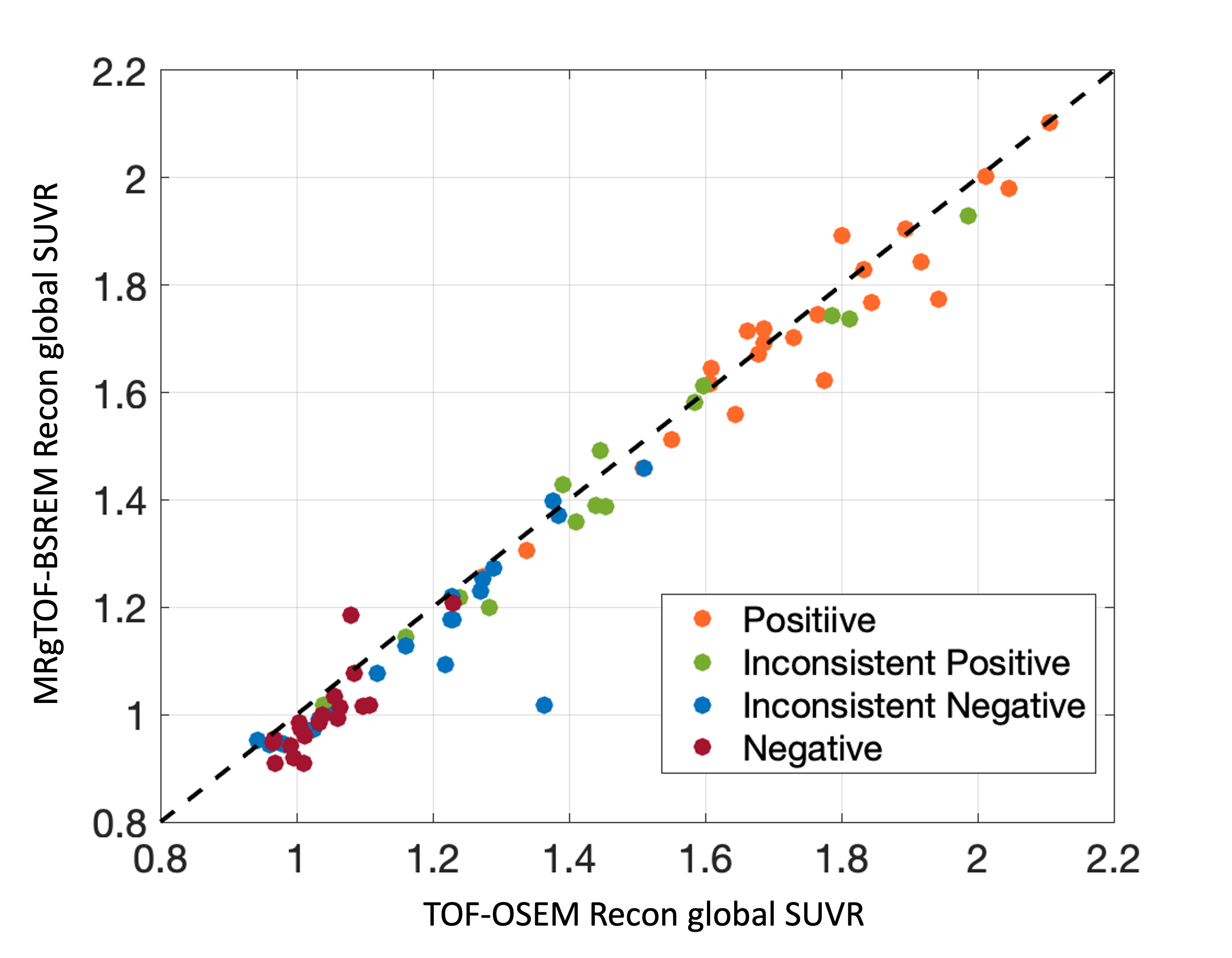
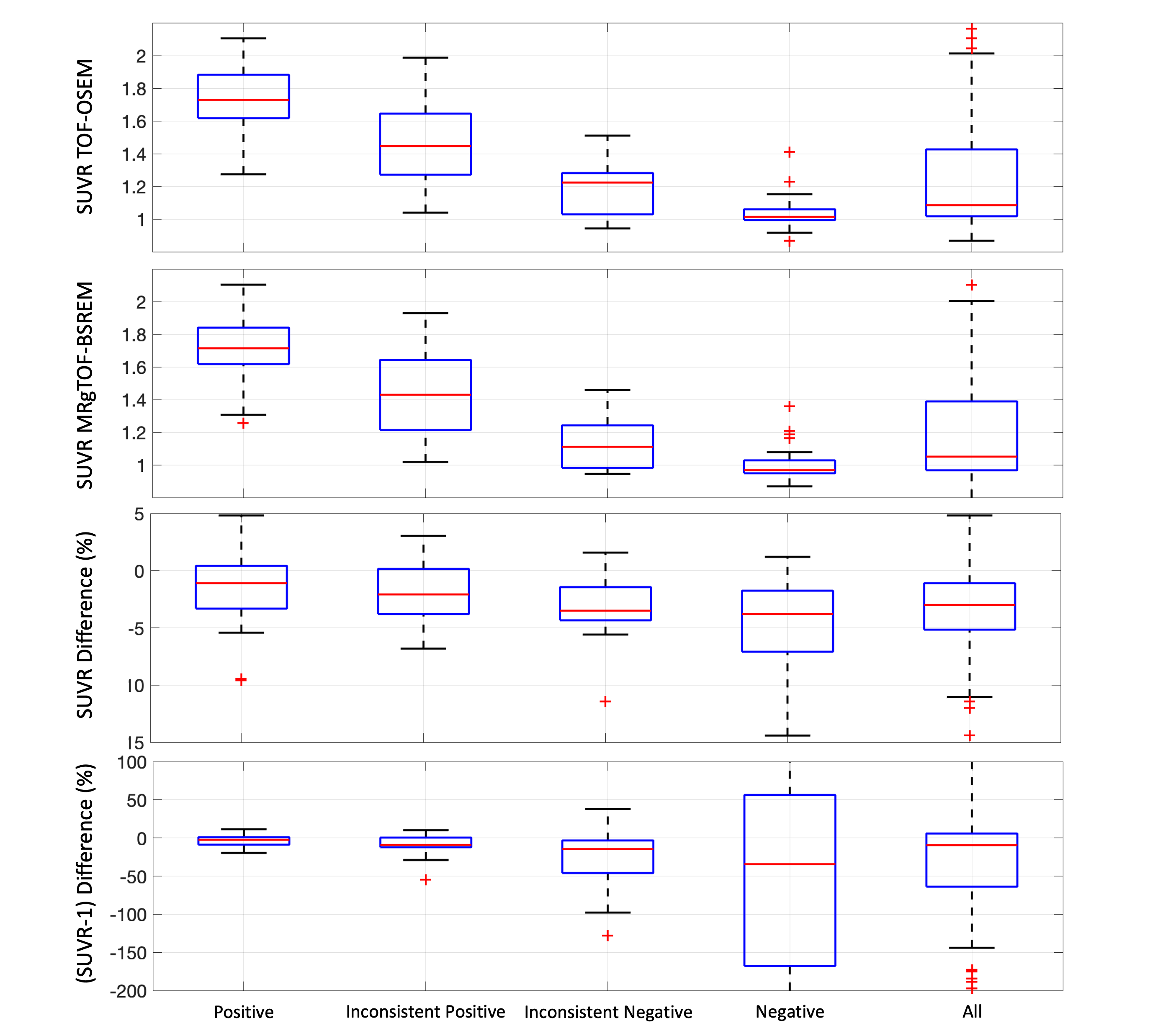
Of the 139 florbetaben scans obtained, the 112 that were rated by 3 trained readers were included in the study. Subjects were 72 ± 9 years old and 47% were female. Of the 112 rated exams, 34 were rated inconsistently across raters (14 were positive for 2/3 readers; 20 were negative for 2/3 readers). The 34 inconsistent scans, along with 78 scans matched for clinical diagnosis that were either consistently read as positive (N=23) or negative (N=55) were reconstructed (Figure 1) using MR priors (Figure 2) and motion correction. SUVRs using standard recon and recon with MR priors were highly correlated (Figure 3). SUVR differences (Figure 4) between standard recon (TOF-OSEM) and PET recon with MRI priors (MRgTOF-BSREM) differed by group. Although the SUVR values measured by MRgTOF-BSREM decreased for all exams due to the spill-in correction from white-matter, the change had a different impact between similarly rated groups. The SUVR difference between all groups is shown in Figure 5. The highest impact is observed in consistently or majority rated negative groups. Because the spill-in effect from white-matter is mitigated with MR priors, it is easier to see the normal gray-matter in these two groups and therefore minimizes potential false positive cases.Discussion
MRI-based recons improve quantitative measures of amyloid burden in ambiguously rated cases by resolving the spill-in effects to gray-matter from adjacent white-matter voxels. This can make identifying negative cases more consistent between readers. Given how common inconsistent ratings are for amyloid PET, methods that improve the ability to distinguish intermediate amyloid levels may be valuable for the widespread use of this modality. MRI-based recons improve quantitative measures of amyloid burden in ambiguously rated cases by resolving the spill-in effects to gray-matter from adjacent white-matter voxels. This can make identifying negative cases more consistent between readers. Given how common inconsistent ratings are for amyloid PET, methods that improve the ability to distinguish intermediate amyloid levels may be valuable for the widespread use of this modality.Acknowledgements
No acknowledgement found.References
1. Khalighi MM, Deller T., et al., “High Resolution PET/MR Imaging Using Anatomical Priors & Motion Correction”, ISMRM 2021.
2. Khalighi MM, Deller T., et al., “High Quality Isotropic Whole-body PET Imaging Using MR Priors”, J Nucl Med May 1, 2020 vol. 61 no. supplement 1 1477
3. Spangler-Bickell M., Hurley S., Pirasteh A., Perlman S., Deller T., McMillan A., “Evaluation of Data-Driven Rigid Motion Correction in Clinical Brain PET Imaging,” Journal of Nuclear Medicine, October 2022, 63 (10) 1604-1610.
Figures