1324
Deep Learning-based Disambiguation for Multiple AD Radiotracers using PET/MRI1Radiology, Stanford University, Stanford, CA, United States, 2Neurology, Stanford University, Stanford, CA, United States, 3Biomedical Engineering, National Taiwan University, Taipei, Taiwan
Synopsis
Keywords: PET/MR, PET/MR
Motivation: AD patients must undergo repeated visits for amyloid and tau radiotracer imaging, leading to high costs and dose concerns due to PET's inability to simultaneously acquire multiple radiotracers during a single session.
Goal(s): Using PET/MRI scans, we used deep learning to create separate amyloid and tau PET images from a simulated combined dual-tracer image.
Approach: We simulated a combined amyloid-tau image by blending co-registered list-mode data and employed a 2.5D U-Net architecture for effective separation.
Results: Mixed-dose models, incorporating physics-inspired data augmentation and MR information, exhibited enhanced anatomical preservation and reduced variability in quantitative metrics.
Impact: The demonstrated separation of a simulated combined amyloid and tau PET/MRI study into its individual components using DL may allow for simultaneous injection of multiple radiotracers in a single acquisition, streamlining the imaging process for AD patients.
Introduction
Positron Emission Tomography (PET) has started to facilitate clinical Alzheimer's Disease (AD) diagnosis, with the ability to detect the neuropathological hallmarks of the disease using radiotracers like 18F-Florbetaben (FBB) and 18F-PI-2620 (Tau) respectively1. However, simultaneous dual-tracer acquisition creates indistinguishable 511-KeV photon pairs2, necessitating multiple visits for AD patients and caregivers.Various simulation techniques aim to distinguish individual components in a combined dual-tracer PET image and model simultaneous injection of both radiotracers2-4. However, inherent challenges persist in capturing underlying biology and modeling dual-tracer kinetics in a simulated combined image. To facilitate the creation of more realistic combined mixed-dose amyloid and tau PET images, we simulated combining static list-mode data from individual FBB and Tau scans in the same patients by co-registered list-mode data5. We here propose to use deep learning (DL), specifically using a 2.5D U-Net, to disambiguate the combined radiotracer image into its separate amyloid and tau components.
Methods
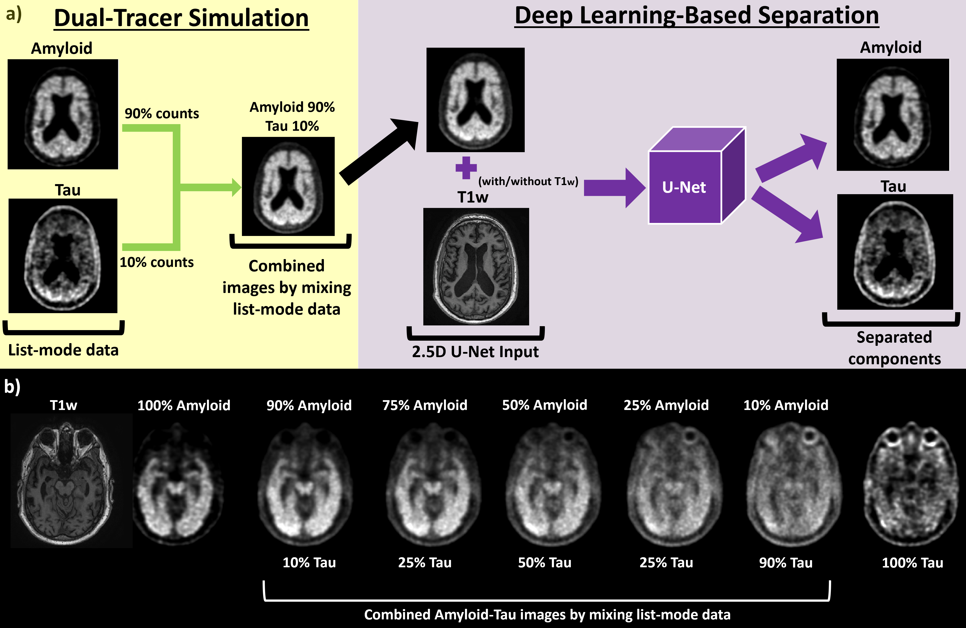
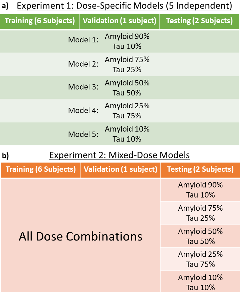
Dataset and Pre-ProcessingIn this IRB-approved study, 9 subjects were identified and split into training (subjects: 6; age: 70.7±12.4 yrs; 5 female, Amyloid+: 4; Tau+: 3), validation (subjects: 1; age: 73.2 yrs; male; Amyloid+:, Tau–), and testing (subjects: 2; age: 75.8±1.7 yrs; 1 female; Amyloid+: 2; Tau+: 1). T1-weighted MR imaging, 18F-FBB (amyloid), and 18F-PI-0620 (tau) PET data were acquired on an integrated 3T PET/MR (Signa; GE Healthcare). The original amyloid and tau images were created using standard doses and static reconstruction. Amyloid and tau PET images were acquired between 90-110 min and 45-75 min after injection. The raw list-mode PET data from the FBB and Tau acquisition for each patient underwent rigid motion correction based on lava-water MRI images, under-sampling according to simulated dose mixing, and incorporation of random estimates, average scatter, and sensitivity maps for the combined study (Figure 1a)5. The combined images were reconstructed using five empirically selected dose blends, ranging from 90/10 to 10/90 amyloid/tau simulated dose (Figure 2).
These mixed-dose combined radiotracer images (Figure 1b) will be used as training input for the model. The ground truth for the model will be both pure amyloid and tau images.
CNN Models and Approach Overview
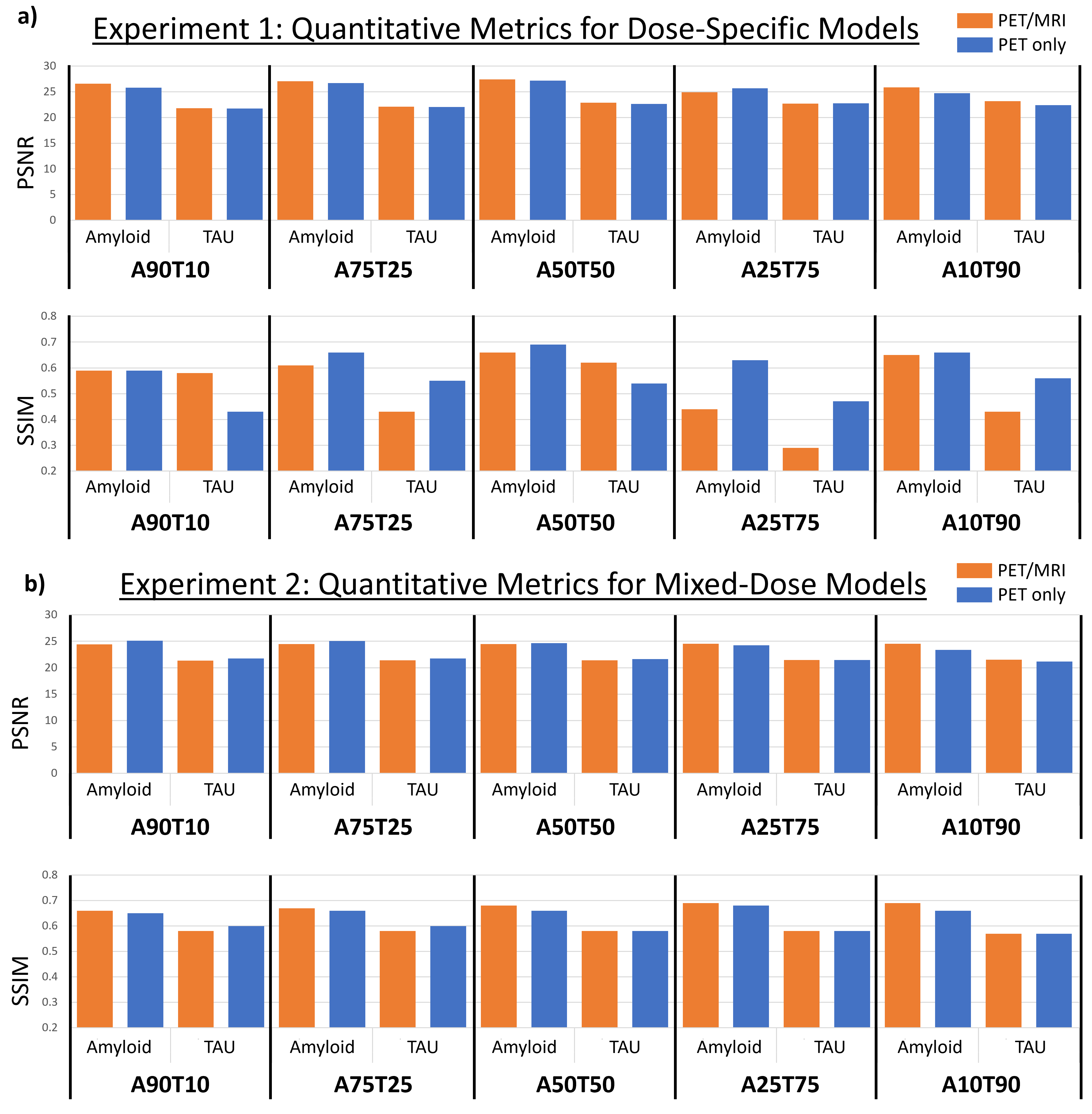
We trained 2.5D models using MONAI's U-Net6 (Figure 1a). This approach allowed us to effectively aggregate multiple slices and leverage larger batch sizes, potentially enhancing separation performance. The 2.5D U-Net model was set up using the following parameters: 3/6 input channels (PET versus PET/MR; 3 slice sliding window), 2 output channels (amyloid and tau), channels sequence of (32, 64, 128, 256, 512), 4 residual units, and 0.2 dropout probability. These models were then compared to ground truth amyloid and tau scans and evaluated on image quality metrics such as peak signal-to-noise ratio (PSNR) and structural similarity index (SSIM).
Results
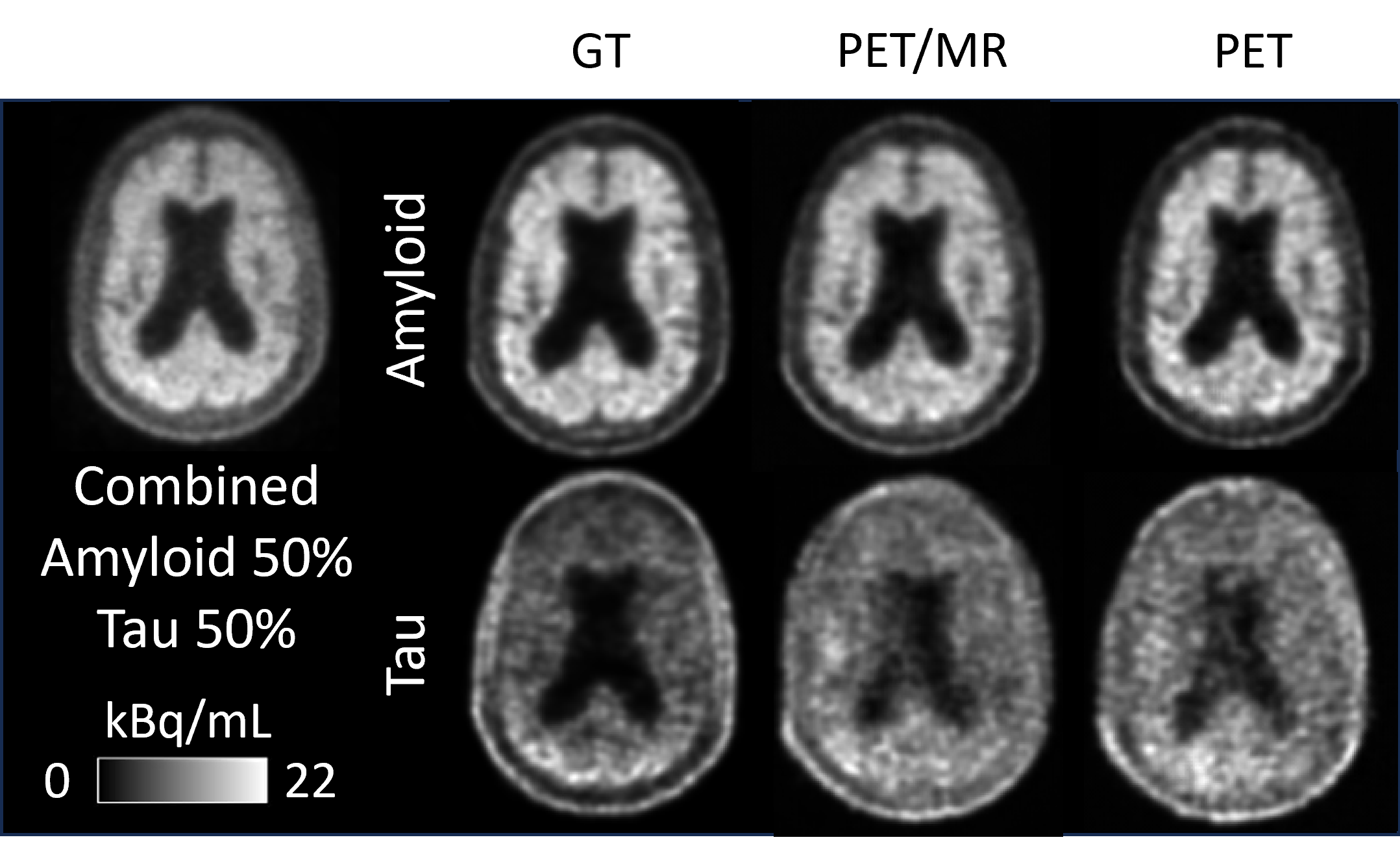
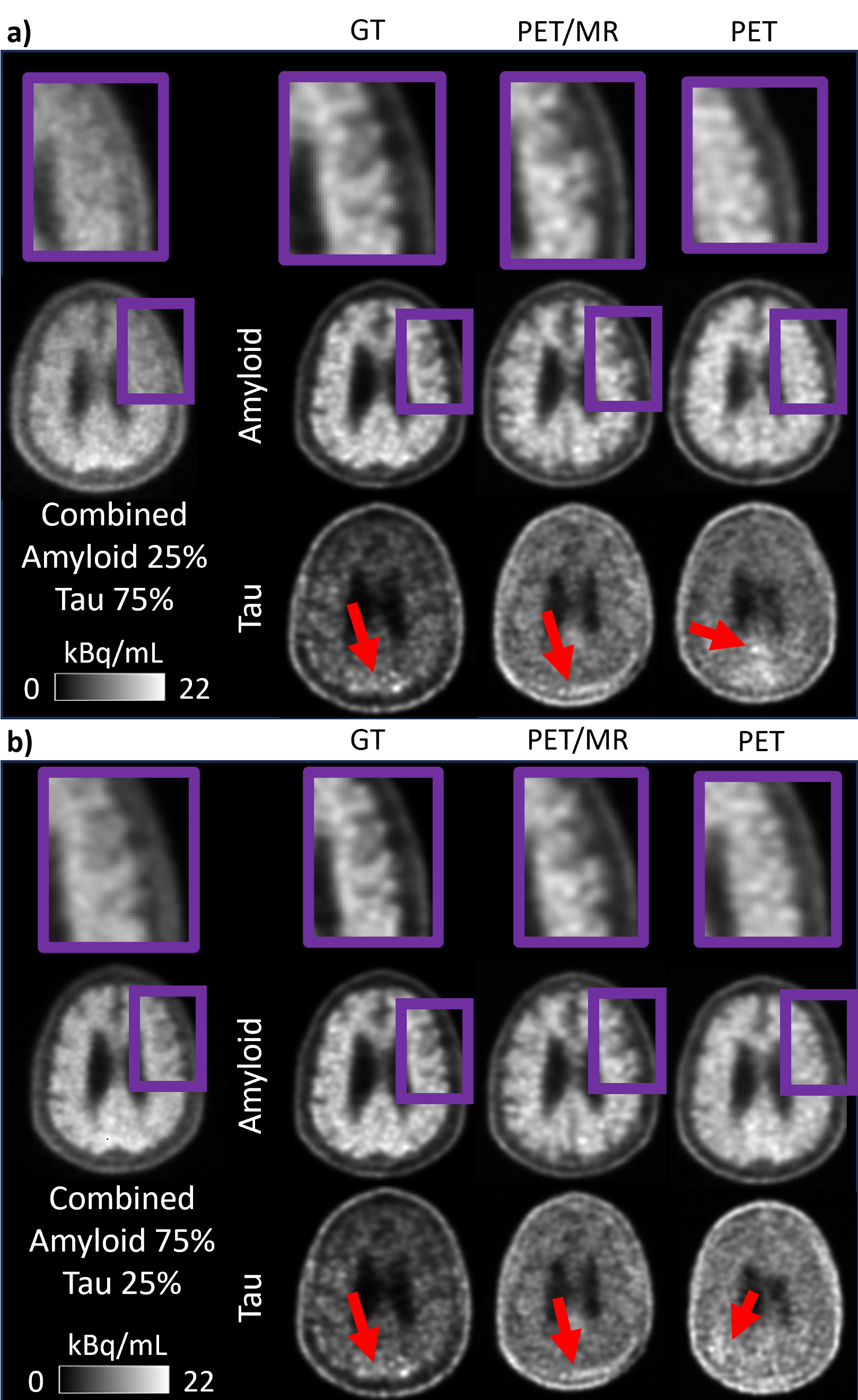
Dose-specific PET/MR and PET models exhibited relatively consistent PSNR values for separating both amyloid and tau components, though SSIM performance showed inconsistencies (Figure 3a). Qualitative evaluation confirmed that dose-specific models reasonably separated the amyloid component, but exhibited limitations in capturing the tau distribution (Figure 4).To address this, we investigated training on all dose combinations, effectively implementing physics-inspired data augmentation during training. This approach aimed to enhance model consistency and improve dose-specific separation (Figure 2b). The results showed more consistent quantitative model performance across mixed-doses (Figure 3b). Additionally, we observed enhanced anatomical preservation and consistent tau positivity with MR information (Figure 5).
Discussion
This study successfully demonstrated the separation of a simulated dual-tracer amyloid/tau image into its amyloid and Tau components using DL. The implementation of a physics-inspired data augmentation pipeline significantly enhanced the robustness of component estimation, reducing variability across mixed-dose combinations. This augmentation approach shows promise, particularly given that the optimal dose combinations for acquiring amyloid and tau for DL separation are still being investigated.Considering that Tau PET images, especially in early disease stages7, may have lower SNR, this could contribute to the observed variability in dose-specific model performance. Since each dose-specific model was trained on a fifth of the mixed-dose model training data, the mixed-dose model may have benefited from noise averaging.
Previous studies on non-AD radiotracers have employed deep belief networks to separate Monte Carlo simulated dual-tracer images2, and multi-task networks to separate dynamic dual-tracer images3. Our study demonstrates that combining static list-mode data may provide a method to simulate realistic training data for effective separation.
Conclusion and Future Work
The 2.5D U-Net, augmented with physics-based data and MR information, holds promise for effectively disambiguating combined amyloid/tau images. This study suggests the translation feasibility of simultaneous dual injection of AD radiotracers in a single acquisition. Future studies would likely benefit from additional patient data.Acknowledgements
This work was support by NIH R56AG071558. A.K. was supported by the Tau Beta Pi and Stanford Knight-Hennessy fellowship.References
- Paul A Rowley, Alexey A Samsonov, Tobey J Betthauser, Ali Pirasteh, Sterling C Johnson, and Laura B Eisenmenger. Amyloid and tau pet imaging of alzheimer disease and other neurodegenerative conditions. In Seminars in Ultrasound, CT and MRI, volume 41, pages 572–583. Elsevier, 2020.
- Jinmin Xu and Huafeng Liu. Deep-learning-based separation of a mixture of dual-tracer single-acquisition pet signals with equal half-lives: A simulation study. IEEE Transactions on Radiation and Plasma Medical Sciences, 3(6):649–659, 2019.
- Todd Macdonald, Kevin Chen, Mary Ellen Koran, Michael Moseley, and Greg Zaharchuk. DualNet: a deep neural network to predict individual tau and amyloid pet images from a combined dose image using the disambiguation of dual dose amyloid-tau pet scans using the ADNI dataset, 2020.
- Bolin Pan, Paul K Marsden, and Andrew J Reader. Dual-tracer pet image separation by deep learning: A simulation study. Applied Sciences, 13(7):4089, 2023.
- Mehdi Khalighi, Kevin Chen, Timothy Deller, Floris Jansen, Elizabeth Mormino, and Greg Zaharchuk. Dual tracer brain pet simulation from two separate exams, 2021.
- M Jorge Cardoso, Wenqi Li, Richard Brown, Nic Ma, Eric Kerfoot, Yiheng Wang, Benjamin Murrey, Andriy Myronenko, Can Zhao, Dong Yang, et al. Monai: An open-source framework for deep learning in healthcare. arXiv preprint arXiv:2211.02701, 2022.
- Colin Groot, Sylvia Villeneuve, Ruben Smith, Oskar Hansson, and Rik Ossenkoppele. Tau pet imaging in neurodegenerative disorders. Journal of Nuclear Medicine, 63(Supplement 1):20S–26S, 2022.
Figures