1323
Characterization of a Pseudo-Tissue Liver Flow Phantom for Use in Magnetic Resonance Elastography Validation Experiments1Mechanical Engineering, University of Wisconsin-Madison, Madison, WI, United States, 2Radiology, University of Wisconsin-Madison, Madison, WI, United States, 3Medical Physics, University of Wisconsin-Madison, Madison, WI, United States, 4University of Wisconsin-Madison, Madison, WI, United States, 5Biomedical Engineering, University of Wisconsin-Madison, Madison, WI, United States
Synopsis
Keywords: Phantoms, Elastography, MRE, Hydrogel, Phantoms
Motivation: MRE is utilized to assess tissue stiffness in vivo, however, robust validation in tissue-mimicking phantoms is needed.
Goal(s): Create hydrogel liver phantoms of varying stiffness, characterize them with MRE and compare MRE with a mechanical reference standard. Utilize pulsatile flow to mimic cardiac motion.
Approach: Flow phantoms of varying stiffness were created. Stiffness from mechanical test was compared to MRE. Cardiac tagging visualized motion inside each phantom.
Results: MRE shear stiffness showed agreement with indenter stiffness and cardiac motion decreased with stiffness.
Impact: Advances in MRE have led to its increasing use to determine tissue stiffness in vivo, however, robust validation in tissue mimicking phantoms has yet to be achieved. The proposed methodology may help to assess the quantitative performance of MRE.
Introduction
Recent advances in magnetic resonance elastography (MRE) have led to its expanded use as a non-invasive technique to quantify tissue stiffness in vivo1-3. MRE aims to evaluate the mechanical properties of tissues by measuring the speed at which shear waves propagate in them4. While numerous studies have validated MRE in vivo, this validation has been limited. To bridge this gap, anthropomorphic, in vitro liver flow phantoms have been developed using hydrogel materials to reproduce the mechanical properties of the liver and systematically assess and validate MRE performance. Hydrogels can be used to create MRI-compatible, tissue-mimicking phantoms with tissue-like stiffness for organs such as the liver5. In this study, we first use MRE to quantify the stiffness of various hydrogel formulations used to create pseudo-tissue liver phantoms. Using indenter testing we validated our MRE stiffness measurements. Additionally, different hydrogel formulations were used to create liver phantoms of known stiffness and investigate the effects of cardiac pulsation on the non-rigid motion profiles inside each phantom.Methods
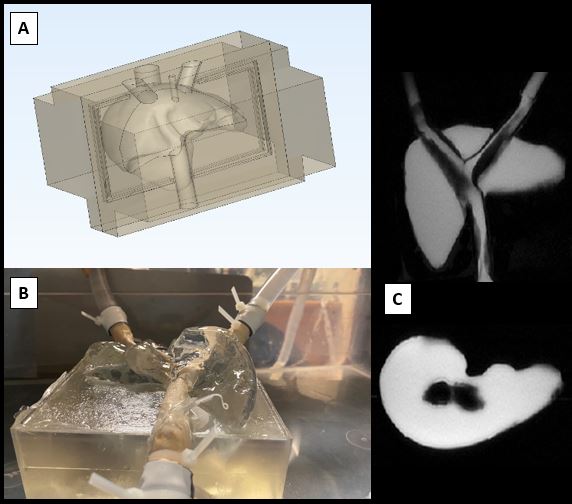
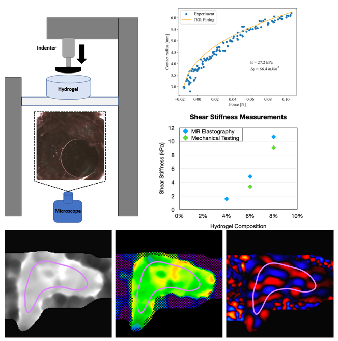
Three tissue-mimicking liver-shaped hydrogel phantoms were constructed using a hybrid additive manufacturing process5-6. Each hydrogel was fabricated using different concentrations of -acrylamide/bis-acrylamide polymers, resulting in gel concentrations of 4%, 6% and 8% (Figure 1). To quantify stiffness using a mechanical testing reference, indentation tests were performed and used to extract the Young’s modulus of each hydrogel sample. Assuming linear elastic, isotropic behavior the shear stiffness of the hydrogel samples can be estimated. A detailed schematic of the mechanical testing setup is shown in Figure 2. Phantoms were scanned on a clinical 3.0T MRI system (Signa Premier, GE Healthcare) with high density receive array coils (Air Coil, GE Healthcare). 2D MRE was used to characterize the shear stiffness in coronal cross-sections of each phantom using a EPI-based MRE method (Resoundant-GE) following the Quantitative Imaging Biomarkers Alliance (QIBA) guidelines for MRE acquisitions involving phantoms7. Additionally, for each phantom, the driver amplitude was held constant at 30% while the driver frequency was altered until visual inspection confirmed no phase wrapping was present in the acquired phase images. This resulted in driver frequencies of 160, 180 and 200 Hz for the 4%, 6% and 8% gel compositions, respectively. Shear stiffness obtained with MRE were directly compared to shear stiffness measurements derived from the elastic modulus obtain from mechanical testing for samples that overlapped in gel percentage. Additionally, each phantom was designed to be incorporated into a mock circulatory loop capable of reproducing pulsatile flow. Using the pump, the flow characteristics were changed to induced pulsatile motion (0.5, 1.0 and 1.5 liter/min (LPM)). To visualize motion, CINE-based tagging sequences were acquired throughout the flow cycle using a tag spacing of 8mm.Results
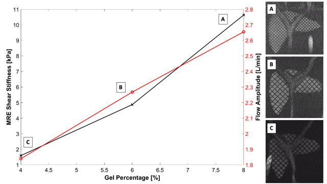
Hydrogel based phantoms were successfully created for 4%, 6% and 8% gel formulations. Stiffness estimates for the 6% and 8% hydrogel formulations were successfully acquired using both mechanical experimentation and MRE. Each show good agreement with a slight overestimation of MRE-derived shear stiffness compared to mechanical estimates assuming linear elastic and isotropic behavior of the hydrogel samples. Figure 3 shows MRE-derived shear stiffness and flow amplitude at 0.5, 1.0 and 1.5LPM. Flow amplitude of the measured waveform for each mean flow rate, which can be thought of as highest degree of cardiac-mimicking pulsation, decreased with increasing hydrogel stiffness. Cine images confirm more motion induced inside the softest phantom and are used to qualitatively assess vessel motion in each phantom.Discussion
MRE in liver tissue-mimicking phantoms was validated using optical-based mechanical indentation testing. We demonstrated that under the assumption of isotropic, linear elasticity measured shear stiffness from MRE agrees well with stiffness derived from mechanical testing data. This is further confirmed by examining the pulsatile motion profiles at a nominal flow rate of 1.5 LPM. As the stiffness of the hydrogel decreased, the pulsatility of the flow waveform downstream of the phantom exit decreased. This suggest that softer tissues absorb energy and dampen pulsatility. Unfortunately, mechanical estimates could not be determined for the 4% hydrogel sample due to difficulties in extracting the contact area for the softest hydrogel. This is likely due to adhesive forces gripping the indenter. Limitations of this study include the assumption of linear elasticity of the hydrogel materials to determine the shear stiffness from mechanical indentation testing. In reality, polyacrylamide-based hydrogels display, some viscoelasticity that may complicate this assumption. Overall, this study represents a novel step in robust validation of MRE for shear stiffness estimation using anthropomorphic, tissue-mimicking phantoms.Acknowledgements
We would like to acknowledge GE Healthcare, who provides research support to the University of Wisconsin-Madison and from the NIH (R01 EB030497).References
[1] Venkatesh SK, Yin M, Ehman RL. Magnetic resonance elastography of liver: Technique, analysis and clinical applications. J Magn Reson Imaging. 2013;37(3):544-555.
[2] Bensamoun SF, Wang L, Robert L, et al. Measurement of liver stiffness with two imaging techniques: Magnetic resonance elastography and ultrasound elastometry. J Magn Reson Imaging. 2008;28:1287-1292.
[3] Ichikawa H, Yasuda E, Kumada T, et al. Intra-individual comparison of liver stiffness measurements by magnetic resonance elastography and two-dimensional shear-wave elastography in 888 patients. Ultrasonography. 2023;42(1):65-77.
[4] Mariappan, YK, Glaser KJ, Ehman, RL. Magnetic resonance elastography: A review. Clin. Anat. 2010;23:497–511.
[5] Rice J, Geng R, Hernando D, Roldán-Alzate A. Fabrication of an anthropomorphic, in-vitro liver flow phantom for use in motion-robust MRI sequence validation experiments. Proceedings of the 2022 ISMRM & SMRT Annual Meeting & Exhibition; 2022; London, UK.
[6] Geng R, Zhang Y, Rice J, et al. Motion-robust, blood-suppressed, reduced-distortion diffusion MRI of the liver. Magn Reson in Medicine
[7] Quantitative Imaging Biomarkers Alliance. QIBA Profile: Magnetic Resonance Elastography of the Liver. 2018. https://qibawiki.rsna.org/images/a/a5/MRE-QIBAProfile-2018-05-02-CONSENSUS.pdf. Accessed December 2, 2020.
[8] Johnson K L, Kendall K and Roberts A D 1971 Surface energy and the contact of elastic solids Proc. R. Soc. Lond. A 324 301–13.
Figures