1322
An Anthropomorphic Cardiac Phantom based on MR-Visible Additive Manufacturing for Development and Training of Interventional CMR Procedures1Magnetic Detection and Imaging group, TechMed Centre, University of Twente, Enschede, Netherlands, 2Robotics and Mechatronics group, University of Twente, Enschede, Netherlands, 3Department of Cardiology, Amsterdam University Medical Center, Amsterdam, Netherlands
Synopsis
Keywords: Phantoms, Phantoms, Phantom, 3D Printing, Cardiac Interventions
Motivation: Both technological development as well as procedural training of interventional cardiac MR (iCMR) procedures require anatomically realistic models. Animal studies are undesirable and becoming virtually impossible due to legal and ethical restrictions.
Goal(s): To develop a realistic cardiac phantom that can be 3D printed using standard MR-visible support material.
Approach: Material properties were determined and an anatomically realistic cardiac model was developed and constructed using 3D printing. Imaging features were assessed and an MR-guided cardiac intervention was simulated experimentally.
Results: A simple and realistic MR-visible cardiac phantom has been presented for technological development and training purposes, to improve MR-guided cardiac interventions.
Impact: The proposed phantom facilitates the development, testing and validation of novel technologies to improve MR-guided cardiac interventions, can be used for procedural training purposes, and may ultimately contribute to the improvement of clinical outcome.
Introduction
MR guidance of cardiac interventions offers several advantages over conventional X-ray fluoroscopy, including detailed 3D visualization of the anatomy. However, due to the different way structures are visualized, cardiac interventionalists (i.e., electrophysiologists) must retrain their hand-eye coordination to navigate devices to the target area in 3D space.To facilitate training and to improve pre-procedural preparation, there has been a growing interest in the use realistic patient-specific models.1 Additive manufacturing, also known as “3D printing”, provides excellent capabilities to design and construct anatomically realistic structures and include patient-specific challenges to simulate cardiac interventional procedures. Recently several MR-visible materials have become commercially available which are suitable to create realistic models, although being considerably more costly.
In this project, we aim to exploit the MR-visible properties of standard support material as a more accessible and cheaper alternative for printing an anatomically realistic cardiac phantom. As the support material is water-soluble, water-tightness of a MR-invisible layer is evaluated and the imaging features of the phantom are studied in the simulation of an MR-guided cardiac intervention.
Methods
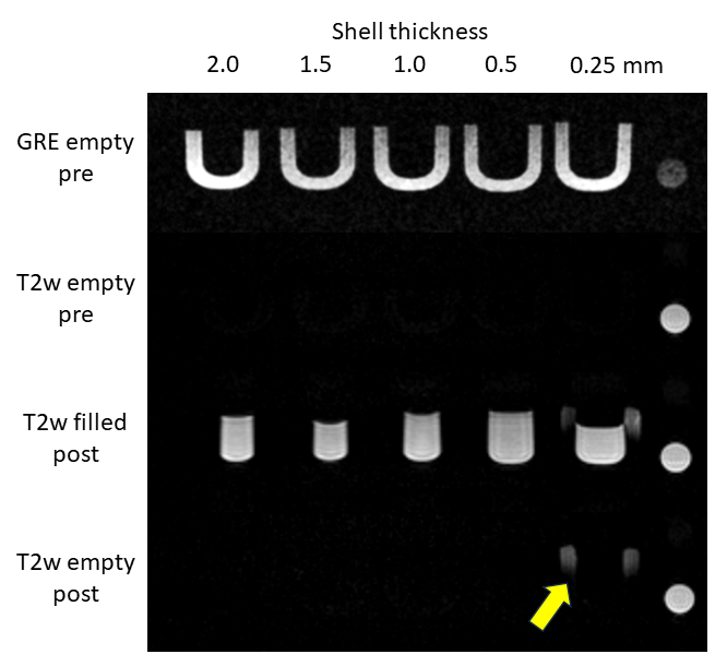
All MR-experiments were performed using a 1.5T MRI system (Aera, Siemens Healthineers, Erlangen, Germany) equipped with an 18-element cardiac coil array and 12-element spine array. Two MR-visible 3D printer materials (RGD525 and SUP706, Stratasys, Eden Prairie, MN) were analyzed and compared to regular silicone rubber (Ecoflex 00-30, Smooth-On, Macungie, PA) in terms of SNR, T1 and T2, using basic inversion-recovery and multi-echo spin echo sequences.2To confirm water-tightness of the non-soluble layer, a series of ‘U’-shaped objects covered with a MR-invisible layer of 2.0 mm down to 0.25 mm thickness were printed. Scans were performed before and after having the structures filled with tapwater over a 3-day period, to assess material alterations.
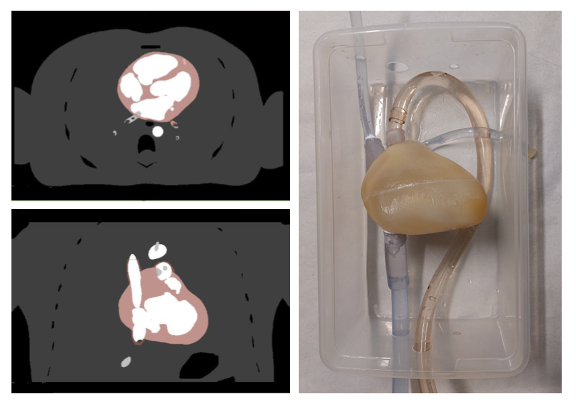
An anatomically realistic cardiac model was derived from the extended cardiac-torso (XCAT) digital phantom using 3D Slicer.3,4 Small detailed anatomical features such as the papillary muscles and coronary arteries were simplified to improve the fabrication process. A shell structure was created by dilating the resulting model by 1 mm on the inside to ensure water-tightness of the lumen compartment and by 1.5 mm on the outside to provide mechanical strength. The shell design was split mid-way to facilitate easy removal of the internal support material. The shell was printed using standard printing materials (Vero Clear and SUP706, Stratasys, Eden Prairie, MN). After closing the structure, connections to the main vasculature such as the inferior vena cava were implemented using simple tubing. The design and final phantom configuration are shown in figure 1.
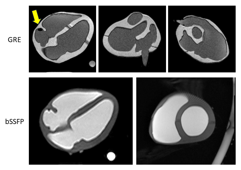
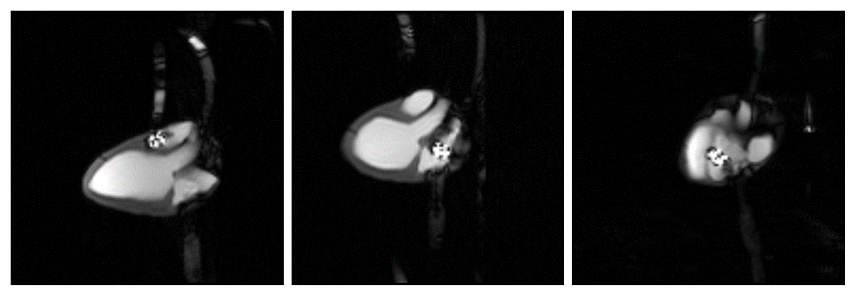
The anatomical features were evaluated using a basic untriggered spoiled 3D GRE sequence and 2D bSSFP sequence, after filling the configuration with tapwater. Finally, an MR-guided intervention was simulated using an ablation catheter (Vision-MR, Imricor, Burnsville, MN). A real-time 2D bSSFP sequence with interleaved active catheter tracking was used with the following parameters: TR/TE = 4/2 ms, in-plane resolution = 2 mm2, slice thickness = 10 mm, field-of-view = 256 mm, flip angle = 70°, parallel imaging acceleration factor = 2.
Results
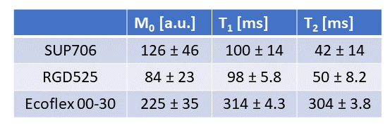
Relaxometry results are shown in figure 2, showing that the T1 of SUP706 is quite a bit lower than physiological values (~1100 ms) while the T2 is comparable (~50 ms). The SUP706 had a ~50% higher overall SNR compared to the conventional MR-visible RGD525.Figure 3 shows the evaluation of water tightness for different thicknesses of the MR-invisible shell material. The shell material can be reduced to a thickness of 0.5 mm without risking water seepage and saturation of the SUP706 material.
MR images obtained in the anatomical phantom are shown in Figure 4. Finally, real-time imaging snapshots corresponding to the simulated MR-guided intervention are shown in Figure 5.
Discussion
An anatomically realistic cardiac phantom was developed by using only standard 3D printing materials. This offers an accessible and easy alternative to conventional approaches, which require for example multiple casts in intermediate stages of the production process. The current cardiac phantom can serve as an anatomically realistic testing and training object, which can help both in technological developing as well as procedural training. Future extensions may include anatomical targets or inclusions with specific MR contrast.5Conclusion
An easy to produce and realistic MR-visible cardiac phantom has been presented for technological development and training purposes to improve MR-guided cardiac interventions.Acknowledgements
The authors would like to thank Dr. P. Segars for providing the digital phantom model and Dr. H. Mirgolbabaee for his assistance.References
1. Laing J, Moore JT, Vassallo R, Bainbridge D, Drangova M, Peters TM. Patient-specific cardiac phantom for clinical training and preprocedure surgical planning. https://doi.org/101117/1JMI52021222 2018;5:021222. doi: 10.1117/1.JMI.5.2.021222.
2. Thangavel K, Saritaş EÜ. Aqueous paramagnetic solutions for MRI phantoms at 3 T:A detailed study on relaxivities. Turkish Journal of Electrical Engineering and Computer Sciences 2017;25:2108–2121. doi: 10.3906/elk-1602-123.
3. Segars WP, Sturgeon G, Mendonca S, Grimes J, Tsui BMW. 4D XCAT phantom for multimodality imaging research. Med Phys 2010;37:4902–4915. doi: 10.1118/1.3480985.
4. Fedorov A, Beichel R, Kalpathy-Cramer J, et al. 3D Slicer as an image computing platform for the Quantitative Imaging Network. Magn Reson Imaging 2012;30:1323–1341. doi: 10.1016/J.MRI.2012.05.001.
5. Valladares A, Oberoi G, Berg A, Beyer T, Unger E, Rausch I. Additively manufactured, solid object structures for adjustable image contrast in Magnetic Resonance Imaging. Z Med Phys 2022;32:466–476. doi: 10.1016/J.ZEMEDI.2022.03.003.
Figures