1321
Modular open-source imaging phantom1Melbourne Brain Centre Imaging Unit, The University of Melbourne, Melbourne, Australia, 2Department of Biomedical Engineering & Graeme Clark Institute, The University of Melbourne, Melbourne, Australia, 3School of Science and Engineering, The University of Dundee, Dundee, United Kingdom
Synopsis
Keywords: Phantoms, Phantoms
Motivation: Overcome the high cost, lack of customisation and low global uptake of standard MRI imaging phantoms.
Goal(s): Design an affordable open-source MRI imaging phantom, imaging protocols and analysis tools demonstrated, showcasing their efficacy through the evaluation of slice profile and spatial resolution assessment.
Approach: A complex, multilayered phantom is designed with careful material selection aimed at low cost (US$500 worth of materials) and accessible construction principles. Slice profile and resolution measurements were performed using a double-wedge and a coarse resolution grid, respectively.
Results: This study has successfully produced an affordable open-source imaging phantom with detailed MRI assessment protocols.
Impact: This project's affordable, open-source MRI phantom and specialised assessment protocol benefit researchers by reducing costs and encouraging collaboration. The findings on slice profiles and resolution parameters enhance MRI research, improving imaging outcomes and accessibility.
Introduction
Many general-purpose and specialised variants are available in the realm of imaging phantoms, including a commercial version of the ISMRM-designed standard phantom1. Nevertheless, these offerings are often characterised by high cost and insufficient customisability concerning their internal composition, a critical feature in the research context. The present study addresses these issues by designing an economical, open-source imaging phantom and analysis software. Furthermore, we aimed to develop a comprehensive MRI acquisition and analysis quality assurance (QA) protocol tailored explicitly for assessing slice profile and resolution measurements.Methods
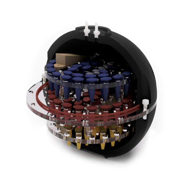
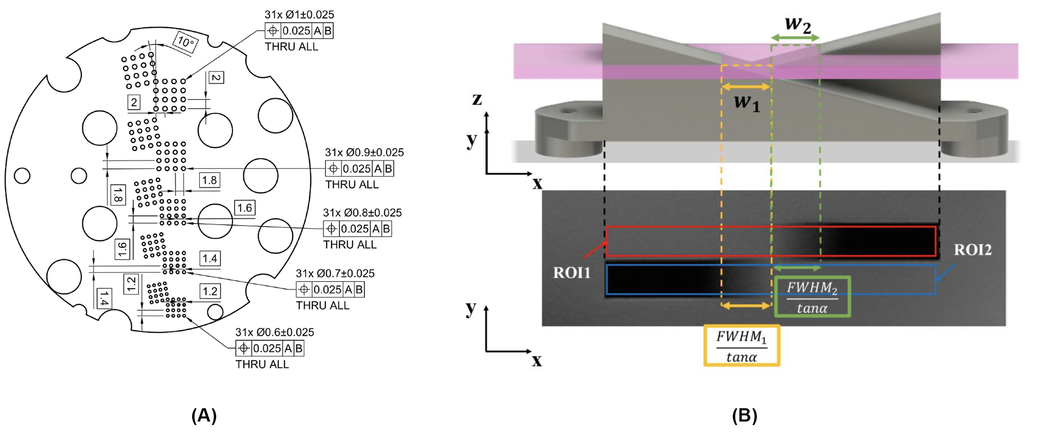
The imaging phantom developed within this project's scope comprises a complex, multilayered structure enclosed within a spherical shell measuring 170 mm in diameter (Figure 1). The phantom consists of three distinct layers, each equipped with multiple mounting holes designed to accommodate various modules, complemented by a coarse-resolution grid on each layer (Figure 2A). The top and bottom layers are securely affixed to a central disc between the two hemispheres. Two silicone O-rings are strategically placed between the intermediate disc and the hemispheres to ensure a fluid-tight seal.The materials for this imaging phantom were carefully selected, considering critical factors such as magnetic susceptibility, water absorption properties, mechanical strength, cost-effectiveness, and ease of machinability for each prospective candidate material. Consequently, the discs are fabricated from acrylic sheets, while the spherical shell is machined from an acetal rod. The phantom was filled with 0.9% NaCl solution to facilitate suitable coil loading and to create an appropriate background signal.
The slice profile in MRI is defined as a representation of the MR signal orthogonal to the plane of the imaging slice2. In this context, the slice profile's full width at half maximum (FWHM) value is denoted as the slice thickness. The slice profile was measured using the 3D-printed double-wedge module comprised of two crossed wedges oriented at a 15-degree angle to each other (Figure 2B) as outlined in the NEMA MS 5:2018 standard2. Specifically, the measurement protocol involved defining the axis and a region of interest (ROI) within each wedge to compute two edge response functions (ERFs). Two widths, w1 and w2, were calculated, and the values were corrected for rotational error.
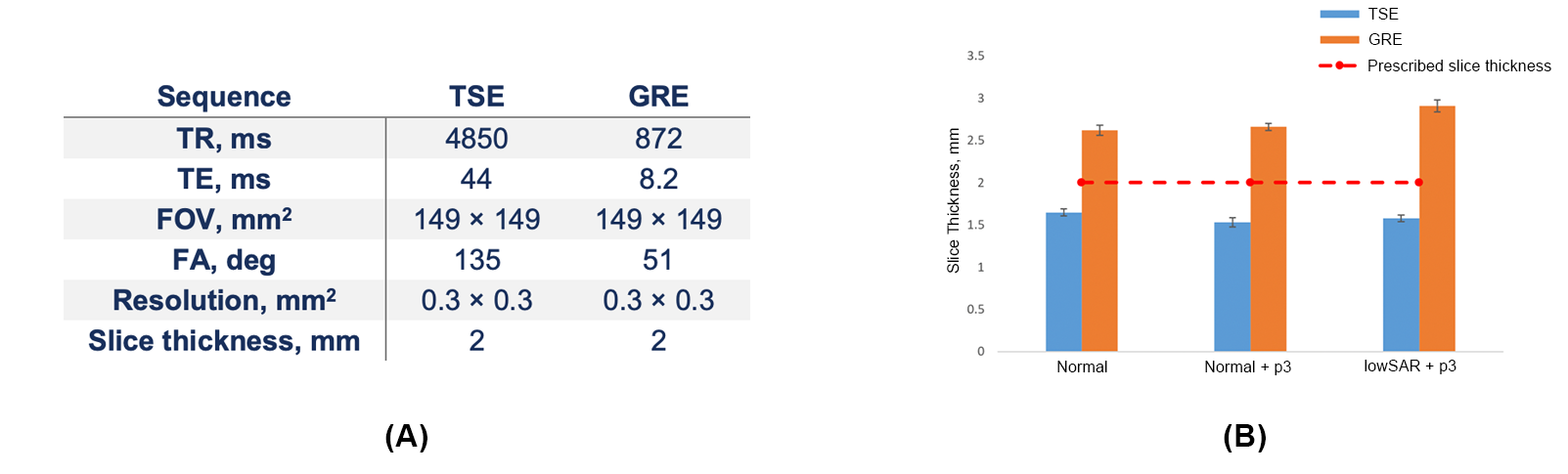
To demonstrate the phantom’s applicability for slice profile QA, we assessed commercially available 2D turbo spin echo (TSE) and gradient echo (GRE) sequences with different slice selection pulses, such as 'normal' and 'lowSAR' options. The sequence parameters are outlined in Figure 3A. Experiments were performed on a Magnetom 7T Plus MRI scanner (Siemens Healthcare, Erlangen, Germany). The phantom was positioned on a phantom holder in a 1Tx/32Rx head coil (Nova Medical, Wilmington, MA).
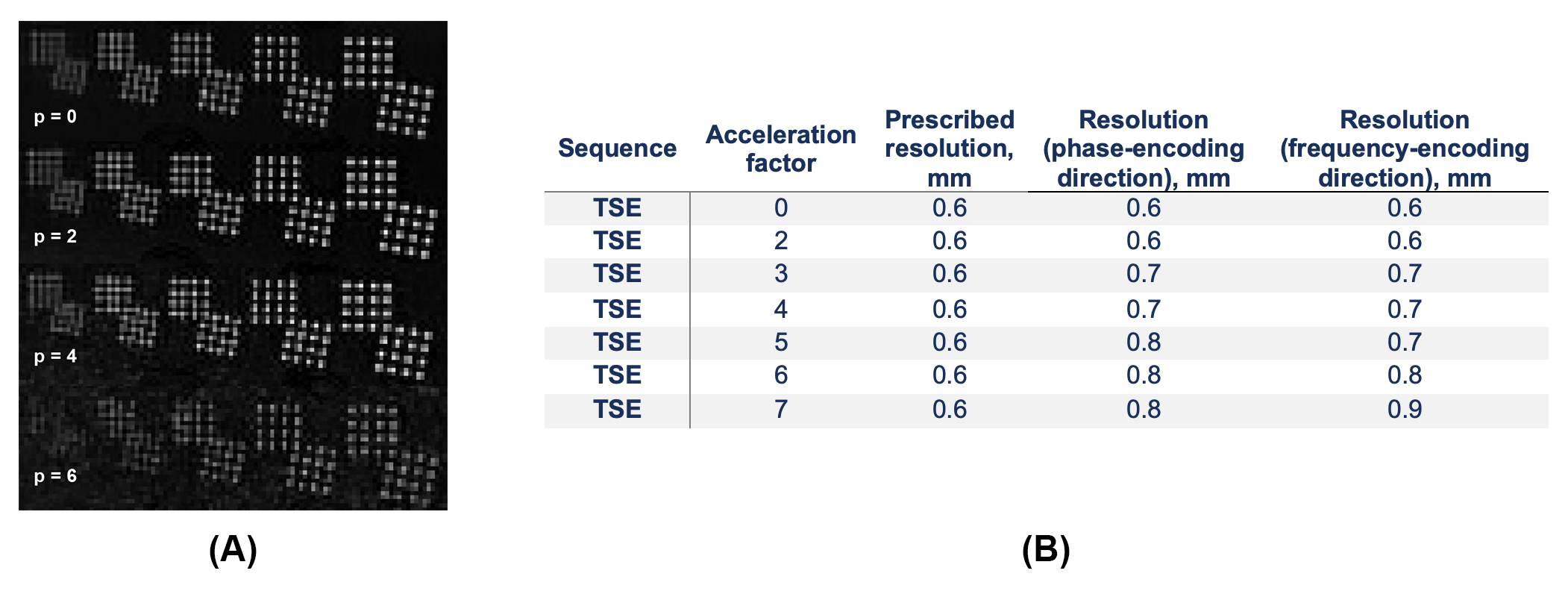
Resolution assessments were conducted on a coarse resolution grid (Figure 2B) using the TSE sequence, with varying GRAPPA acceleration factors ranging from 0 to 7, while maintaining constant parameters: TR = 4850 ms, TE = 44 ms, ETL = 11, FOV = 150×150 mm², flip angle = 135 deg, resolution = 0.3×0.3 mm², and slice thickness = 2 mm.
Results & Discussion
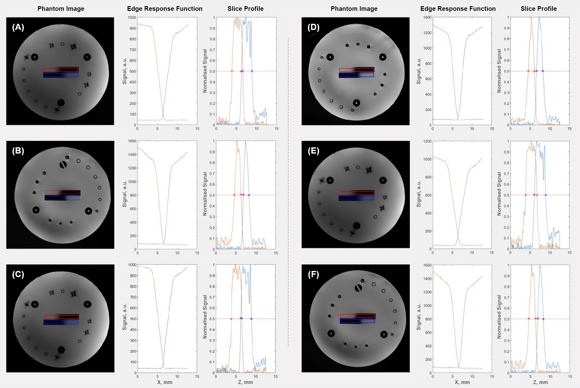
Figure 4 illustrates the phantom images, as well as measured slice thicknesses. Notably, the images acquired with the TSE sequence exhibited slice thicknesses narrower than the prescribed values. In contrast, the GRE sequence produced thicker slices than the specified thickness. Measurements obtained from four consecutive slices are presented as a bar chart (Figure 3B). No significant difference in slice thickness was observed when GRAPPA was enabled. Additionally, the 'lowSAR' mode appeared to further increase the thickness of slices generated by GRE, but it did not notably affect those obtained through TSE.Analysing the spatial resolution results, it was evident that up to 4-fold acceleration can be achieved without significant degradation of image resolution (Figure 5A). Furthermore, visual inspection suggests that applying GRAPPA tends to compromise image resolution by reducing the signal-to-noise ratio (SNR) of the image rather than blurring the image's edges. The measurements for different GRAPPA factors suggested that the prescribed in-slice resolution was maintained up to a factor of 3 (Figure 5B)
Conclusion
Performing QA of slice profile and spatial resolution is not readily available, as typically only homogeneous phantoms are provided by scanner vendors, while specialised phantoms are available at high cost. We have designed an affordable and open-source imaging phantom with a specialised acquisition and analysis protocol for assessing slice profile and resolution measurements. The design files, as well as scripts, are made available in our GitHub repository: https://github.com/igoresso/Phantom-DeathStar. This type of phantom is widely applicable for enabling MRI centres to evaluate the effects of common pulse sequence parameter settings on real spatial resolution and slice thickness.Acknowledgements
The authors acknowledge the facilities and scientific and technical assistance of the National Imaging Facility, a National Collaborative Research Infrastructure Strategy (NCRIS) capability, at the Melbourne Brain Centre Imaging Unit, University of Melbourne.References
- Stupic KF, Ainslie M, Boss MA, et al. A standard system phantom for magnetic resonance imaging. Magn Reson Med. 2021;86(3):1194-211.
- National Electrical Manufactures Association. Determination of slice thickness in diagnostic magnetic resonance imaging MS5-2008. Rosslyn, VA: NEMA Standards Publication; 2018.
Figures