1318
A novel 3D printed anthropomorphic phantom for evaluation of MR image characteristics1Mayo Clinic, Jacksonville, FL, United States
Synopsis
Keywords: Phantoms, Phantoms, image quality evaluation
Motivation: The typical MRI phantoms with simple geometric inserts in homogenous background do not allow in-depth evaluation of MR image characteristics necessary for assessing advanced imaging and reconstruction techniques.
Goal(s): To develop phantoms that can produce images with realistic anatomical structure that are more suitable for thorough evaluation of MR image characteristics.
Approach: We developed an approach to construct novel anthropomorphic phantoms using 3D-printing technique and use an example to demonstrate the utilization of this phantom for image quality evaluation.
Results: The phantom created with this approach can produce images resembling original MRI images acquired on human subjects.
Impact: The phantom generated using the proposed approach allows in-depth evaluation of MR image characteristics utilizing images with realistic anatomical structure, which may be especially beneficial for developing and evaluating advanced data acquisition and reconstruction techniques.
Background
MRI phantoms are widely used for various purposes in clinical practice and research studies, such as assessing equipment performance, evaluating image characteristics, and comparing different imaging techniques. A typical MRI phantom, such as the phantom used in the American College of Radiology MRI accreditation program [1], consists of only simple geometric inserts (e.g., high/low-contrast resolution dots/disks/bars or slice thickness ramps) in a homogeneous background. Such phantoms are only suitable for basic evaluations like routine system performance monitoring and simple image characteristics assessment since they cannot produce images with appropriate anatomical structure to perform in-depth evaluation of imaging techniques. For example, it was shown that some advanced reconstruction algorithms, e.g., compressed sensing [2], can well-preserve high-contrast objects like large vessels in contrast-enhanced angiography, but might smooth out low-contrast, fine details in anatomical MR images such as soft tissues that is absent in simple MRI phantoms [3]. Therefore, MR phantoms that can produce images with realistic anatomical structure is more suitable for thorough evaluation of image characteristics [4]. In this work, we describe the use of 3D-printing technique to create MRI phantoms and show that this phantom can produce images resembling original MRI images acquired on human subjects.Methods
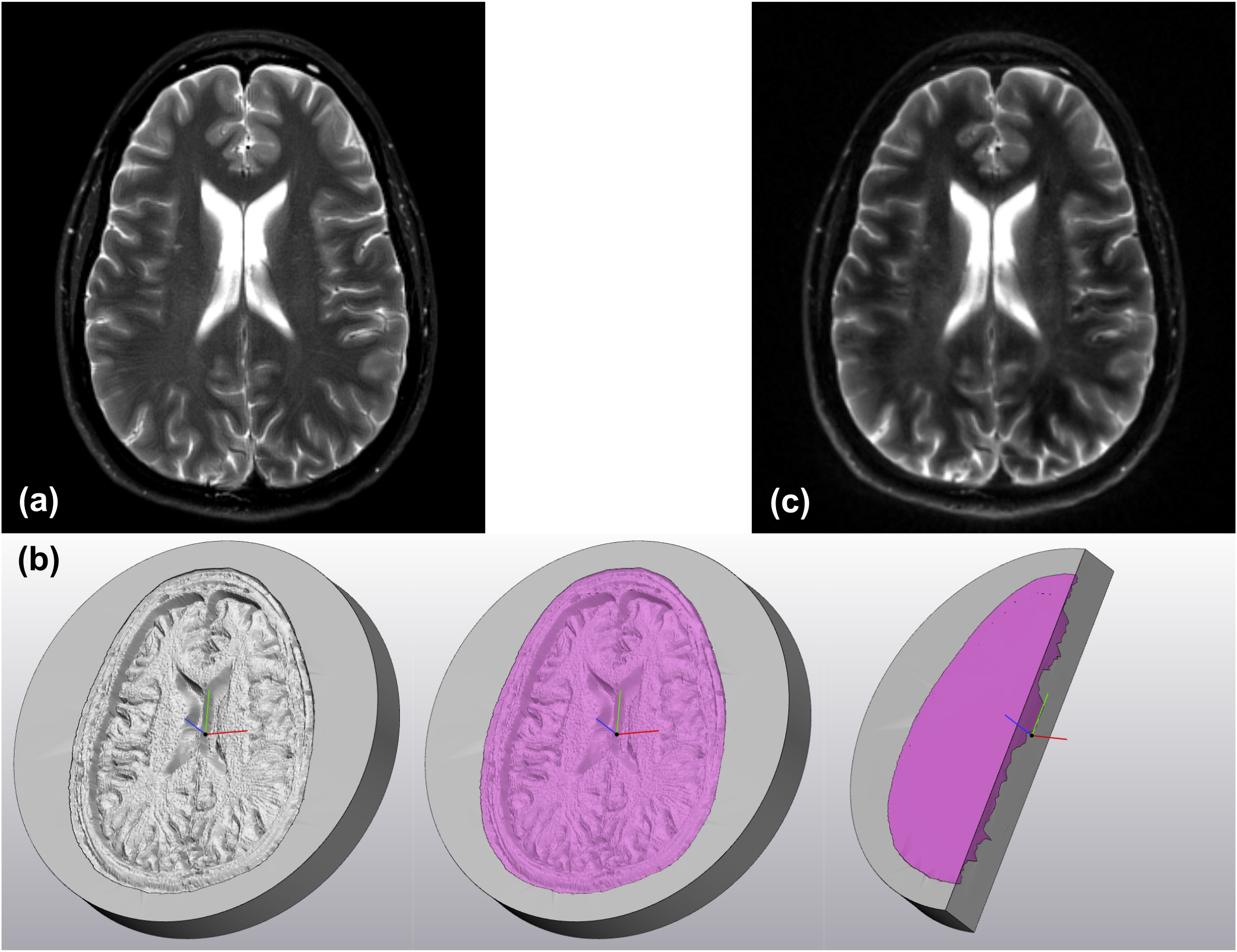
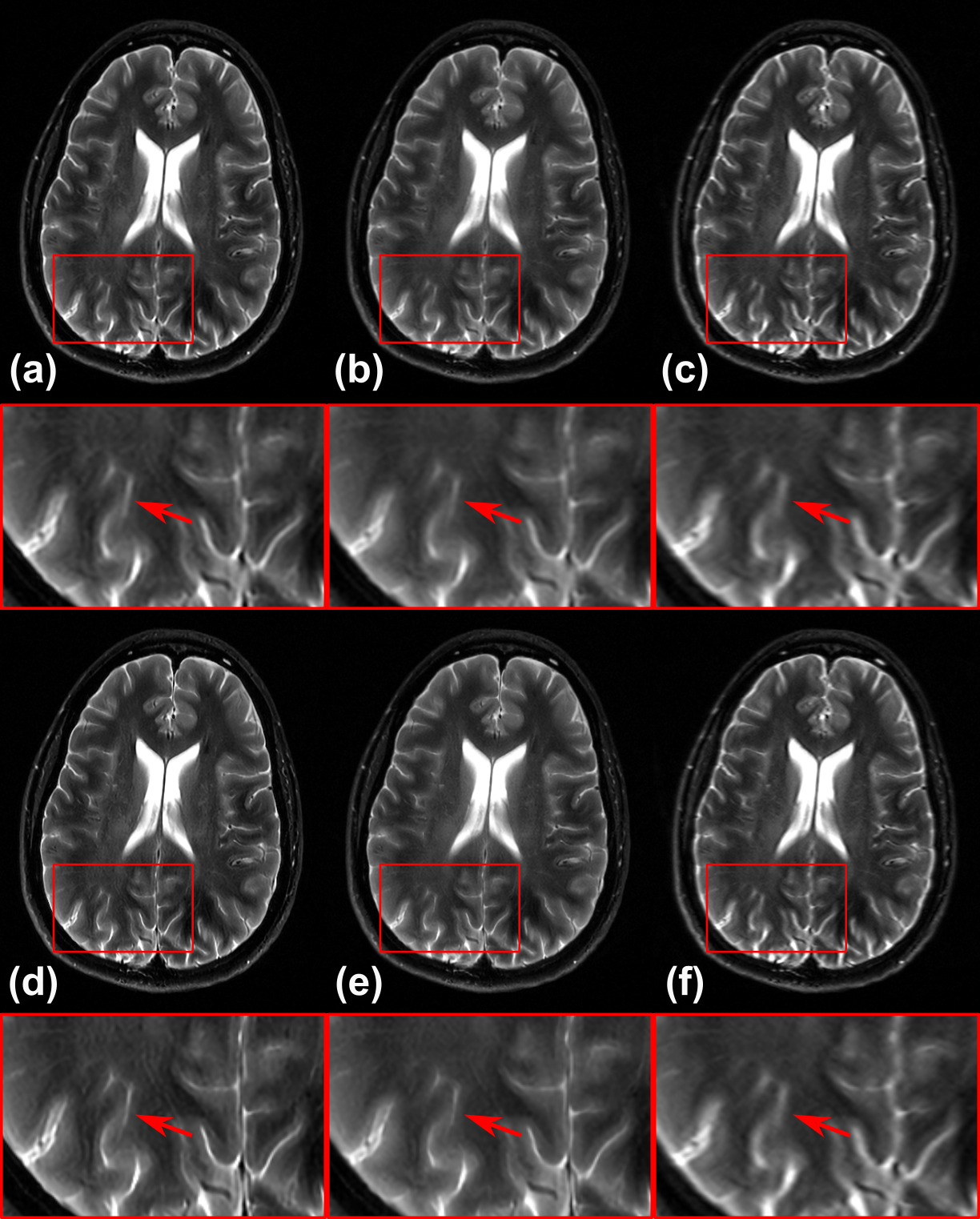
With the proposed approach, a 2D MRI image acquired on a human subject (Fig. 1a) was used to create the 3D model (Fig. 1b). This model was 3D printed (Stratasys J750) as a single assembly with MR visible “GelMatrix” material encased in solid material without detectable MR signal. The thicknesses of the two printing materials at different locations in the model were modulated by the pixel intensity of the source 2D MRI image. The phantom produces the same image contrast as in the source image by utilizing partial volume effect. When the phantom is imaged with a 2D sequence, and when the entire gel region is covered within the imaging slice, the partial-volume effect in the slice direction can produce variable signal intensities at different locations determined by the relative amount of MRI visible/invisible materials at that location. To test this approach, a T2-weighted brain image acquired on a Siemens 3T MRI scanner was used to create the 3D-printed phantom. To demonstrate how this phantom may be used for evaluating the effect of acquisition parameters and advanced processing techniques, we scanned it using the same sequence but with different echo train lengths (ETL=11/21/31), and reconstructed without and with a DL-based image sharpening technique (“Deep Resolve Sharp”, or DRS, on Siemens scanners) [5].Results
Figure 1a-c show: the original image, a rendering of the 3D-printed model, and the image acquired from the 3D-printed phantom, respectively. Note how the phantom structure and the thicknesses of MR visible/invisible printed materials are modulated by the pixel intensity of the original image. Also note how the phantom image resembles the original image. Figure 2 shows the phantom images acquired using different ETLs, and reconstructed without DRS. It is well-known that longer ETL can introduce blurring effects, which is demonstrated in Fig. 2. Images acquired with the same ETL as a-c but reconstructed with DRS (d-f) are visually sharper compared to a-c. DRS appears more effective for shorter ETL=11/21, where fine anatomical structures are sharpened, but less so for longer ETL=31, which demonstrates the efficacy and limitation of this technique.Discussion
We described the design of an MR phantom using 3D printing techniques and demonstrated how the phantom may be used to evaluate image quality of acquisition parameters (ETL) and processing algorithms (DRS). Although scanning human volunteers for image evaluation is a common practice, human subjects do not necessarily have the pathologies of interest and may be subject to various artifacts such as motion. Additionally, imaging human subjects is subject to various regulations and requires appropriate Institutional Review Board approval. Scanning phantoms, however, allows more flexibility. The proposed method has a few limitations. Because it uses partial volume effect in the slice direction, it can only be used with a 2D sequence. However, 2D imaging is still widely used in routine MRI. Secondly, the phantom was imaged soon after printing, so does not allow evaluation of long-term stability, which is to be determined in a follow-up test.Conclusion
We constructed an MR phantom using 3D printing techniques which allows generation of images resembling real MRI images acquired on human subjects and demonstrated its applications in a simple example.Acknowledgements
No acknowledgement found.References
[1] Phantom test guidance for the ACR MRI accreditation program. Reston: The American College of Radiology, Reston, Virginia, 2005.
[2] Lustig M, Donoho D, and Pauly JM. Sparse MRI: The application of compressed sensing for rapid MR imaging. Magn Reson Med 2007;58:1182-1195.
[3] Trzasko JD et al. Sparsity and Low-Contrast Object Detectability. Magn Reson Med 2012;67:1022-1032.
[4] Conzelmann J et al. Comparison of low‑contrast detectability between uniform and anatomically realistic phantoms—influences on CT image quality assessment. European Radiology 2022;32:1267-1275.
[5] https://www.siemens-healthineers.com/en-us/magnetic-resonance-imaging/options-and-upgrades/clinical-applications/deep-resolve-sharp
Figures