1312
Blood-brain barrier breakdown in Alzheimer's disease and dementia with Lewy Bodies based on water exchange DCE-MRI1Center for Biomedical Imaging Research, School of Medicine, Tsinghua University, Beijing, China, Beijing, China, 2Department of Neurology, Beijing Friendship Hospital, Capital Medical University, Beijing, China, Beijing, China, 3Department of Neurology, Tianjin Huanhu Hospital, Tianjin, China, Tianjin, China
Synopsis
Keywords: Microstructure, DSC & DCE Perfusion, blood-brain barrier; Alzheimer's disease; dementia with Lewy bodies
Motivation: Transfer rate of contrast agent and water plays complementary role in assessing the integrity of blood-brain barrier, however there is currently no means of evaluating them simultaneously in dementia patients.
Goal(s): We aimed to develop a new pharmacokinetic model to comprehensively evaluate the blood-brain barrier damage in dementia patients.
Approach: Transfer rate of contrast agent and water was calculated based on a new pharmacokinetic model by simultaneous fitting of the Bloch–McConnell equation and the Patlak model.
Results: The proposed model was able to exhibit distinct patterns of blood-brain barrier damage in different types of dementia, which was significantly associated with cognitive impairment.
Impact: The transfer rate of contrast agent and water based on water exchange DCE-MRI demonstrated distinct patterns of blood-brain barrier damage in patients with Alzheimer’s disease and dementia with Lewy bodies, providing promising new non-invasive imaging biomarkers for dementia.
Introduction
Alzheimer’s disease (AD) and dementia with Lewy bodies (DLB) are the two most prevalent causes of dementia1,2. Although their pathological mechanisms are controversial, several evidence suggested involvements of the blood-brain barrier (BBB) dysfunction in both AD and DLB3,4. Dynamic contrast-enhanced MRI (DCE-MRI) provides in vivo accessibility of quantitatively evaluating BBB leakage to contrast agent (CA) and has been reported to be associated with cognitive decline in AD5,6. Additionally, arterial spin labeling (ASL) has emerged as a new method to measure the water exchange rate across the BBB7,8, providing complementary information on BBB integrity with DCE-MRI9. So in this study, we aim to comprehensively evaluate BBB damage in AD and DLB patients by simultaneously measuring BBB leakage to CA and water.Methods
Subjects: This study was approved by the Ethical Review Board and written informed consents were obtained from all participants. Patients with probable AD diagnosed according to the National Institute on Aging and Alzheimer’s Association criteria10, patients with probable DLB according to the criteria McKeith established in 201711 and age-matched healthy controls (HC) were recruited. Essential demographic and clinical information were recorded.Imaging Protocol: All participants were scanned on a 3T MR scanner with a 64-channel head coil. DCE images were acquired with the following parameters: TR/TE = 5.2/1.8 ms, resolution = 1.25×1.25×3 mm3; imaging time = 30 × 12s, a bolus of 0.1mmol/kg of Gd-DTPA was injected intravenously at a rate of 2mL/s. Pre-contrast magnetization prepared-rapid gradient echo (MPRAGE), T1 mapping and B1 mapping were also acquired.
MR Imaging Analysis: Global cerebral cortex, frontal lobe, temporal lobe, parietal lobe and occipital lobe were regarded as regions of interest (ROIs) according to previous studies12,13. ROIs were manually segmented on MPRAGE images by two experienced radiologists who were blinded to the result of BBB leakage.
Pharmacokinetic Model: We assume that the CA can only be distributed in blood (b) and extracellular extravascular space (o), while the water molecule is well mixed in the blood plasma and blood cells (i). CA extravasation rate constant Ktrans and plasma volume fraction vp were analyzed using the Patlak model14 and the arterial input function was extracted from those pixels with rapid enhancement in internal carotid artery15. Then intracellular-to-extracellular water exchange rate constant kio and intra-to-extravascular water exchange rate constant kbo were analyzed based on Bloch–McConnell equations16.
$$ \frac{d\left(\begin{matrix}{\ M}_b\\{\ M}_o\\{\ M}_i\\\end{matrix}\ \right)}{dt}=\left(\begin{matrix}-\left(\frac{1}{T_{1b}}+k_{bo}\right)&k_{ob}&0\\k_{bo}&-\left(\frac{1}{T_{1o}}+k_{ob}+k_{oi}\right)&k_{io}\\0&k_{oi}&-\left(\frac{1}{T_{1i}}+k_{io}\right)\\\end{matrix}\right)\left(\begin{matrix}{\ M}_b\\{\ M}_o\\{\ M}_i\\\end{matrix}\ \right)+\left(\begin{matrix}\frac{M_{b0}}{T_{1b}}\\\frac{M_{o0}}{T_{1o}}\\\frac{M_{i0}}{T_{1i}}\\\end{matrix}\right)$$
where Mx (x=b, o, i) represents the longitudinal magnetization, T1x represents the longitudinal relaxation time and “0” represents the steady state. Water exchange rate constant kxy and kyx (x,y = b, o, i) are mutually related based on the volume fraction vx and vy. Processing pipeline for water exchange DCE-MRI was shown in Figure 1.
Statistical Analysis: Analysis of variance (ANOVA) or Kruskal-Wallis H test or chi-squared test was performed to compare water exchange DCE parameters among the three groups. Spearman’s correlation was used to analyze the correlation between the significant DCE parameters and clinical characteristics in all patients. P values of less than 0.05 were considered significant.
Results
A total of 25 patients with AD, 18 patients with DLB and 24 HC were included in this study. The demographic and clinical characteristics of the three groups are presented in Table 1. DLB group showed significantly higher Ktrans in the cerebral cortex compared to both AD group and HC group. Specifically, DLB demonstrated significantly higher Ktrans in the temporal lobe than AD, as well as higher Ktrans in the parietal lobe and occipital lobe than HC. AD patients, on the other hand, demonstrated significantly lower kio and kbo in the cerebral cortex than HC, but no significant differences were found between DLB and HC groups in the cerebral cortex (Figure 2). Spearman’s correlation analysis revealed significant associations between Ktrans in the cerebral cortex and parietal lobe with plasma Aβ1-42/Aβ1-40 ratio across all patients (Figure 3). Moreover, Ktrans in the parietal lobe, kio in the temporal and occipital lobes showed significant associations with mini-mental state examination (MMSE) in all patients (Figure 4).Discussion and Conclusions
To our best known, our study firstly reported the BBB leakage of CA and water molecule simultaneously in dementia patients. Based on the proposed new pharmacokinetic model, we found that patients with AD and DLB exhibited different patterns of BBB damage. AD patients demonstrated a significant decline in their ability to actively transport water, while DLB patients showed a significantly increase in BBB leakage to CA. Furthermore, the water exchange DCE parameters demonstrated significant correlations with cognitive impairment, thus providing promising new non-invasive imaging biomarkers for dementia.Acknowledgements
None.References
1. Blennow K, de Leon MJ, Zetterberg H. Alzheimer's disease. Lancet. 2006 Jul 29;368(9533):387-403.
2. Sanford AM. Lewy Body Dementia. Clin Geriatr Med. 2018 Nov;34(4):603-615.
3. Benarroch EE. Neurovascular unit dysfunction: a vascular component of Alzheimer’s disease? Neurology. 2007 May 15;68(20):1730-2.
4. Wong YY, Wu CY, Yu D, et al. Biofluid markers of blood-brain barrier disruption and neurodegeneration in Lewy body spectrum diseases: A systematic review and meta-analysis. Parkinsonism Relat Disord. 2022 Aug;101:119-128.
5. Montagne A, Barnes SR, Sweeney MD, et al. Blood-brain barrier breakdown in the aging human hippocampus. Neuron. 2015 Jan 21;85(2):296-302.
6. van de Haar HJ, Burgmans S, Jansen JF, et al. Blood-Brain Barrier Leakage in Patients with Early Alzheimer’s disease. Radiology. 2016 Nov;281(2):527-535.
7. Gold BT, Shao X, Sudduth TL, et al. Water exchange rate across the blood-brain barrier is associated with CSF amyloid-β 42 in healthy older adults. Alzheimers Dement. 2021 Dec;17(12):2020-2029.
8. Shao X, Zhao C, Shou Q, et al. Quantification of blood-brain barrier water exchange and permeability with multidelay diffusion-weighted pseudo-continuous arterial spin labeling. Magn Reson Med. 2023 May;89(5):1990-2004.
9. Voorter PHM, van Dinther M, Jansen WJ, et al. Blood-Brain Barrier Disruption and Perivascular Spaces in Small Vessel Disease and Neurodegenerative Diseases: A Review on MRI Methods and Insights. J Magn Reson Imaging. 2023 Sep 1.
10. McKhann GM, Knopman DS, Chertkow H, et al. The diagnosis of dementia due to Alzheimer's disease: recommendations from the National Institute on Aging-Alzheimer's Association workgroups on diagnostic guidelines for Alzheimer's disease. Alzheimers Dement. 2011 May;7(3):263-9.
11. McKeith IG, Boeve BF, Dickson DW, et al. Diagnosis and management of dementia with Lewy bodies: Fourth consensus report of the DLB Consortium. Neurology. 2017 Jul 4;89(1):88-100.
12. Poulose SM, Miller MG, Scott T, et al. Nutritional Factors Affecting Adult Neurogenesis and Cognitive Function. Adv Nutr. Nov 2017;8(6):804-811.
13. Liu S, Wang XD, Wang Y, et al. Clinical and neuroimaging characteristics of Chinese dementia with Lewy bodies. PLoS One. 2017;12(3):e0171802.
14. Buckley DL, Shurrab AE, Cheung CM, et al. Measurement of single kidney function using dynamic contrast-enhanced MRI: comparison of two models in human subjects. J Magn Reson Imaging. 2006 Nov;24(5):1117-23.
15. Chen H, Cai J, Zhao X, et al. Localized measurement of atherosclerotic plaque inflammatory burden with dynamic contrast-enhanced MRI. Magn Reson Med. 2010 Aug;64(2):567-73.
16. Bai R, Wang B, Jia Y, et al. Shutter-Speed DCE-MRI Analyses of Human Glioblastoma Multiforme (GBM) Data. J Magn Reson Imaging. 2020 Sep;52(3):850-863.
Figures
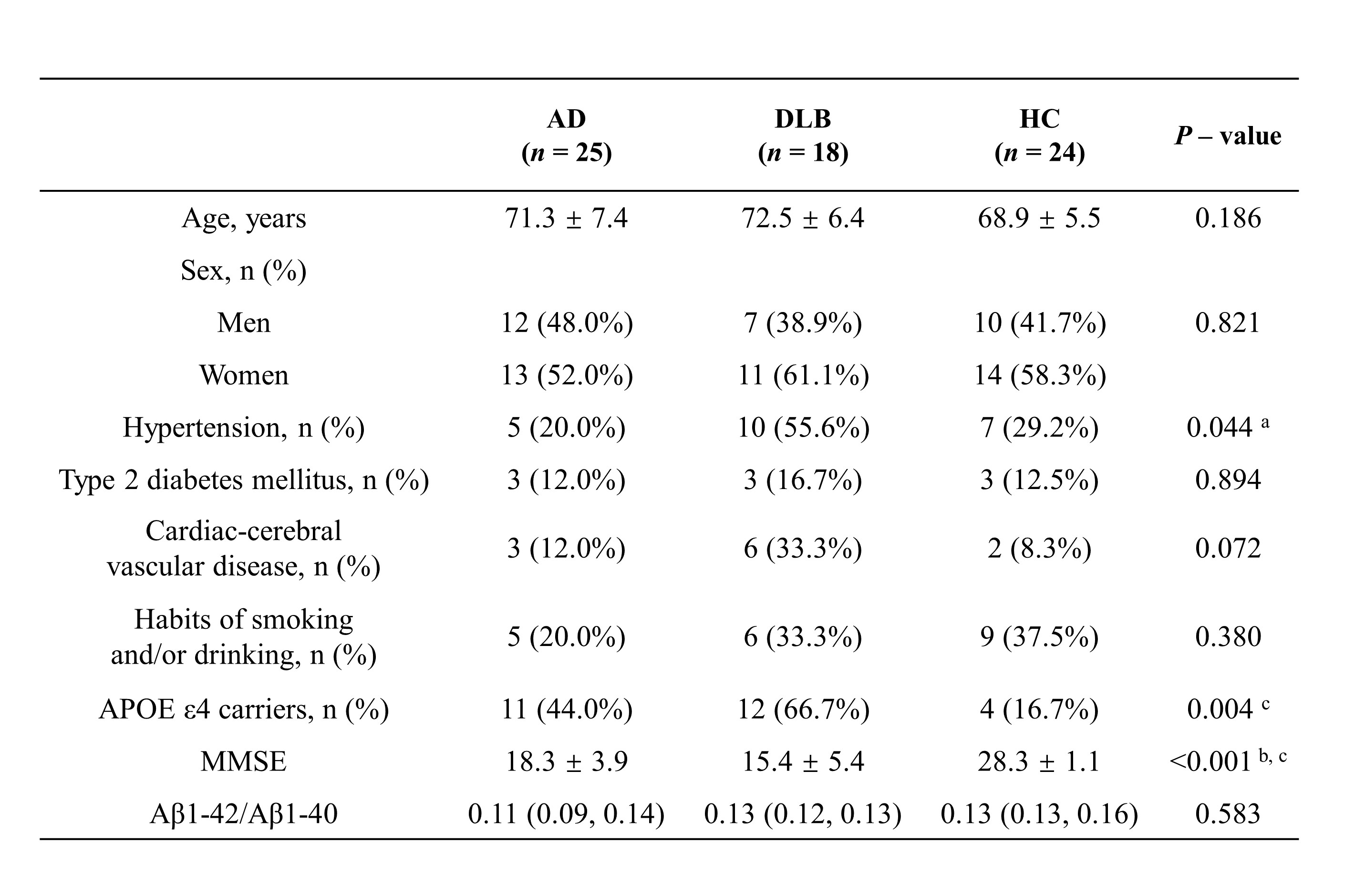
Table 1 Demographic and clinical information in all participants. Data are expressed as mean ± SE or median (quartiles) or n (%). a For the comparison between the AD and DLB (Bonferroni-corrected P < 0.05). b For the comparison between the AD and HC (Bonferroni-corrected P < 0.05). c For the comparison between the DLB and HC (Bonferroni-corrected P < 0.05). AD: Alzheimer’s disease; DLB: dementia with Lewy body; HC: healthy controls; APOE ε4: apolipoprotein E ε4 allele; MMSE: mini-mental state examination; Aβ: amyloid-beta.
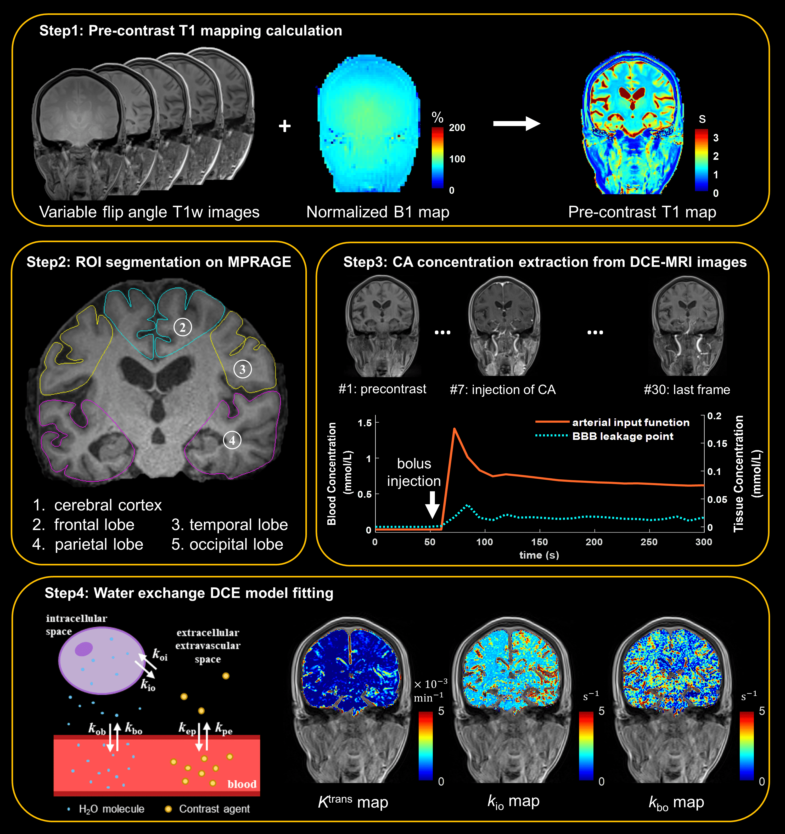
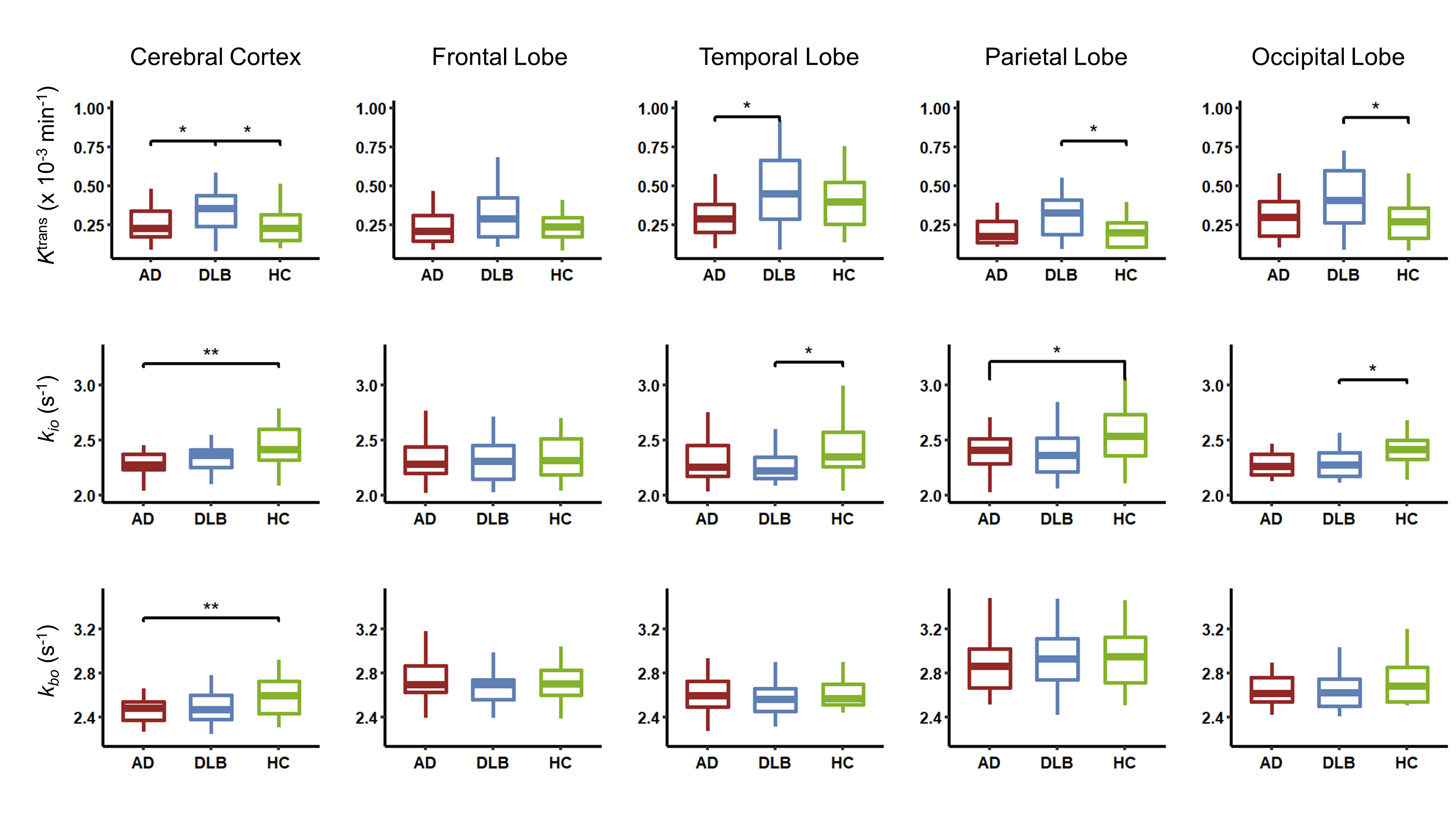
Figure 2. Comparison of Ktrans, kio and kbo among patients with AD, DLB and HC in cerebral cortex, frontal lobe, temporal lobe, parietal lobe and occipital lobe. Significance was tested by one-way analysis of variance (ANOVA) or Kruskal-Wallis H test. *: P < 0.05; **: P < 0.01. Ktrans: contrast agent extravasation rate constant; kio: water molecule cellular efflux rate constant; kbo: water molecule extravasation rate constant; AD: Alzheimer’s disease; DLB: dementia with Lewy body; HC: healthy controls.
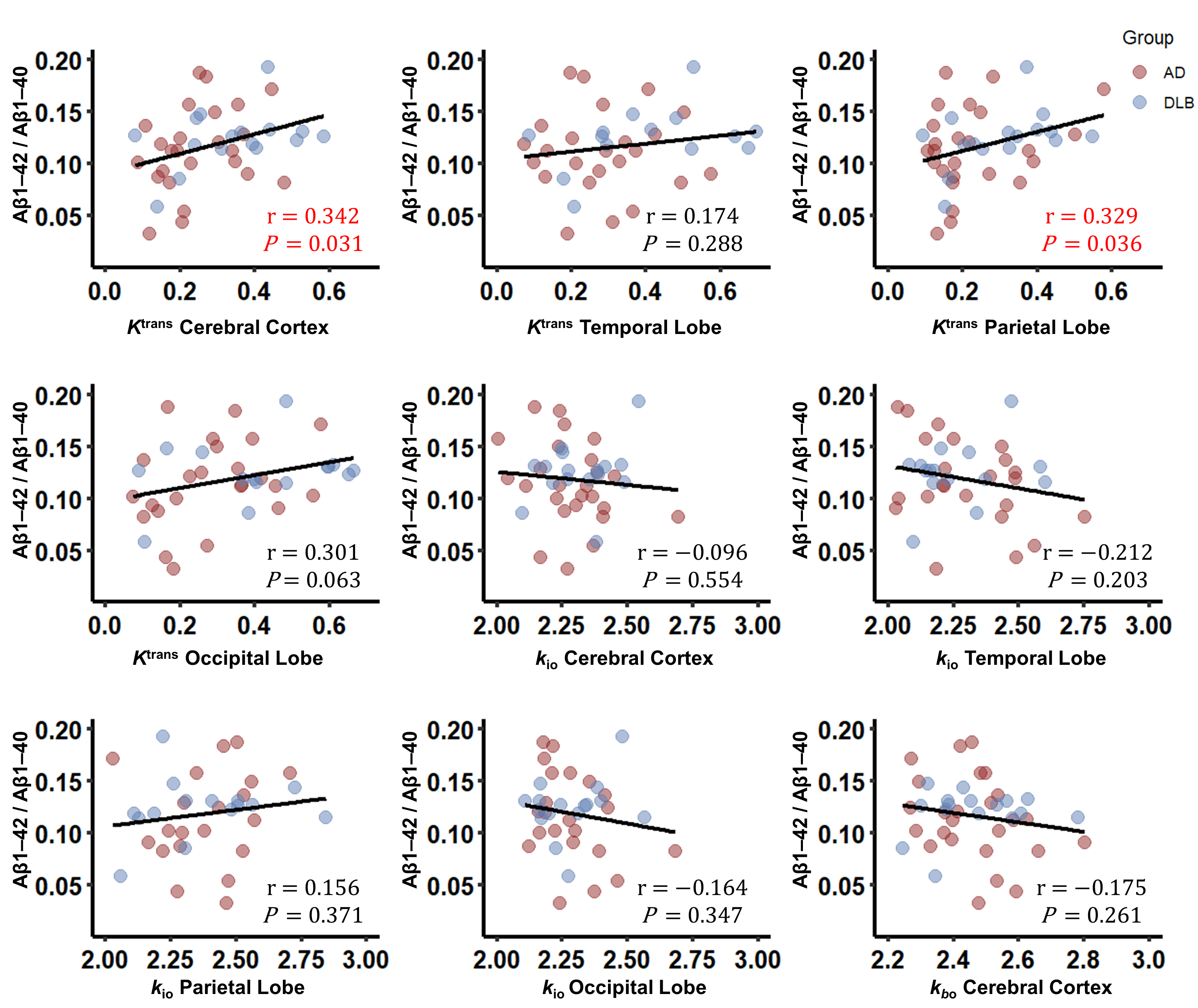
Figure 3. Correlations between significant Ktrans, kio, kbo and plasma Aβ1-42/Aβ1-40 ratio in all patients. Significance was tested by Spearman’s correlation. Aβ: amyloid-β; Ktrans: contrast agent extravasation rate constant; kio: water molecule cellular efflux rate constant; kbo: water molecule extravasation rate constant; AD: Alzheimer’s disease; DLB: dementia with Lewy body.
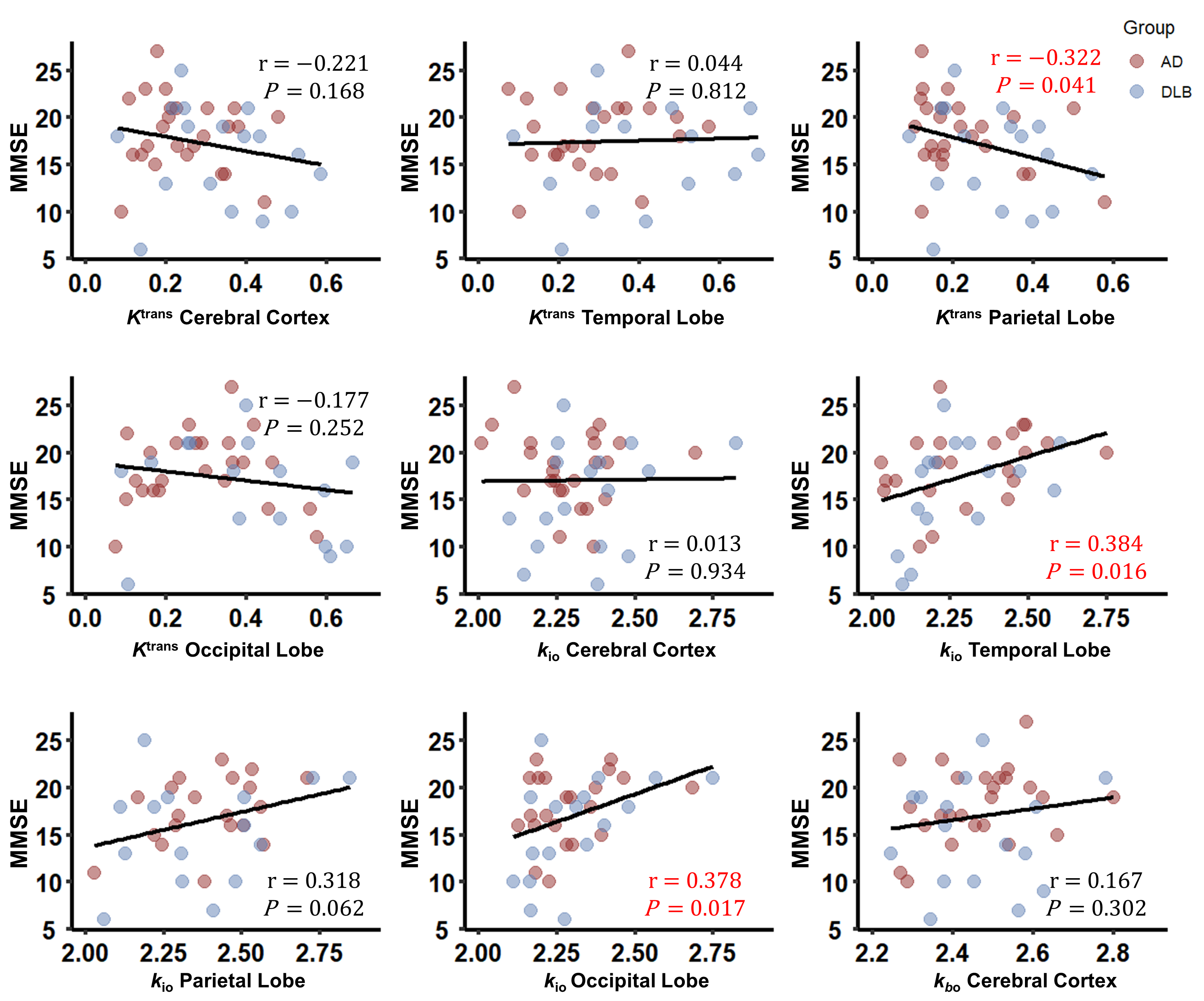
Figure 4. Correlations between significant Ktrans, kio, kbo and MMSE score in all patients. Significance was tested by Spearman’s correlation. MMSE: mini-mental state examination; Ktrans: contrast agent extravasation rate constant; kio: water molecule cellular efflux rate constant; kbo: water molecule extravasation rate constant; AD: Alzheimer’s disease; DLB: dementia with Lewy body.