1305
Quantifying Rotational Correlation Time in Clinical MRI Scanners: A Novel Framework for Enhanced Tissue Characterization1Department of Radiology, University Hospitals Cleveland Medical Center, Cleveland, OH, United States, 2Department of Biomedical Engineering, Case Western Reserve University, Cleveland, OH, United States, 3Case Western Reserve University School of Medicine, Cleveland, OH, United States, 4Department of Pediatrics, Case Western Reserve University, Cleveland, OH, United States, 5Seidman Cancer Center and Case Comprehensive Cancer Center, Cleveland, OH, United States
Synopsis
Keywords: Relaxometry, Contrast Mechanisms, Paramagnetic Relaxation Enhancement; Rotational Correlation Time; Clinical MR Relaxometry
Motivation: This study exploits the underexplored potential of paramagnetic relaxation enhancement (PRE) in clinical MR scanners to characterize molecular interactions and tissue microenvironments in vivo, leveraging Gadolinium-based contrast agents (GBCAs).
Goal(s): Develop and validate methodology for estimating Gadolinium-based contrast agent (GBCA) correlation time via relaxivity ratio measurements at 1.5 and 3 Tesla.
Approach: Applying relaxation models, we devised a dictionary-matching framework correlating GBCA relaxivities with correlation times, and validated our methods using phantom experiments.
Results: Our framework accurately estimates GBCA correlation times at a single field strength showing <5% error (1.5T to 3T) and <11% (3T to 1.5T) in cross-field relaxivity predictions.
Impact: This framework leverages MR Relaxometry for precise estimation of GBCA rotational correlation time at single field strengths, offering insights in tissue characteristics. There is significant potential to improve tumor imaging and diagnosis through insights into pH, viscosity, and protein interactions .
INTRODUCTION
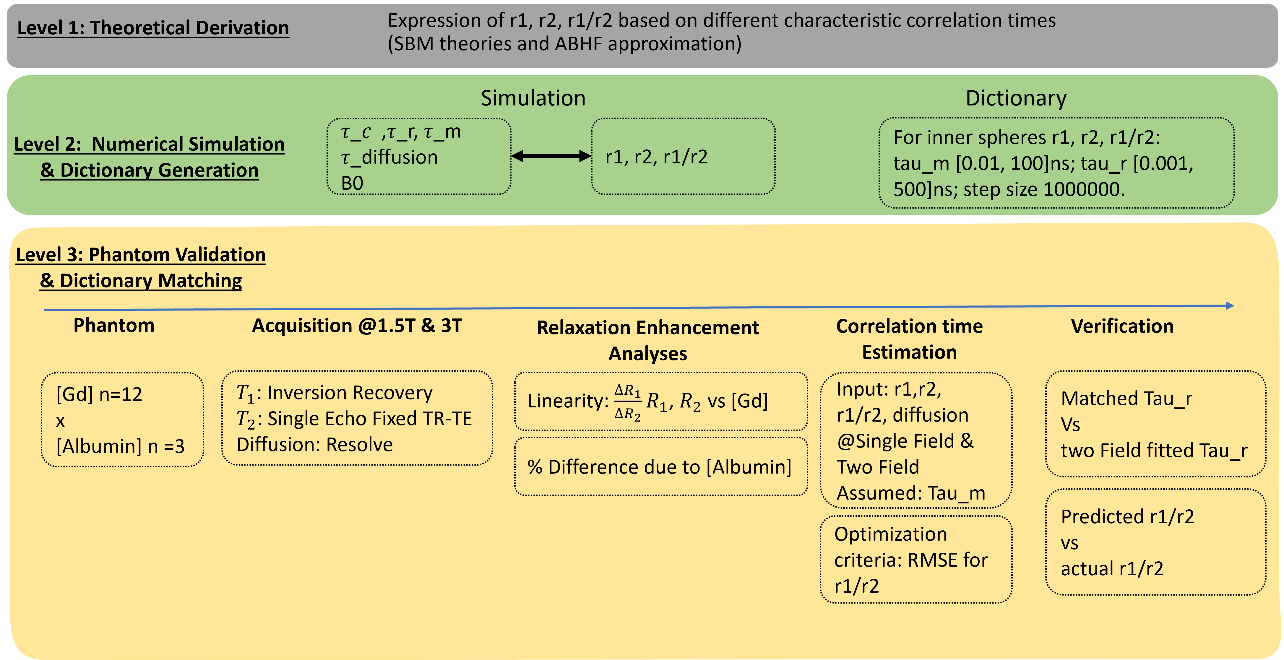
Paramagnetic Relaxation Enhancement (PRE) by exogenous contrast agents impacts solution proton relaxivity mainly via dipole-dipole interactions, quantified by characteristic correlation times 1. Correlation time, pivotal in field-varying NMR analysis for molecular interactions and tissue microenvironments 2,3,4, has been extrapolated to clinical MR field strength for potential in vivo insights 5,6. There is no method that estimates correlation time in clinical MR scanners, which will facilitate enhanced tissue characterization and tumor imaging.We developed a framework (Fig 1) for estimating heteronuclear spin-spin interaction correlation time via MR relaxometry ratios at 1.5 T and 3T, applying it to gadolinium-based contrast agents (GBCA) and validating via dictionary matching.
THEORY
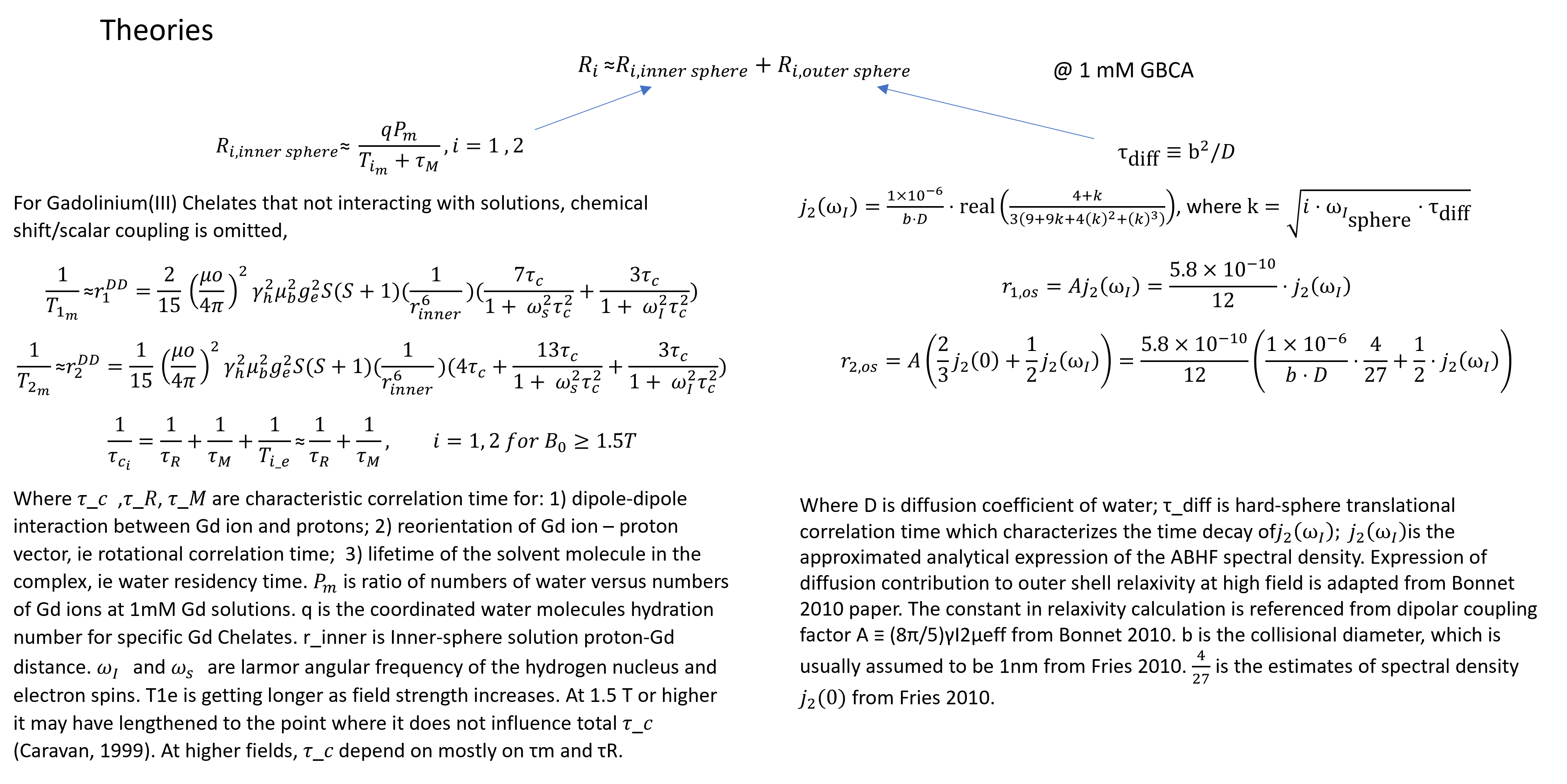
We employed the Solomon-Bloembergen-Morgan theory 6,7 for inner sphere interactions and the Ayant-Belorizky-Hwang-Freed model 8 for outer sphere interactions in modeling GBCA-solution interactions. Clinical GBCAs are designed to minimize tissue interaction, thus we can derive constant expressions for relaxivities (r1, r2, r1/r2) incorporating characteristic correlation times (τ_r, rotational correlation time; τ_m, water residency time; τ_diff, rotational diffusion correlation time) relevant for clinical MRI.METHODS
Numerical Simulation and Dictionary Generation:Simulations demonstrated the impact of characteristic correlation times on relaxivities. We assembled a relaxivity-correlation time dictionary with τ_m [0.01, 200]ns; τ_r [0.01, 100]ns; step size 1000000, utilizing field strength and diffusion coefficient as inputs.
Phantom Validation:
Validation employed T1 (SE), T2 (SE), and Diffusion (RESOLVE) protocols on clinical scanners (Siemens Vida and Aera), optimizing B1 homogeneity. Eleven gadolinium concentrations (0.04-1.2mM; Gd-DOTA, Guerbert) in buffered saline with varying albumin were imaged promptly to minimize albumin degradation.
Data Analysis:
The contrast-specific relaxivities (r1, r2, r1/r2) were calculated as (Ri-Ri_origional) / [Gd], with Ri and Ri_origional measured in a 100mm³ VOI. Dictionary matching was performed with both single field data and two field data, with r1/r2, and ADC as inputs. Assumed constants included hydration number, electron spin relaxation correlation time, and τ_m. Matched criteria included an RMSE between predicted and actual r1/r2. τ _r that qualified were pooled for the experimental mean estimated τ_r.
To validate the algorithym accuracy, non-linear fitting was performed. To validate the single field dictionary match τ_r results, predicted τ_r was used to calculate predicted r1/r2 of the other field, with %error to the actual r1/r2.
RESULTS
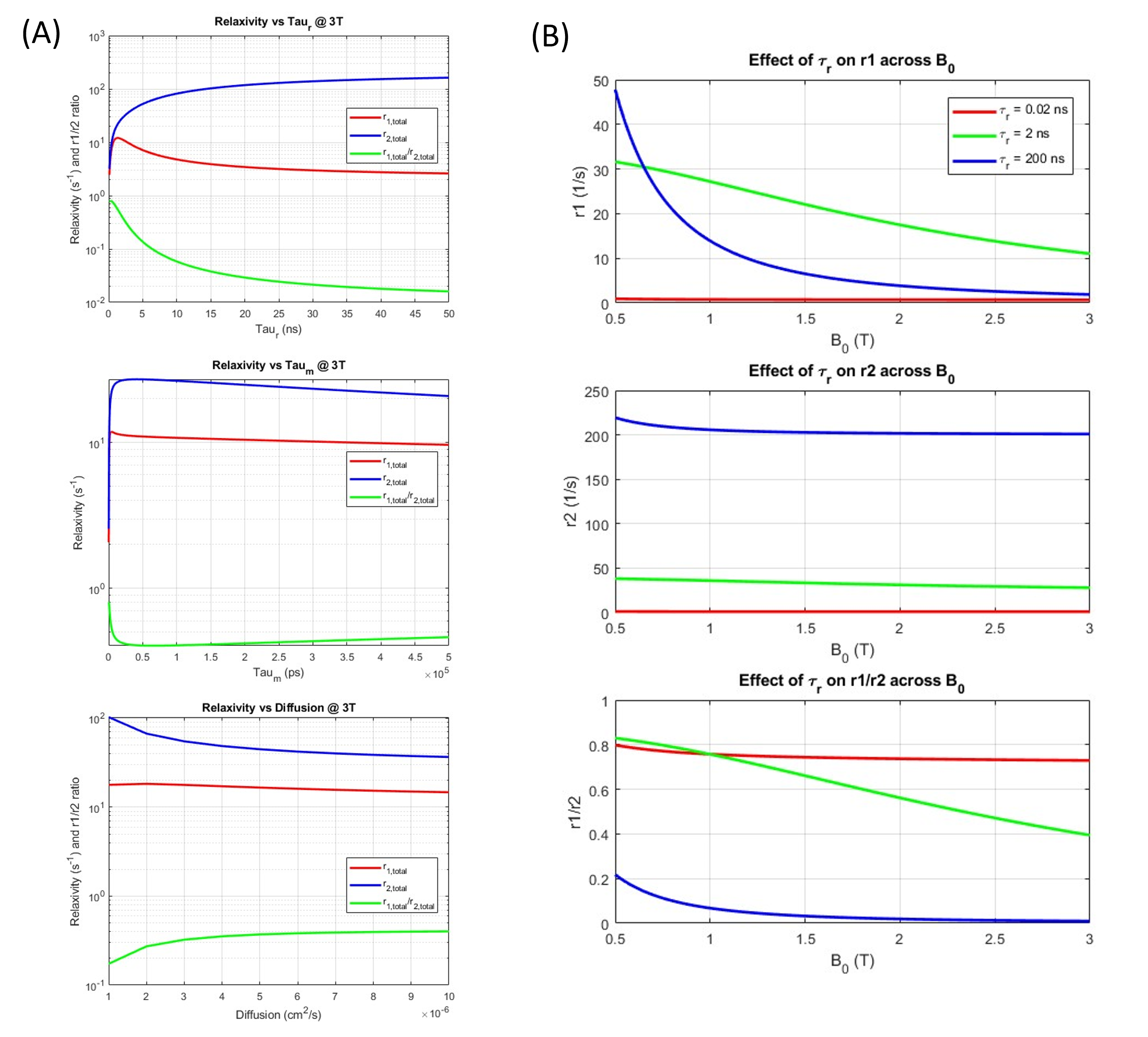
Theoretical Models and Simulations::Simulation illustrated the dominant effect of τ_r on r1,r2, and r1/r2 at 3T (Fig 3A), which provided the rationale for calibrating or assuming other correlation time constants (τ_m and τ_diff) and the design of cross-field validation. Specifically, diffusion scarcely impacted r1/r2 within the physiological range (~1x10⁻³ mm2/s). Varied magnetic field strengths altered the effect of rotational correlation time on relaxivities (Fig. 3B), with a notably larger impact at intermediate correlation time (2ns) compared to fast (0.02ns) and slow (200ns) values. These latter times represented scenarios of Gd complex in pure water and binding with very large macromolecules, respectively.
Phantom Validation:
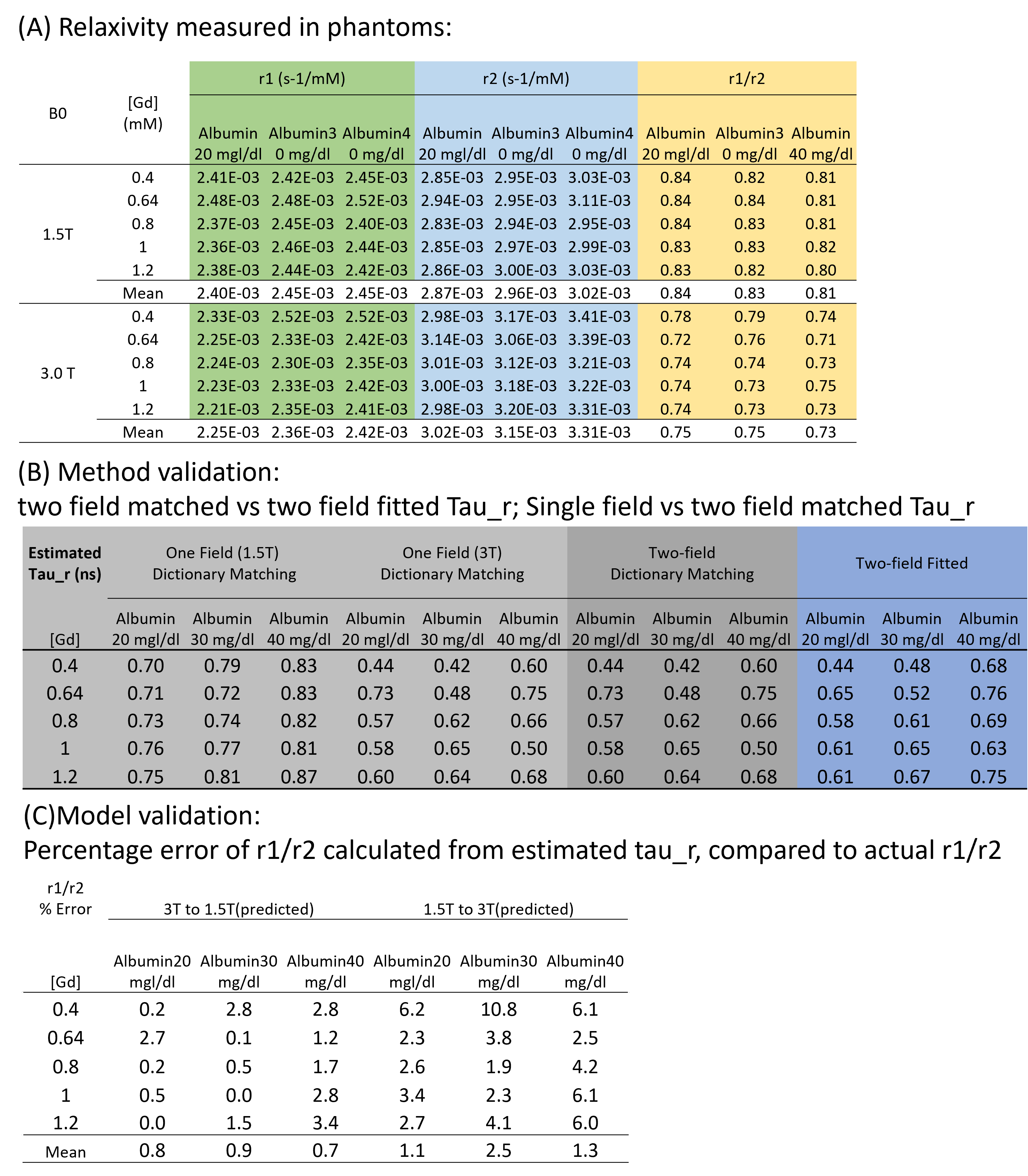
We highlighted good rotational correlation time estimation at 3T (<5%error, Fig 4C). The results was backed up by validation of relaxivity measurements (Fig 4A), and the validation of algorithm (Fig 4B).
Specifically, r1 and r2 were consistent in [Gd] between 0.4 to 1.2 mM (Fig 4A). The r1/r2 ratios decreased with a lower B0 and increased [albumin]. For algorithmic validation, two-field dictionary matching results were in good agreement with two-field non-linear fitted results. (Fig 4B) For model validation (Fig 4C), predicted r1/r2 using dictionary matched τ_r at 3T showed a <5% error, while <11% error at 1.5T. This indicated the model describes relaxation process at 3T better than 1.5T, potentially due to additional electron-spin relaxation mechanisms at lower fields.
DISCUSSION
Our methodology enables precise estimation of rotational correlation time within clinical MRI systems, adaptable to various contrast agents. Accurate relaxivity measurements are crucial for this estimation, yet they pose a challenge within the clinical setting due to unknown voxelwise GBCA concentrations in vivo9. As an alternative approach, concentration-independent relaxivity ratios (r1/r2), derived from Delta Relaxometry and Magnetic Resonance Fingerprinting10, can simplify the correlation time estimation process.The current phantom design manipulates rotational correlation time through varying albumin concentrations. The congruence of observed relaxivity with estimated rotational correlation time endorses the model's validity. The resulting data aligns with the predicted outcomes, reinforcing the reliability of our measurement techniques and supporting the framework's applicability.
CONCLUSION
We have introduced a framework for quantifying spin-spin interactions via MRI relaxometry in a single clinical magnetic field. Our preliminary results suggest that rotational correlation time can act as a contrast agent to illuminate microenvironment changes such as pH shifts, viscosity variations, and protein binding, paralleling the function of paramagnetic relaxation enhancement in NMR.Acknowledgements
This work was supported by Siemens Healthineers, NIH grants R01 CA269604, T32 EB007509, T32 GM007250, and TL1 TR000441, the Imaging Devices and AI Technologies Track Funding Agency (Jobs Ohio), an American Cancer Society Institutional Research Grant, and the Radiation Oncology Institute. We thank Mike Kavran for his support in providing laboratory space access.References
1. Clore, G.M. and Iwahara, J., 2009. Theory, practice, and applications of paramagnetic relaxation enhancement for the characterization of transient low-population states of biological macromolecules and their complexes. Chemical reviews, 109(9), pp.4108-4139.
2. Lenard, A.J., Mulder, F.A. and Madl, T., 2022. Solvent paramagnetic relaxation enhancement as a versatile method for studying structure and dynamics of biomolecular systems. Progress in Nuclear Magnetic Resonance Spectroscopy.
3. Baroni, S., Ruggiero, M.R., Aime, S. and Geninatti Crich, S., 2019. Exploring the tumour extracellular matrix by in vivo Fast Field Cycling relaxometry after the administration of a Gadolinium‐based MRI contrast agent. Magnetic Resonance in Chemistry, 57(10), pp.845-851.
4. Alford, J.K., Rutt, B.K., Scholl, T.J., Handler, W.B. and Chronik, B.A., 2009. Delta relaxation enhanced MR: improving activation‐specificity of molecular probes through R1 dispersion imaging. Magnetic Resonance in Medicine: An Official Journal of the International Society for Magnetic Resonance in Medicine, 61(4), pp.796-802.
5. Okuno, Y., Szabo, A. and Clore, G.M., 2020. Quantitative interpretation of solvent paramagnetic relaxation for probing protein–cosolute interactions. Journal of the American Chemical Society, 142(18), pp.8281-8290.
6. Caravan, P., Ellison, J.J., McMurry, T.J. and Lauffer, R.B., 1999. Gadolinium (III) chelates as MRI contrast agents: structure, dynamics, and applications. Chemical reviews, 99(9), pp.2293-2352.
7. Kowalewski, J., Nordenskiöld, L., Benetis, N. and Westlund, P.O., 1985. Theory of nuclear spin relaxation in paramagnetic systems in solution. Progress in nuclear magnetic resonance spectroscopy, 17, pp.141-185.
8. Fries, P.H., Imbert, D. and Melchior, A., 2010. Determination of outer-sphere dipolar time correlation functions from high-field NMR measurements. Example of a Gd3+ complex in a viscous solvent. The Journal of chemical physics, 132(4).
9. Matsumoto, Y., Harada, M., Kanazawa, Y., Taniguchi, Y., Ono, M. and Bito, Y., 2022. Quantitative parameter mapping of contrast agent concentration and relaxivity and brain tumor extracellular pH. Scientific Reports, 12(1), p.2171.
10. Deng, S., Zhao W., Jordan, D.W., Badve, C., Ma, D., 2023. Delta-relaxometry with contrast-enhanced MR Fingerprinting: phantom validation and application to tumor imaging. In: Proceedings of the 31st Annual Meeting of the ISMRM. Toronto, Canada;
Figures