1303
in vivo longitudinal imaging unravels the origin of neuromelanin-MRI contrast in a rat model of Parkinson’s disease1Paris Brain Institute – ICM, MOVIT team, Sorbonne Université, Inserm U1127, CNRS 7225, Hôpital Pitié-Salpêtriere, Paris, France, 2Paris Brain Institute – ICM, Centre de NeuroImagerie de Recherche – CENIR, Sorbonne Université, Inserm U1127, CNRS 7225, Hôpital Pitié-Salpêtriere, Paris, France, 3Paris Brain Institute – ICM, Data Analysis Core, Sorbonne Université, Inserm U1127, CNRS 7225, Hôpital Pitié-Salpêtriere, Paris, France, 4Aix-Marseille Univ, CRMBM, CNRS UMR 7339, APHM, La Timone Hospital, CEMEREM, Marseille, France, 5Neurodegenerative Diseases Research Group, Vall d’Hebron Research Institute (VHIR)-Network Center for Biomedical Research in Neurodegenerative Diseases (CIBERNED), Barcelona, Spain
Synopsis
Keywords: Novel Contrast Mechanisms, Contrast Mechanisms
Motivation: Neuromelanin-MRI contrast is a promising biomarker for Parkinson's disease, but still needs investigation as the biological and physical origins of the contrast are still unclear.
Goal(s): The objective was to unravel the mechanisms behind neuromelanin-MRI contrast, both biologically and physically, and to better understand the role of neuromelanin in Parkinson's disease.
Approach: We performed in vivo longitudinal neuromelanin-MRI coupled with quantitative multiparametric imaging on a rat model of Parkinson's disease based on accumulation of neuromelanin, with histological validation.
Results: Results show that contrast increases with neuromelanin accumulation and decreases with neuronal loss. The contrast arises from T1 reduction due to paramagnetic neuromelanin-iron complexes.
Impact: We provide first in vivo validation and better understanding of neuromelanin-MRI as a biomarker of neuronal loss in Parkinson's disease. Results also suggest a pathogenic threshold of neuromelanin accumulation triggering neurodegeneration. Investigating this hypothesis may lead to new therapeutic window.
Purpose
Neuromelanin MRI (NM-MRI) is a promising biomarker of dopaminergic neuron loss in the substantia nigra (SN) in Parkinson’s disease (PD)1,2. The biological and physical origins of NM-MRI contrast are still debated. It is believed that NM synthesis per se might have a protective role by binding to potentially neurotoxic reactive species such as iron3. Analyses of post-mortem SN tissue described important T1 shortening4, which reportedly involves macromolecular proton fraction (MPF) and paramagnetic ion concentration in these neurons5,6.We quantitatively describe the origin of NM signal and longitudinal changes in vivo using multiparametric qMRI with histological confirmation in a recent induced PD rat model with accumulation of NM in the SN7. Based on a previous study7, we hypothesized that progressive intracellular NM accumulation occurring with age, until occupying most of the neuronal cytoplasm, may be ultimately associated with degeneration in dopaminergic neurons, establishing a threshold for the initiation of PD.
Methods
Preclinical imaging: Forty rats injected in the right SN with adeno-associated viral vector expressing human tyrosinase (AAV-hTyr) were imaged at 11.7T (Bruker, Ettlingen, Germany) before, and 1-, 2-, 4-, and 8-month post injection (mpi) with a protocol for NM-MRI (Figure 1a), and quantitative R1, R2, R2*, MPF, and susceptibility (QSM)8 mapping. Eight rats were euthanized following each imaging session.Image analysis: Images from AAV-hTyr rats brains were coregistered to a study template using ANTs multicontrast registration9 (Figure 1b). Regions of interest (ROI) were segmented manually on ipsilateral SN at 1mpi, registered to the study template and flipped for segmentation of contralateral SN. All parameters were compared with the contralateral SN. Contrast-to-noise ratio (CNR) was computed by measuring standard deviation of the signal in a spheric ROI outside of the brain.
Histology: Rat brains were sliced (thickness=14µm) using a cryostat. Tyrosine Hydroxylase (TH) staining was used to count dopaminergic neurons in ipsi- and contralateral SN. Intracellular NM optical density was measured without staining.
Results
Accumulation of NM: Following injection, intracellular and extracellular NM accumulated in the ipsilateral SN (Figure 2). This resulted in an increased contrast at 1mpi.During this phase, NM-MRI CNR correlated positively with intracellular (r=0.76, p<0.01, Figure 3a) and extracellular NM (r=0.76, p<0.01) accumulation, but not with the number of dopaminergic neurons.
Neurodegeneration: Subsequently, intracellular NM continued to increase, extracellular NM reached a plateau, and the number of dopaminergic neurons decreased in the ipsilateral SN (-60% at 4mpi, p<0.001, Figure 2). This resulted in an exponential decrease of NM-MRI contrast between 1 and 8mpi.
During this phase, NM-MRI CNR correlated positively with the number of dopaminergic neurons (r=0.55, p<0.05, Figure 3b). The number of dopaminergic neurons correlated negatively with intracellular NM (r=-0.51, p<0.05). Indirectly, this led to negative correlation between NM density and CNR during this degenerative phase.
qMRI: R1 showed a similar curve to that of NM-MRI, with initial increase (+15% at 1mpi, p<0.001, Figure 4) followed with decrease. Magnetic susceptibility showed continuous increase (p<0.001 after 2mpi). MPF, R2, and R2* did not show significant changes.
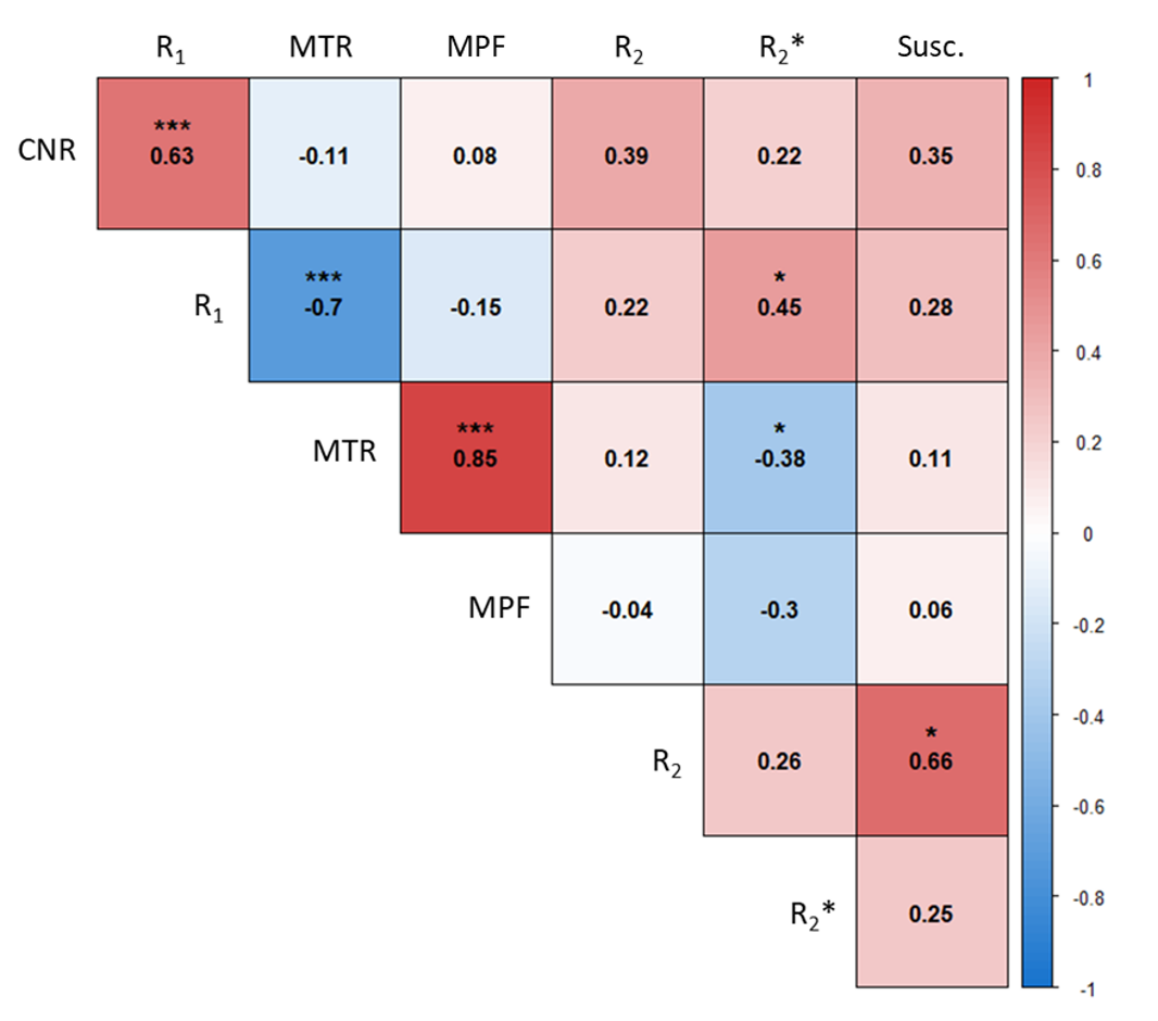
NM-MRI CNR showed a strong positive correlation with R1 (r=0.63, p<0.001, Figure 5) and non-significant positive correlations with R2, R2*, and susceptibility.
Discussion
Biological origin of the contrast:The results clearly demonstrate that the increase in NM signal is due to the increase in intra and extracellular NM up to a certain threshold beyond which the NM signal drops due to the appearance of neurodegeneration of melanized neurons. In the AAV-hTyr rat model, neurodegeneration is associated with accumulation of Lewy bodies and motor symptoms reminiscent of PD7. These data support our previously established hypothesis that NM concentration reaches a pathogenic threshold in PD which results in neurodegeneration7.
Physical origin of the contrast:
The results are in line with the hypothesis that the NM signal is due to paramagnetic effects of NM-iron complexes which significantly reduce T1 and increase susceptibility10. This is also consistent with iron accumulation found in the SN of PD patients11. In this model, MPF was not associated with NM-MRI contrast. This suggests that the increase in contrast observed with magnetization transfer preparation is due to the T1 reduction in NM-rich tissue.
Conclusion
This first in vivo longitudinal imaging study of NM accumulation in the rat SN confirms i) that NM-MRI is highly sensitive to nigral neuronal loss, ii) that the physical origin of NM-MRI contrast is the accumulation of paramagnetic NM-iron complexes. The results are also in agreement with the hypothesis that there would exist a deleterious concentration threshold of intracellular NM beyond which PD neurodegeneration would appear.Acknowledgements
This project was funded by Agence Nationale de la Recherche (ANR JPND NIPARK). The authors would like to thank the HISTOMICS and PHENOPARC platforms of the Paris Brain Institute for their support.References
1. Biondetti, E. et al. Spatiotemporal changes in substantia nigra neuromelanin content in Parkinson’s disease. Brain J. Neurol. 143, 2757–2770 (2020).
2. Gaurav, R. et al. Longitudinal Changes in Neuromelanin MRI Signal in Parkinson’s Disease: A Progression Marker. Mov. Disord. Off. J. Mov. Disord. Soc. 36, 1592–1602 (2021).
3. Double, K. L. et al. Influence of neuromelanin on oxidative pathways within the human substantia nigra. Neurotoxicol. Teratol. 24, 621–628 (2002).
4. Sulzer, D. et al. Neuromelanin detection by magnetic resonance imaging (MRI) and its promise as a biomarker for Parkinson’s disease. NPJ Park. Dis. 4, 11 (2018).
5. Trujillo, P. et al. Contrast mechanisms associated with neuromelanin-MRI. Magn. Reson. Med. 78, 1790–1800 (2017).
6. Priovoulos, N. et al. Unraveling the contributions to the neuromelanin-MRI contrast. Brain Struct. Funct. 225, 2757–2774 (2020).
7. Carballo-Carbajal et al. Brain tyrosinase overexpression implicates age-dependent neuromelanin production in Parkinson’s disease pathogenesis. Nat. Commun. 10, 973 (2019).
8. Perot, J.-B. et al. Quantitative multiparametric MRI describes neuromelanin-linked pathogenesis in the AAV-hTyr rat model of Parkinson’s disease. Proc. Intl. Soc. Mag. Reson. Med. (2023).
9. Avants, B. B. et al. A reproducible evaluation of ANTs similarity metric performance in brain image registration. NeuroImage 54, 2033–2044 (2011).
10. Trujillo, P., Aumann, M. A. & Claassen, D. O. Neuromelanin-sensitive MRI as a promising biomarker of catecholamine function. Brain awad300 (2023) doi:10.1093/brain/awad300.
11. Pyatigorskaya, N. et al. Iron Imaging as a Diagnostic Tool for Parkinson’s Disease: A Systematic Review and Meta-Analysis. Front. Neurol. 11, 366 (2020).
Figures
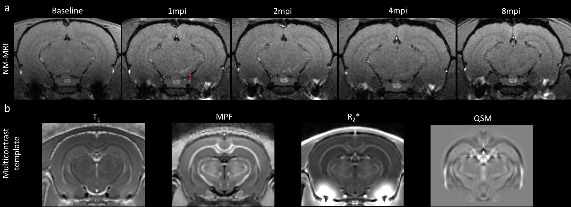
Figure 1 : a) NM-MRI image of one individual rat at every timepoint showing progression of contrast in the ipsilateral substantia nigra (highlighted with red arrow). b) Multicontrast template built from quantitative T1, MPF, R2*, and QSM images from 24 animals at baseline.
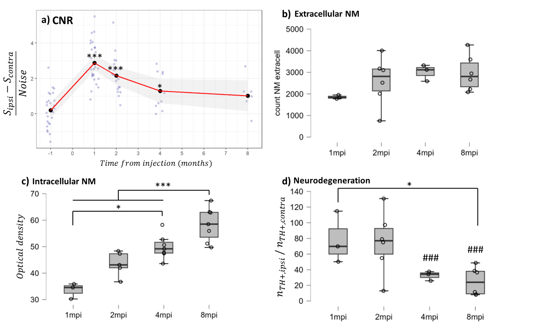
Figure 2: a) NM-MRI Contrast-to-noise ratio in ipsilateral SN of AAV-hTyr rats over time. Black dots represent mean value per timepoint. Red line and grey zone represent mean and confidence intervals. *p<0.05 *** p<0.001, Linear Mixed Model + Tukey post-hoc test with FDR correction. b-d) Histological quantification in ipsilateral SN of extracellular NM (b), Intracellular NM (c) and TH+ neurons in compared with contralateral (d) *p<0.05, ***p<0.001 (between timepoints), ###p<0.001 (ipsilateral vs contralateral). ANOVA + Tukey post-hoc.
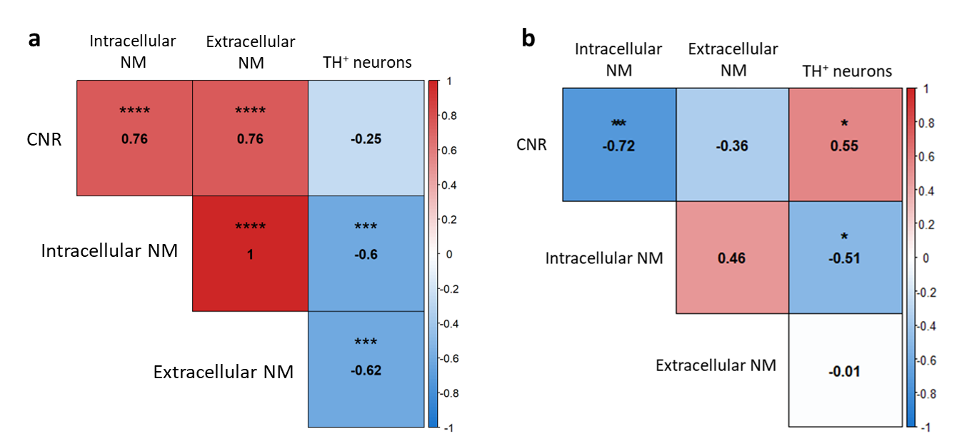
Figure 3 : Heatmap correlation matrix of MR parameters with histological quantification. a) Correlations between baseline and 1mpi. Intracellular and extracellular NM were considered zero and TH+ ratio was considered 100% at baseline, before any injection. b) Correlations between 1 and 8mpi. Spearman’s rho correlation coefficient. Significant correlations are highlighted. *p<0.05, **p<0.01, ***p<0.001, ****p<1e-4
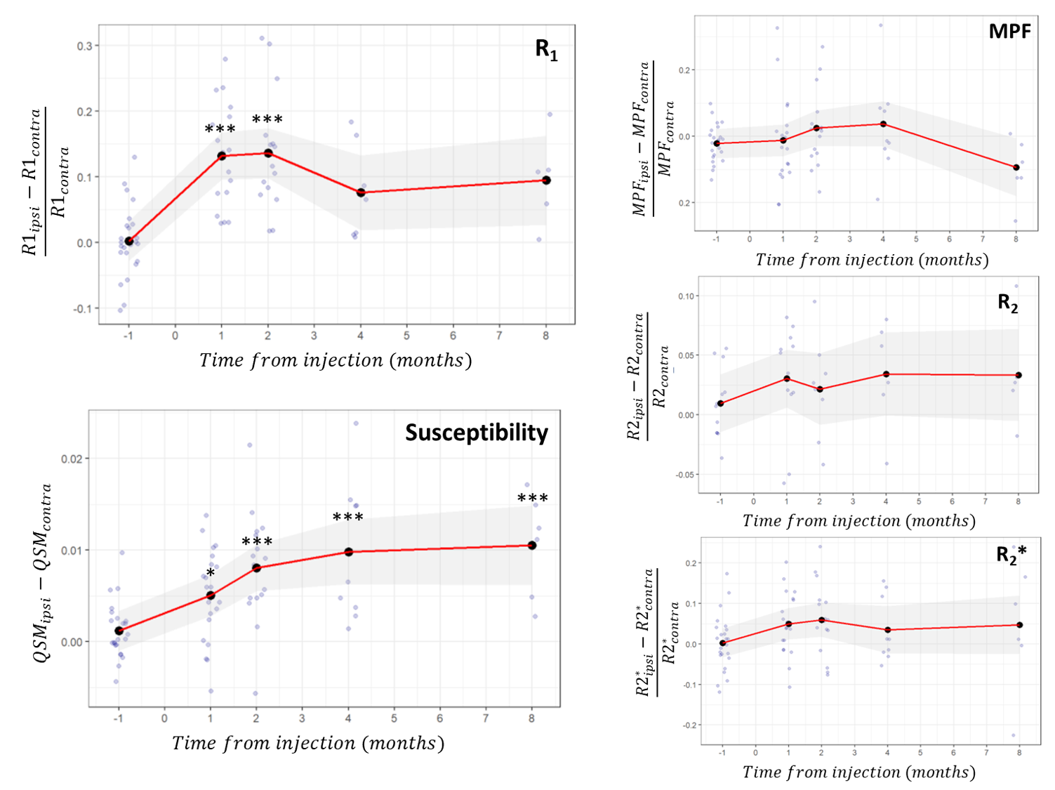
Figure 4: Longitudinal progression of quantitative MR parameters. Black dots represent mean value per timepoint. Red line and grey zone represent mean and confidence intervals *p<0.05 *** p<0.001, Linear Mixed Model + Tukey post-hoc test with FDR correction.