1302
Lung infection alters perivascular aquaporin-4 in rat model of Alzheimer’s Disease detected by filter exchange imaging (FEXI)1Division of Psychology, Communication and Human Neuroscience, University of Manchester, Manchester, United Kingdom, 2Geoffrey Jefferson Brain Research Centre, University of Manchester, Manchester, United Kingdom, 3Division of Neuroscience, University of Manchester, Manchester, United Kingdom, 4Medical Physics and Biomedical Engineering and Department of Neuroinflammation, UCL, London, United Kingdom, 5Division of Informatics, University of Manchester, Manchester, United Kingdom, 6Bioxydyn Limited, Manchester, United Kingdom, 7iBrain, Université de Tours, Tours, France
Synopsis
Keywords: Novel Contrast Mechanisms, Alzheimer's Disease, Blood-brain barrier
Motivation: Pneumonia is more prevalent in Alzheimer’s Disease (AD) patients than in healthy elderly people, which may be due to blood-brain barrier (BBB) vulnerability.
Goal(s): We assess whether filter exchange imaging (FEXI) can be used to understand the comorbid mechanisms occurring at the BBB with pneumonia and AD.
Approach: We apply the FEXI technique to a TgF344-AD rat model of AD with induced Streptococcus pneumoniae lung infection.
Results: FEXI detects significantly higher BBB water exchange in infected rats, with greater increase in the AD group, which significantly correlates to upregulation of hippocampus aquaporin-4 water channels, demonstrating the sensitivity of non-invasive FEXI to BBB alterations.
Impact: This work could be translated to a clinical study using filter exchange imaging to assess whether Alzheimer’s Disease patients suffering with pneumonia also exhibit worse blood-brain barrier alterations than patients without pneumonia and healthy elderly people.
Introduction
Blood-brain barrier (BBB) dysfunction occurs early in the pathogenesis of Alzheimer’s Disease (AD) [1-3], and importantly it seems to drive cognitive decline [4-6]. Pneumonia lung infection is more commonly found in people suffering with AD than healthy elderly people, but there are few non-invasive tools able to probe the mechanisms associated with infection and neurodegeneration. An emerging hypothesis is that infection has a more profound effect on the AD brain because of pre-existing BBB vulnerability [7]. In this study, we induce a Streptococcus pneumoniae lung infection in a rat model of AD (TgF344-AD) and use dynamic contrast enhanced (DCE) MRI and filter exchange imaging (FEXI) to assess the comorbid impact on BBB function.Materials & Methods
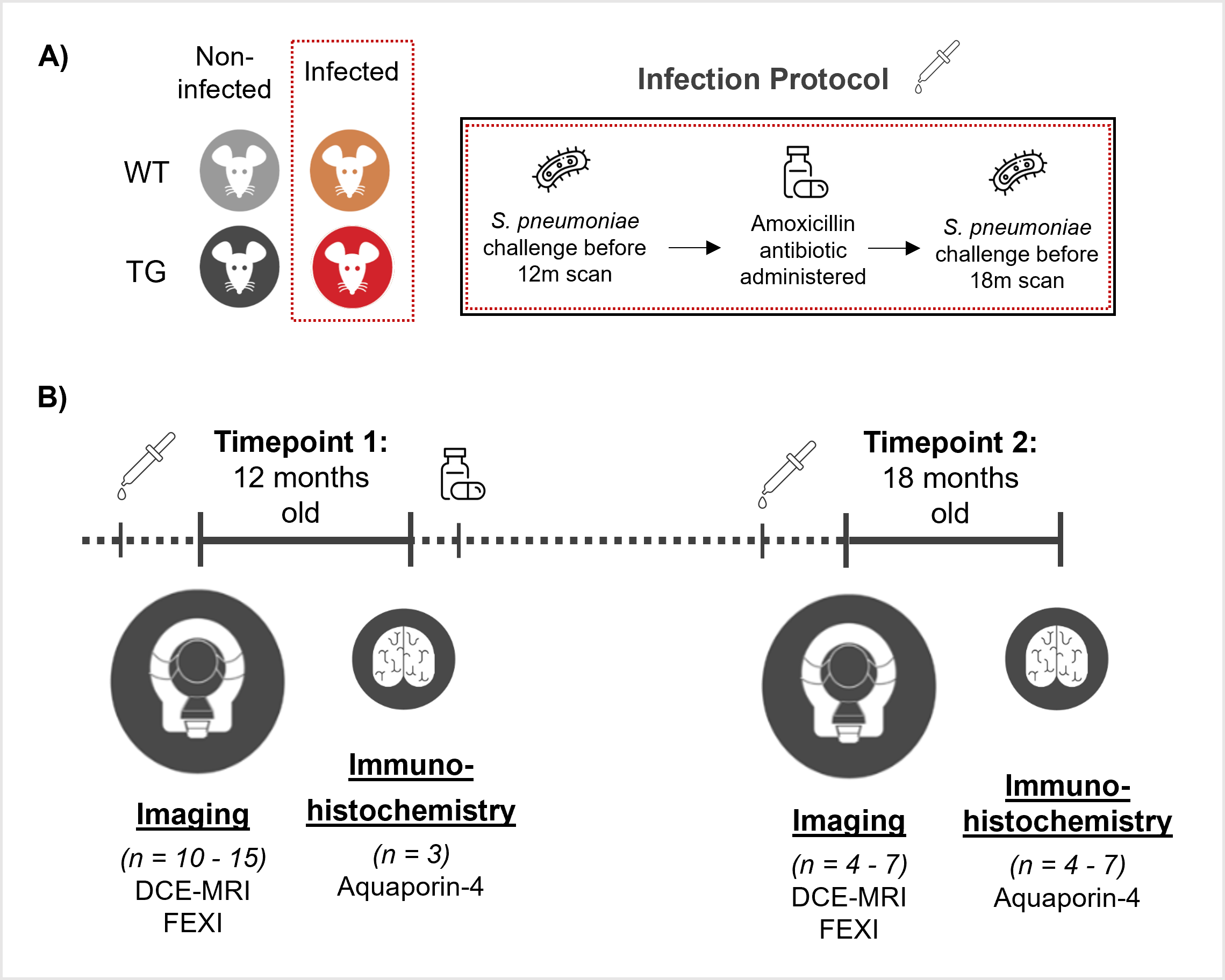
Male TgF344-AD and wildtype littermate rats were split into four groups: non-infected and infected wild-type (NI-WT, n = 11; Inf-WT, n = 15) and non-infected and infected TgF344-AD (NI-TG, n = 11; Inf-TG, n = 11) and were assessed at 12-months and 18-months old. Infection was induced in animals using an ascending Streptococcus pneumoniae infection challenge over 7 days at 12- and 18-months of age. Infection was resolved after the 12-month timepoints by amoxicillin antibiotic, then animals were re-infected at 18-months old. Brain tissue was collected from a subset of animals at 12-months (n = 3/ group) and the remaining animals at 18-months. Full study design: Figure 1.Imaging data was acquired with a Bruker Avance III console interfaced with an Agilent 7T 16-cm bore magnet. DCE-MRI parameters (OSIPI CAPLEX compliant [8]): R1,0 was estimated using a variable flip angle (VFA) 3D spoiled gradient echo (SPGR) scans with acquisition parameters: flip angle = 2°, 5°, 12° and 20°; TR/TE = 6.4/2.0 ms, voxel size = 0.27 × 0.27 × 1.0 mm3, matrix size: 128 × 128 × 20 and 4 signal averages. Dynamic 3D SPGR images were acquired at a single flip angle = 20° before and during intravenous injection (i.v.) of contrast agent, Gd-DOTA (Dotarem, Guerbet) with 0.1 mmol kg−1 dose. Analysis was performed using Madym software to measure BBB permeability to contrast agent (Ktrans) [9]. FEXI parameters: filter b-values (bf) = 0, 250 s/mm2; detection b = 0, 250 s/mm2; mixing times (tm) = 0.025, 0.05, 0.1, 0.2 and 0.3 s; TR = 5000 s; matrix size = 64 x 64; FOV = 32 x 32 mm2; single slice, resolution = 0.5 x 0.5 x 4.0 mm3; repetitions = 10, with a spin-echo EPI readout. BBB water exchange was calculated from manually segmented whole brain region in imaging slice [10].
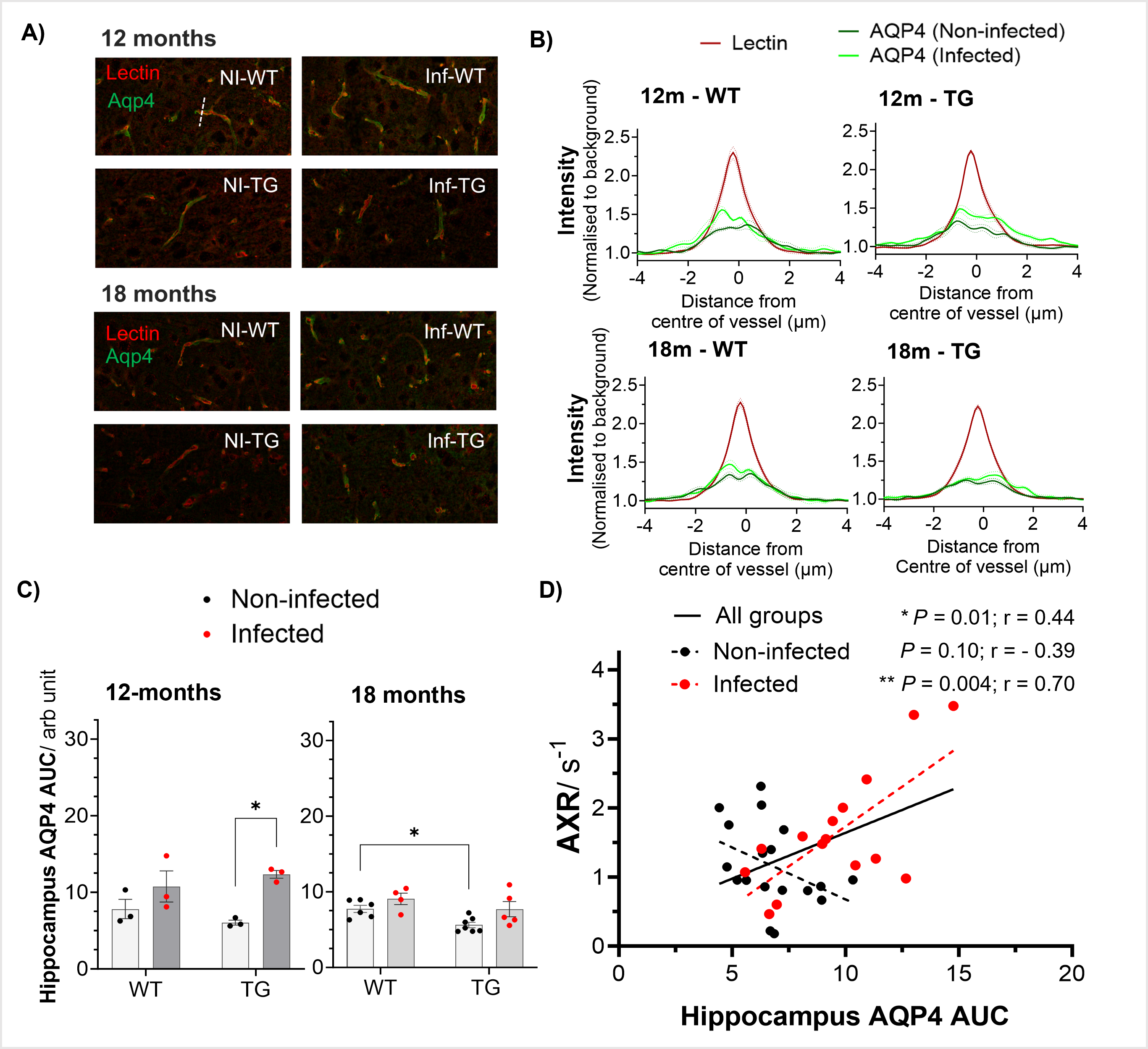
Immunohistochemistry: Brain tissue was used for assessment of aquaporin-4 (AQP4) water channel protein at the BBB. Vessel profiles were manually drawn on lectin images in ImageJ (Fiji), and the area-under curve (AUC) of AQP4 were taken and averaged across 15 line-profiles in the hippocampus region for each animal.
Results presented as mean ± s.e.m for all studies.
Results
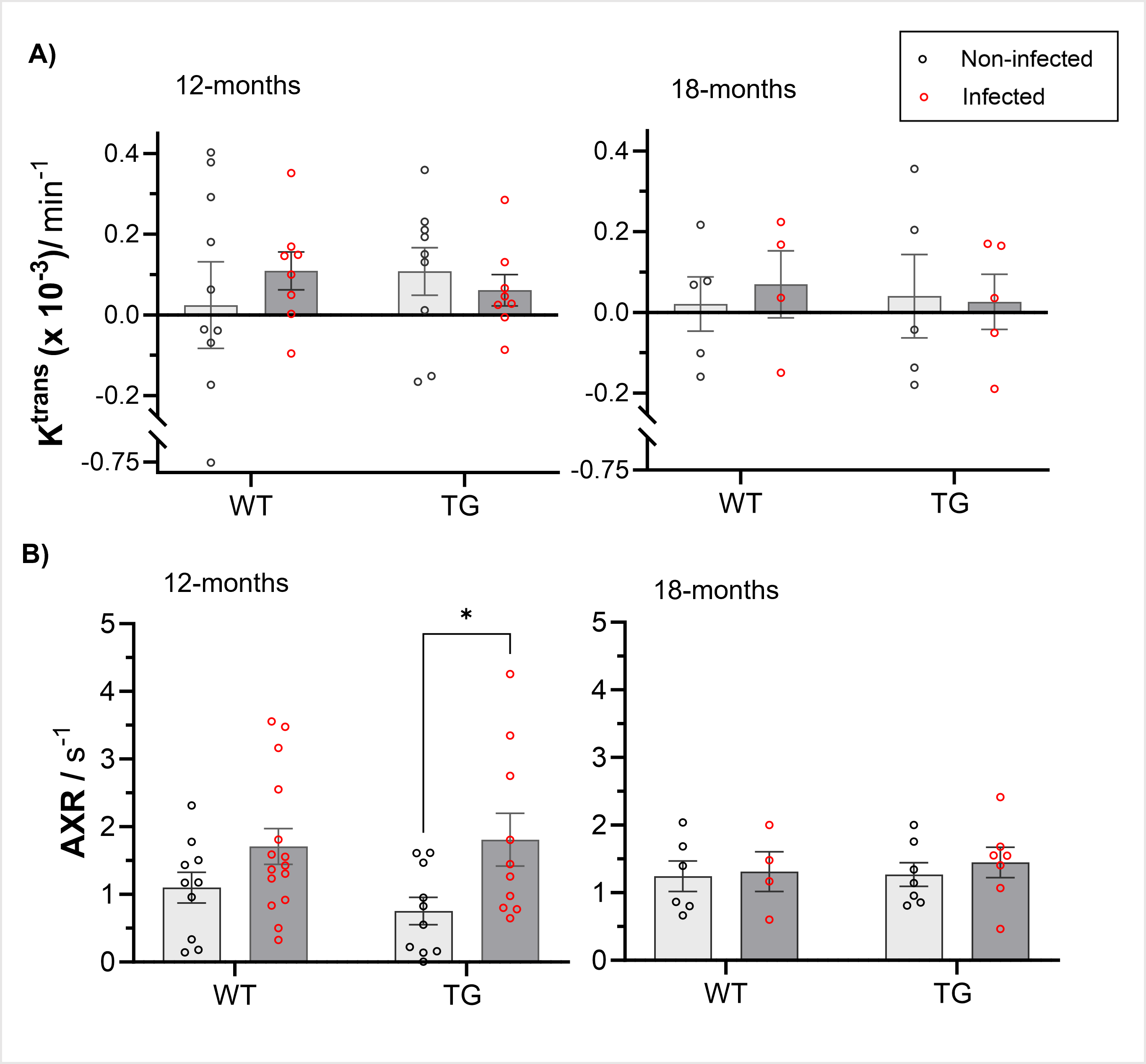
We detected no differences in BBB permeability with Gd-DOTA contrast agent (Ktrans) measured by DCE-MRI at either timepoints, Figure 2A. Whereas, FEXI detected a 138% higher BBB water exchange rate in infected Tg344-AD rats compared to their non-infected counterparts (adjusted P = 0.03), following the initial infection, Figure 2B. Infected wildtype rats showed a smaller effect (55% higher BBB water exchange), Figure 2B. Upon repeated infection at 18-months, no effect of infection or AD genotype was observed. In the hippocampus, we measured 107% higher AQP4 in infected TgF344-AD, compared to their non-infected counterparts at 12-months (adjusted P = 0.01), Figure 3C. While at 18-months, AQP4 was 27% lower in the non-infected TgF344-AD compared to non-infected wildtype rats (adjusted P = 0.04), Figure 3C. Taken the imaging and immunohistochemistry measurements together, we found a positive correlation between non-invasive BBB water exchange measures and hippocampus AQP4 expression across all animals (P = 0.01), with strongest association in the infected group (P = 0.004), Figure 3D.Discussion & Conclusion
Peripheral infection is known to have an impact on the BBB, particularly in conjunction with AD pathology, but is difficult to measure using non-invasive techniques [7]. Following the initial infection, FEXI was able to detect a higher BBB water exchange rate in infected rats compared to their non-infected counterpart, with the largest difference in TgF344-AD rats. Our results suggest that the non-invasive BBB water exchange measurements, using MRI, are associated with AQP4 protein expression changes occurring during peripheral infection, particularly in the hippocampus region, which is affected early in the pathogenesis of AD [3, 11]. This study demonstrates the sensitivity of FEXI in measuring subtle BBB alterations in response to lung infection and AD pathology.Acknowledgements
We would like to thank the staff at The Biological Service Facility University of Manchester, in particular Ray Hodgkiss for their help maintaining animal welfare and environmental enrichment during these studies, and Lidan Christie for their assistance with the acquisition of the MRI data.References
[1] Sweeney, M.D., A.P. Sagare, and B.V. Zlokovic, Blood-brain barrier breakdown in Alzheimer disease and other neurodegenerative disorders. Nat Rev Neurol, 2018. 14(3): p. 133-150.
[2] Erdő, F., L. Denes, and E. de Lange, Age-associated physiological and pathological changes at the blood–brain barrier: A review. Journal of Cerebral Blood Flow & Metabolism, 2017. 37(1): p. 4-24.
[3] Montagne, A., et al., Blood-brain barrier breakdown in the aging human hippocampus. Neuron, 2015. 85(2): p. 296-302.
[4] Nation, D.A., et al., Blood-brain barrier breakdown is an early biomarker of human cognitive dysfunction. Nat Med, 2019. 25(2): p. 270-276.
[5] Montagne, A., et al., APOE4 leads to blood-brain barrier dysfunction predicting cognitive decline. Nature, 2020. 581(7806): p. 71-76.
[6] Hussain, B., C. Fang, and J. Chang, Blood–Brain Barrier Breakdown: An Emerging Biomarker of Cognitive Impairment in Normal Aging and Dementia. Frontiers in Neuroscience, 2021. 15.
[7] Galea, I., The blood–brain barrier in systemic infection and inflammation. Cellular & Molecular Immunology, 2021. 18(11): p. 2489-2501.
[8] Dickie, B.R., et al., A community-endorsed open-source lexicon for contrast agent-based perfusion MRI: A consensus guidelines report from the ISMRM Open Science Initiative for Perfusion Imaging (OSIPI). Magn Reson Med, 2023.
[9] Berks, M.P., G.J.M.; Little, R.; Cheung, S., Madym: A C++ toolkit for quantitative DCE-MRI analysis. . Journal of Open Source Software, 2021. 6(66): p. 3523.
[10] Ohene, Y., et al., Filter exchange imaging with crusher gradient modelling detects increased blood–brain barrier water permeability in response to mild lung infection. Fluids and Barriers of the CNS, 2023. 20(1): p. 25.
[11] Zhang, Y., et al., Vascular-water-exchange MRI (VEXI) enables the detection of subtle AXR alterations in Alzheimer's disease without MRI contrast agent, which may relate to BBB integrity. NeuroImage, 2023: p. 119951.
Figures