1301
A Hydrogen Peroxide-Responsive Multinuclear 1H and 19F MRI Contrast Agent for Quantitative Application – Preliminary Phantoms Validation1MRI Research Center, Auburn University, Auburn University, AL, United States, 2Chemistry and Biochemistry, Auburn University, Auburn University, AL, United States
Synopsis
Keywords: Multi-Contrast, Preclinical, Multinuclear
Motivation: Multinuclear contrast agents (CAs) may provide improved sensitivity and specificity for the detection/quantification of biomolecular processes.
Goal(s): To develop a multinuclear 1H and 19F agent that shortens the T1 relaxation times for both the 1H and 19F signals only when hydrogen peroxide (H2O2) is present.
Approach: Developed the CA: Fe(II)-F2H4qp4 -- an iron(II) complex with a fluorinated quinol-containing macrocyclic ligand. This T1-shortening CA is designed to activate only in the presence of H2O2.
Results: Initial phantom tests with an 1H/19F frequency-selectable Inversion Recovery Look Locker sequence
demonstrate a dual capability to quantify changes of both 1H and 19F signal T1 values.
Impact: The multinuclear Fe(II)-F2H4qp4 agent's ability to quantify changes of both 1H and 19F signal T1 values in an H2O2 environment improves the sensitivity and specificity to superoxide-related pathologies and may allow expanding to other biomarkers.
Introduction
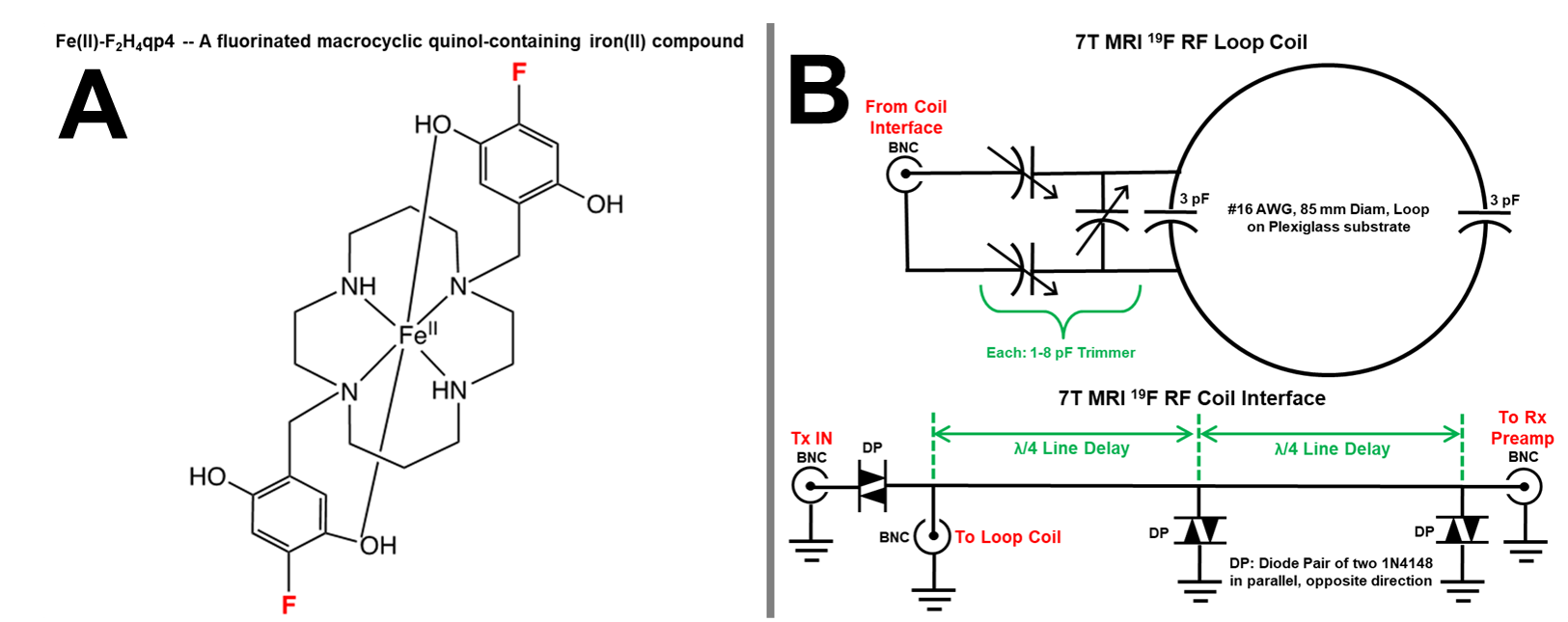
Responsive contrast agents (CAs) that can activate by a specific biochemical marker have the potential to improve MRI sensitivity and specificity to certain pathologies associated with those biomarkers. Most CAs lack such a targeted response and affect only one nuclei signal, such as proton (1H) T1 relaxivity. Recent CA research has expanded to multinuclear agents that can give rise to multiple signals from multiple nuclei, such as 1H and fluorine (19F) 1-3.We developed a multinuclear 1H/19F agent: Fe(II)-F2H4qp4 - an iron(II) complex with a fluorinated quinol-containing macrocyclic ligand (Figure 1A). The Fe(II)-F2H4qp4 molecule is designed to activate only in the presence of hydrogen peroxide (H2O2) to shorten the T1 relaxation times for both 1H and 19F signals. This makes Fe(II)-F2H4qp4 a potentially versatile assay tool for studies involving H2O2 and similar reactive oxygen species.
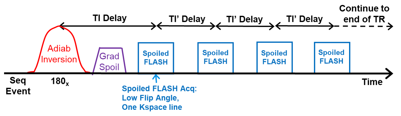
For phantom validation of Fe(II)-F2H4qp4, we constructed a 7T 1H/19F RF coil with custom interface-preamplifier (Figure 1B) and developed a flexible 1H/19F frequency-selectable Inversion Recovery Look-Locker (IRLL) sequence to quantify both 1H and 19F T1 relaxation rates (Figure 2). Although 1H IRLL is common, the application of 19F IRLL is sparse in the literature and has not been applied to study CAs as now 1, 2.
Methods
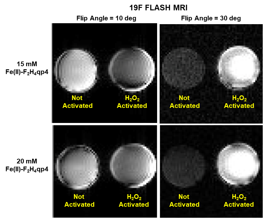
Our 7T 1H/19F RF Tx/Rx coil with interface-preamplifier (Figure 1B) was tuned for 19F peak performance with sodium fluoride (NaF+H2O) phantoms. We then developed a 1H/19F frequency-selectable IRLL sequence that employed a non-selective adiabatic inversion followed by multiple fast low-angle shot (FLASH) readouts of one kspace line with 20 preplanned inversion times (TI) (Figure 2). Additional 19F T1-weighted FLASH at flip angles 10 and 30 deg further demonstrated 19F signal T1 contrast between phantoms.Six 7T Fe(II)-F2H4qp4 phantoms were prepared with 10, 15 and 20 mM concentration and each concentration prepared with or without 10 mM hydrogen peroxide (H2O2) – activated versus non-activated respectively. Similar to these 7T phantoms, ten additional Fe(II)-F2H4qp4 phantoms were prepared for 3T 1H IRLL scans to determine T1 values at 3T. Since the Fe(II)-F2H4qp4 agent is much more T1 responsive with 1H at 3T, the 3T phantoms were prepared at lower concentrations of 0, 0.1, 0.4, 0.7 and 1.0 mM, with and without 0.1 mM H2O2 for activation. All 19F MRI was performed on a 7T human-bore scanner and 1H MRI performed on both 7T and 3T scanners (Siemens, Erlangen, Germany). All 7T and 3T analyses were performed on custom Matlab programs (Mathworks, Natick, MA) using the Neider-Mead Simplex method, and background noise compensation, to curve-fit the IRLL magnitude data into T1 values.
Common MRI parameters: FOV = 64x64 mm; Matrix = 32x32; Slice-thickness = 10 mm; Pixel-size = 2.0x2.0x10 mm. Specific for 7T 1H: TR = 10 sec; 20 TI times ranging from of 10 to 2600 ms; FLASH flip-angle = 3 deg; Manual TxVref = 50 V; Averages = 4. Specific for 7T 19F: TR = 10 sec; 10 TI times ranging from 10 to 2000 ms; Manual TxVref = 50 V; FLASH flip-angle = 5 deg; Averages = 9. Specific for 3T 1H: TR = 10 sec; 20 TI times same as 7T; FLASH flip-angle = 2.5 deg; Averages = 6.
Results
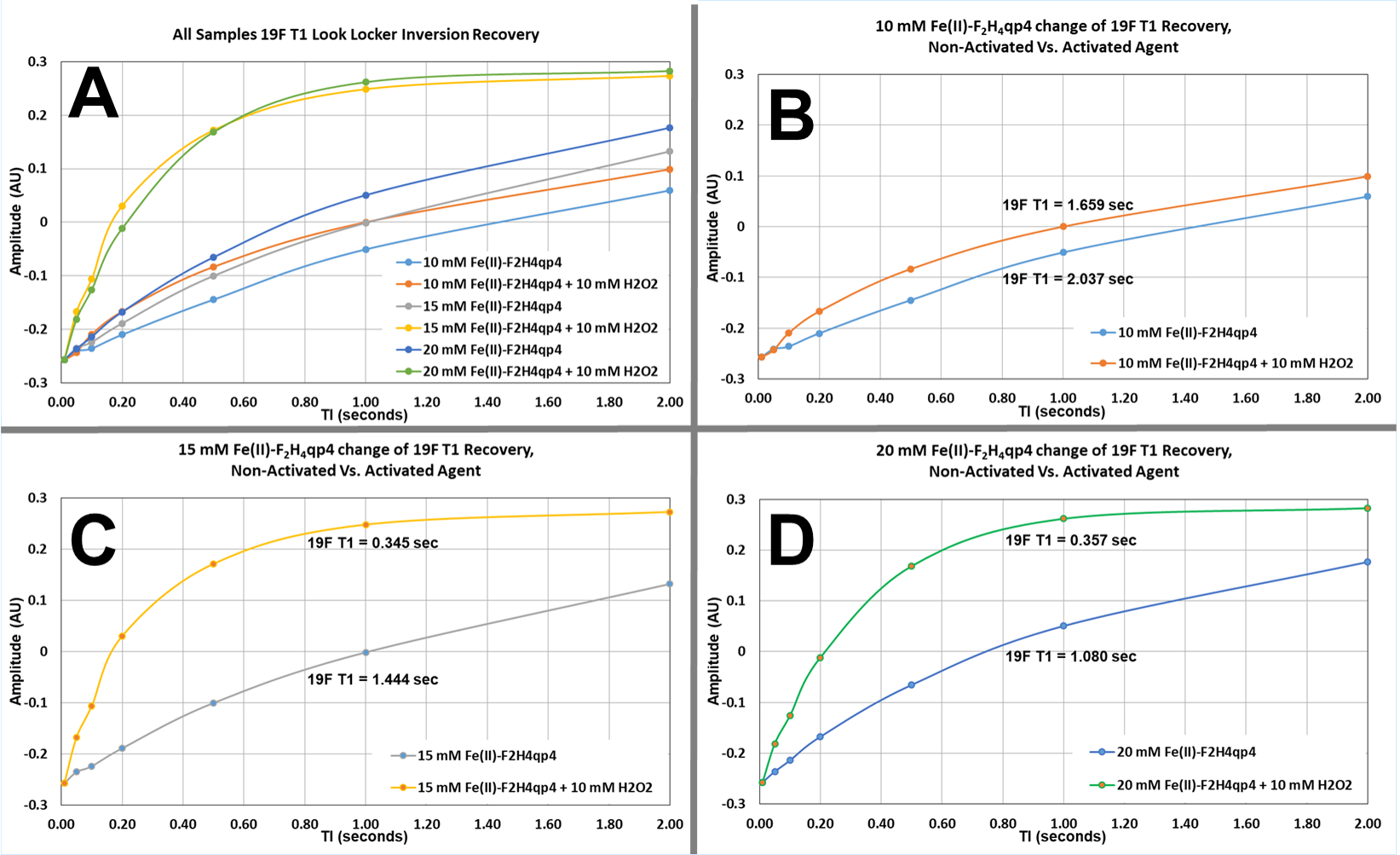
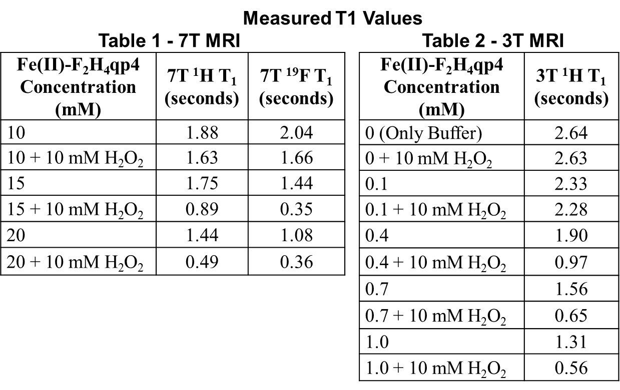
In all 19F scans, the 7T 1H/19F RF coil demonstrated good 19F sensitivity with SNR values ranging from 20 to 150 and 1H sensitivity was acceptable when using multiple signal averages. Figure 3 presents the 19F IRLL T1 recovery graphs. Figure 3A presents a composite of all six 19F T1 curves from all six 19F phantoms. Figure 3B-C-D breakout the 19F T1 curves for each Fe(II)-F2H4qp4 concentration with and without H2O2 activation. Figure 4-Table 1 summarizes the 7T 1H and 19F T1 values and Figure4-Table 2 summarizes the 3T 1H T1 values. The 19F T1 curves and corresponding T1 values clearly show the significant change of 19F T1 with Fe(II)-F2H4qp4 during H2O2 activation. The 1H T1 values also indicate significant change of 1H T1 at both 7T and 3T. Figure 5 presents the 19F T1-weighted FLASH showing the flip angle control for best T1 contrast in activated versus non-activated Fe(II)-F2H4qp4 19F signal.Discussion/Conclusion
Our 1H/19F RF coil combined with 1H/19F IRLL quantified the changes of both 19F T1 and 1H T1 with Fe(II)-F2H4qp4 agent being H2O2 activated versus non-activated. These methods were consistent across both 1H and 19F nuclei at both 7T and 3T. This application of multinuclear 1H and 19F MRI validated the performance of the multinuclear Fe(II)-F2H4qp4 agent under a range of varied concentration and H2O2 activation states. Future efforts will expand to in vivo rodent application of these methods.Acknowledgements
Special thank you for project support goes to Julie Rodiek, Steven Nichols and Clayton Ridner.References
- Adolphi NL, et al “Quantitative mapping of…by 19F MR imaging of T1…”, MRM 2008; 59(4)
- Jordan BF, et al “Rapid monitoring of…by 19F magnetic resonance imaging…“, MRM 2008; 61(3)
- Karbalaei S, et al, “A Highly Water- and Air-Stable Iron-Containing MRI Contrast…” Chem. Eur. J. 2022; 28
Figures