1300
Enhanced prostate imaging in ultra-low field MRI using a passive LC-resonator.1Chongqing University, Chongqing, China
Synopsis
Keywords: Novel Contrast Mechanisms, Contrast Mechanisms
Motivation: As gold standard, MRI indeed matters in prostate imaging. The relative low image quality in ultra-low field MRI precludes its application in prostate imaging.
Goal(s): Our goal was to improve prostate image quality in ultra-low field MRI, particularly emphasizing the target prostate region.
Approach: We utilized an additional specially designed passive LC-resonator in imaging process.
Results: By using the LC-resonator, the prostate image quality in ultra-low field MRI was improved, giving a higher image contrast to the prostate region.
Impact: Adopting passive LC-resonator in imaging process is of highly cost effective, not only highlighting the target region, but offering a new imaging idea for other organs when it comes to bad SNR situations.
Introduction
As a powerful medical imaging tool, MRI plays an increasing role in human diseases and disorders detections. Despite possessing the advantages of non-invasive imaging and flexible imaging modality, current commercial MRI devices suffer from high cost and heavy bulk, which greatly limits its application scenarios. Given the aforementioned situation, more research interests and attentions are paid into ultra-low field MRI, where much lower cost meets movable weight and volume, at the cost of decreased image quality1,2.For prostate imaging, MRI has long time been the gold standard imaging modality for the diagnosis of prostate cancer3, and several mature evaluation criteria have been established for this detection4,5.
MR image quality is fundamental for the accuracy of prostate diseases detection. To make it possible that prostate MR images from ultra-low field work with the widely used criteria, the image quality needs to be improved, as thus providing patients more accessibility to efficient prostate examination. This work was dedicated to enhance the prostate image quality in ultra-low field MRI by using a passive LC-resonator, and explore the feasibility and potentiality of ultra-low field MRI.
Methods
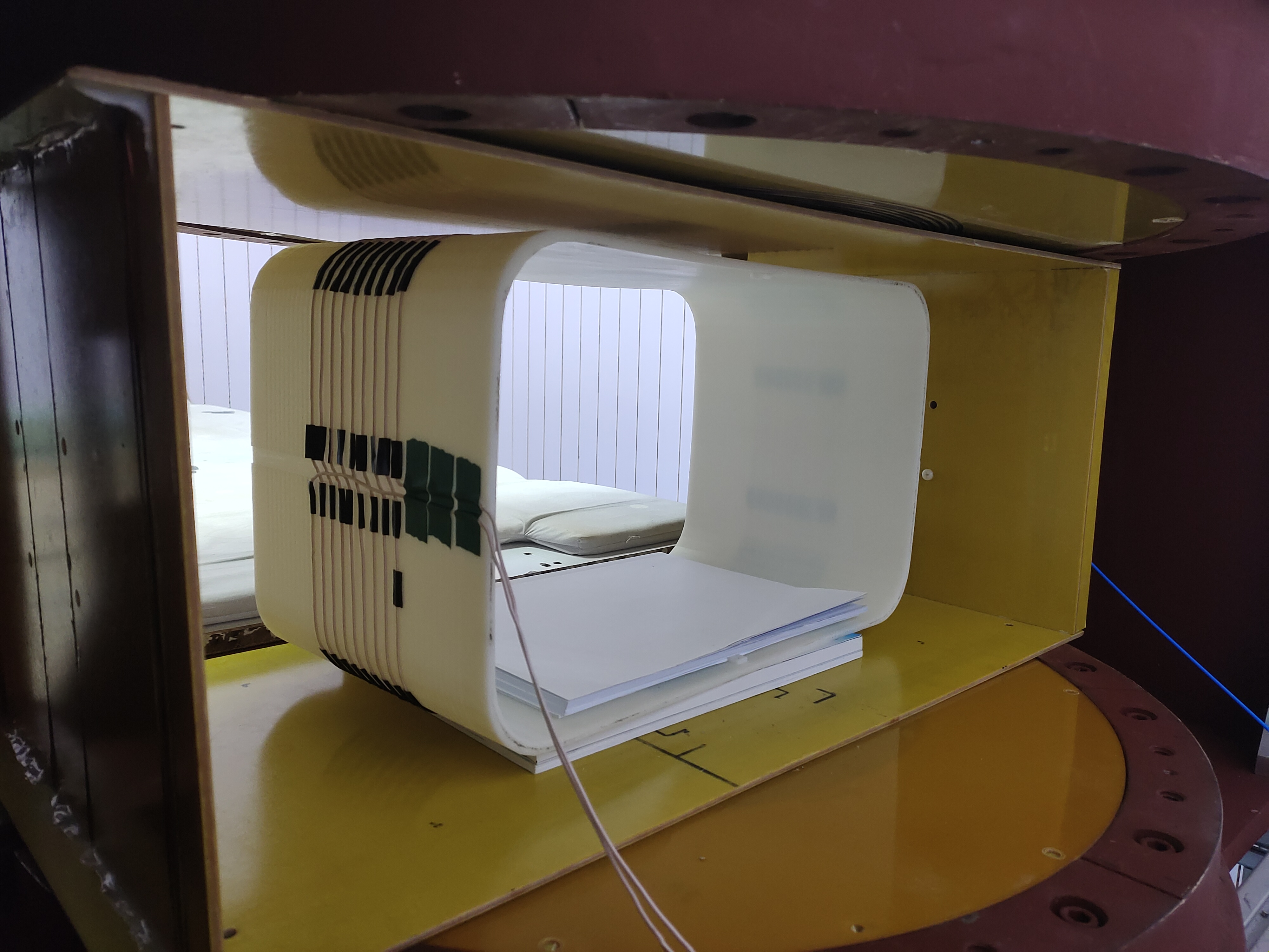
This work was conducted based on the self-developed ultra-low field MRI system in our group1. To conduct prostate examination experiments, we need two more core parts: main receive coil and LC-resonator.For main receive coil design, we utilized target field approach. On the whole, the main receive coil was wound in the form of a solenoid on the 3D printed holder using Litz wire. The holder had a shape of rounded cuboid, with its length, width, and height set to be 40 cm, 22 cm, and 24 cm to fit the body size of adult men. Under the setting, The target magnetic field region was set as a spherical area with diameter in 12 cm, covering the prostate zone when someone lies into the receive coil. According to the emulational optimization results, we chose 9 turns in total with tuan spacing in 4 mm. The designed receive coil is shown as figure 1.
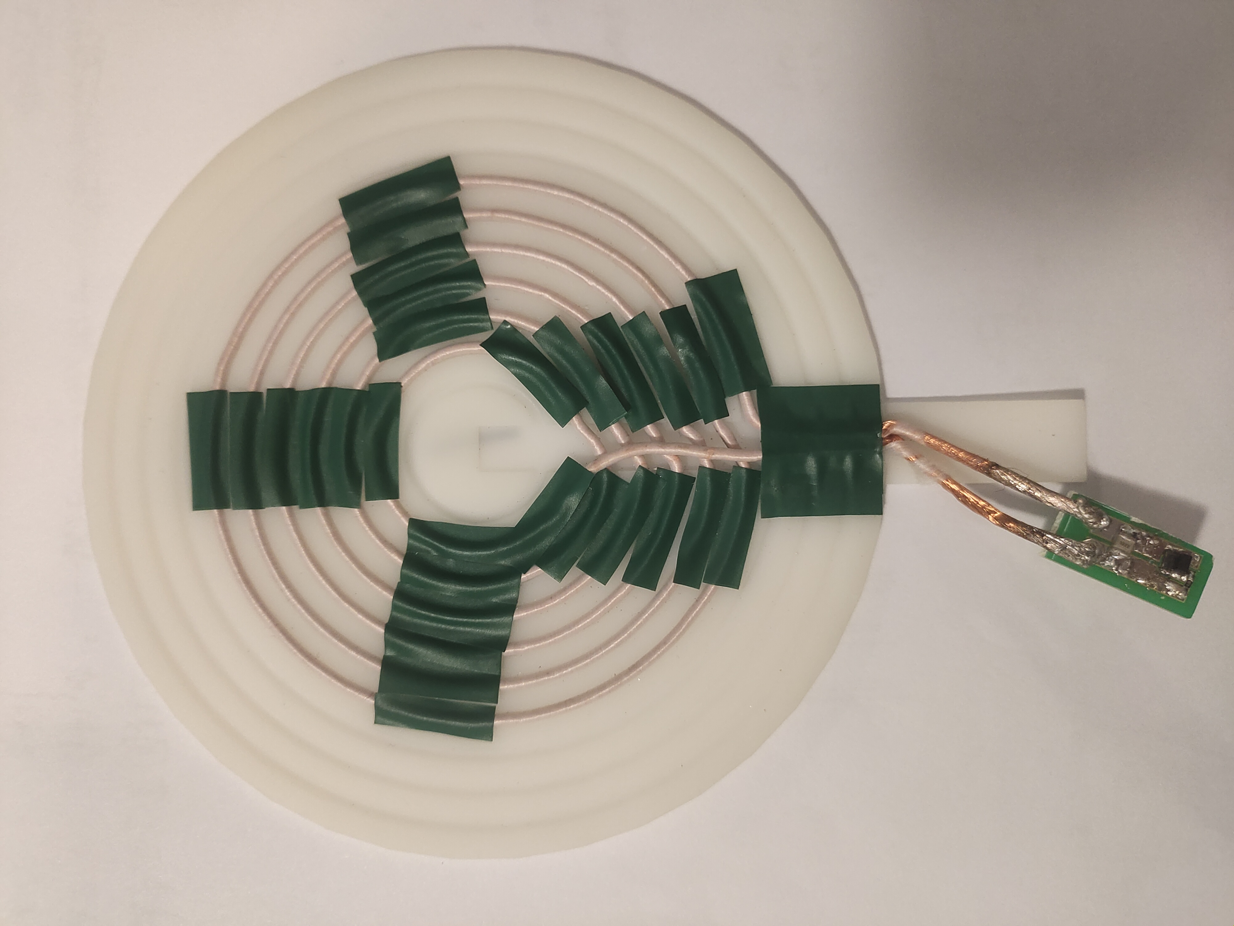
For LC-resonator design, we utilized the simple bisection method. We adopted concentric circle as the basic shape of LC-resonator. The resonator works because of the coupling between receive coil and resonator, and the effect is dependent on the coupling strength. Factors including external diameter, number of turns, and turn spacing can affect the coupling strength. We fixed the external diameter of 10 cm, and turn spacing of 3 mm, regarding number of turns as the only variable to find the optimal resonator structure in emulational experiments. This problem can be easily solved by bisection method, Saving much more computational resource compared to our previous work6, while attaining comparable coupling effects. The resonant frequency is tuned at 2.36 MHz, slightly deviated from 2.32 MHz of our system. The designed LC-resonator is shown as figure 2.
During imaging process, the LC-resonator is sticked close to perineum skin.
Results
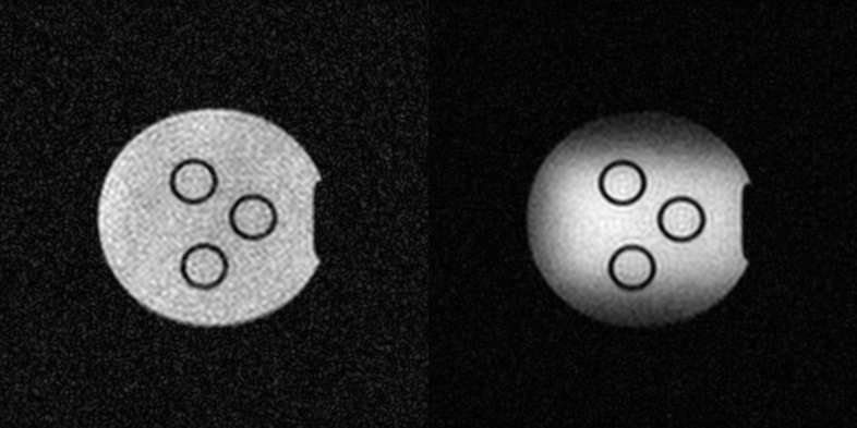
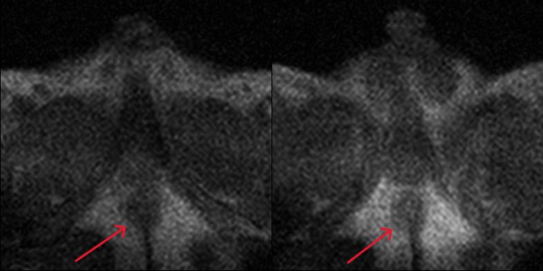
Water phantom experiments and in vivo experiments were conducted respectively with our 54.6 mT ultra-low field MRI system. All the imaging experiments were performed using the same 3D gradient echo pulse sequence with flip angle 60°, TE 18 ms, TR 50 ms, and thickness 8 mm. The total time consuming for 6 slices in single experiment is 36 seconds. The imaging results of water phantom and in vivo are shown in Figure 3 and Figure 4 respectively. In vivo experiments were done with a healthy adult male volunteer.In both comparisons, the improvement of image quality can be observed with LC-resonator, especially in the central region, where brighter pixels and higher contrasts were obtained. What’s more, images generated with LC-resonator demonstrate greater background noise suppression.
Discussion
The above experiments show the feasibility of adopting LC-resonator in ultra-low field MRI to attain better image quality. Even though higher image contrast appeared when using resonator, there is still room for SNR increasing. Our single experiment took only 36 seconds, which means more average scanning can be done to obtain higher SNR within acceptable duration. Besides, we kept only one variable in LC-resonator design to reduce computational burden and simplify the optimization process. While our resonator worked effectively, more elaborately designed ones shall have better performance.Conclusion
We adopted LC-resonator in ultra-low field MRI for prostate imaging, and the results demonstrated its ability in image enhancement. Though only prostate images shown here, other human organ images may benefit from LC-resonator if well designed. This work also provides a new way for human imaging in ultra-low field MRI.Acknowledgements
No acknowledgement found.References
1.He Y, He W, Tan L, et al. Use of 2.1 MHz MRI scanner for brain imaging and its preliminary results in stroke[J]. Journal of Magnetic Resonance, 2020, 319: 106829.
2.Anoardo E, Rodriguez G G. New challenges and opportunities for low-field MRI[J]. Journal of Magnetic Resonance Open, 2022: 100086.
3.Woernle A, Englman C, Dickinson L, et al. Picture Perfect: The Status of Image Quality in Prostate MRI[J]. Journal of Magnetic Resonance Imaging, 2023.
4.Giganti F, Rosenkrantz A B, Villeirs G, et al. The evolution of MRI of the prostate: the past, the present, and the future[J]. American Journal of Roentgenology, 2019, 213(2): 384-396.
5.Padhani A R, Barentsz J, Villeirs G, et al. PI-RADS steering committee: the PI-RADS multiparametric MRI and MRI-directed biopsy pathway[J]. Radiology, 2019, 292(2): 464-474.
6.Zhang Y, Guo Y, Kong X, et al. Improving local SNR of a single-channel 54.6 mT MRI system using additional LC-resonator[J]. Journal of Magnetic Resonance, 2022, 339: 107215.
Figures