1297
Bumetanide drug effect on the transmembrane water exchange by T1 relaxation at ultra-low field1BrainTech Lab, Inserm U1205, Grenoble, France, 2Biomedical Imaging Centre, University of Aberdeen, Aberdeen, United Kingdom
Synopsis
Keywords: Relaxometry, Low-Field MRI, transmemebrane water exchange, cancer invasion/migration, bumetanide drug
Motivation: The transmembrane water exchange (t-Wex) in cancers was demonstrated modulating T1 relaxation at ultra-low field.
Goal(s): Our goal was to demonstrate T1 changes under the administration of drugs that act on cell membrane transports.
Approach: U87 glioma cells sustained H2O2 stimuli before the administration of the bumetanide drug, a NKCC1 inhibitor. T1 changes were measured by FFC-NMR.
Results: At very low field, by comparison to control cells (without H2O2), relaxation rates were found significantly lower under H2O2 stimuli which has been correlated to t-Wex acceleration and higher with the bumetanide addition, that suggests the slowdown of t-Wex.
Impact: Using the bumetanide drug, we show at ultra-low field, the potential of the relaxation T1 as biomarker to evaluate the efficiency of drugs that can target t-Wex, a mechanism that has been connected to cancer invasion/migration pathophysiology
Introduction
Recently we demonstrated that the longitudinal relaxation T1 of water at ultra-low field (<2mT corresponding to ≈105Hz) is highly sensitive to the transmembrane water exchange (t-Wex) and we demonstrated its role in cancer invasion/migration1-2. Also, using immunohistochemistry, we showed that AQP4, a water channel protein of cell membrane is up-regulated in invasion/migration1-2, highlighting the role of this aquaporin which affects t-Wex in T1 relaxation modulation. This work is focused to assess T1 sensitivity under the effect of pharmaceutical drugs that could inhibit pathophysiological processes of invasion/migration; namely hypoxia, H2O2 redox-signaling and the aquaporin AQP4. We propose the bumetanide drug, which is a NKCC1 inhibitor of sodium, potassium and chloride cellular influx3, which already was approved for patients and shown to inhibit the excess expression of AQP4, that should slow down the invasion/migration process. Knowing that AQP4 interacts with several other proteins and channels, we hypothesize that bumetanide should be more beneficial than those that target water channels specifically such as AQP4 SiRNA and/or ShRNA.Methods
Fast Field Cycling (FFC) NMR was used to measure T1 at low and ultra-low fields and to acquire R1-NMRD profiles (R1=1/T1 versus magnetic field or 1H Larmor frequency) as described in our previous work1. This technology was developed to solve the crucial problem of NMR sensitivity at low fields. It is the only NMR technique that permits T1 measurements at low (< 0.2T) and ultra-low fields (< 2mT), covering several decades of the frequency [10 KHz- 40 MHz], with the same relaxometer. The intracellular water lifetime (τIN) that characterizes the kinetic of the t-Wex, was measured in vitro on glioma cells. The method consists to add a paramagnetic contrast agent Gd-DOTA ([Gd]=9mmol/kg) and to use the 2SX model to extract water exchange parameters as described in reference5. Measurements were performed on glioma cell lines: Glio6 and Glio96 of invasion/migration and U87 of high proliferation1. Three different experiments were performed. (1) T1 and τIN of Glio6 and Glio96 were measured and compared to U87. (2) T1 and τIN were measured under hypoxia (3%O2) and compared to normoxia (20%O2) and under H2O2 stimuli (5mM during 20min) and compared to controls (without H2O2 treatment). (3) The last experiment was performed to evaluate the effects of the bumetanide drug on U87 cells. In this case, the bumetanide was added into the cells at a concentration of 1µM, and cells were put in incubation at 37°C for approximately 18H. In a preliminary experiment, a bumetanide dose study was realized using the Boyden chamber assay approach, selecting the dose of 1mM that significantly slowdowns cell migration.Results
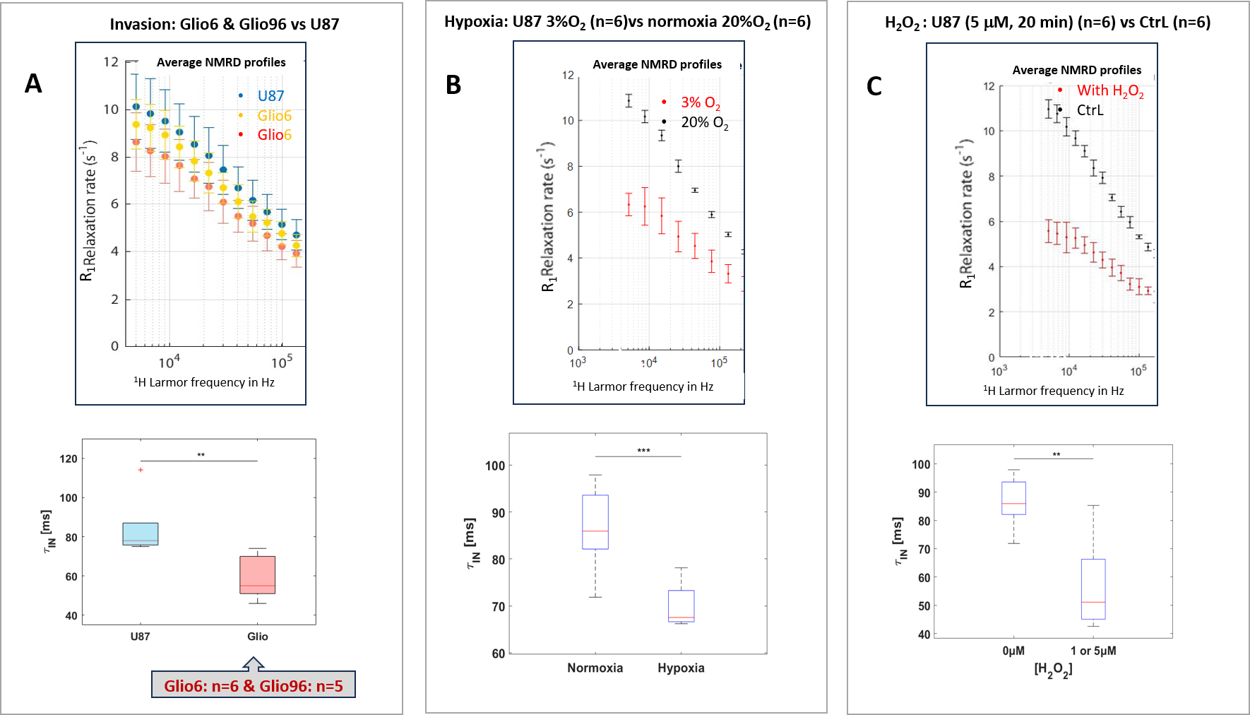
By comparing Glio6 and Glio96 cells to U87, we show that the two parameters R1 at ultra-low fields and τin are sensitive to invasion/migration (Fig.1A). Also, U87 cells they were found lower under hypoxia (Fig.1B) and under H2O2 stimuli (Fig.1C). The proof of the concept of the bumetanide effect was evaluated on U87 cells stressed with H2O2 in order to accelerate t-Wex. R1 relaxation rates at very low field were found higher, attempting to reach control values of U87 cells without H2O2 stimuli (Fig.2), a result that suggests the slowdown of the t-Wex under the effect of the bumetanide drug that is in line with our hypothesis.Discussion
The effect of the bumetanide was evaluated on U87 cells, stressed with H2O2 in order to mimic the characteristics of invasion/migration process and were used because they have a rapid growth than Glio6 and Glio96 (3 weeks versus 3 months). t-Wex measurements on U87 cells and on Glio6 and Glio96 under bumetanide are works in progress and should confirm our findings. Our results stipulate the major role of FFC-imaging (FCI) and ultra-low field MRI to visualize the entire invasion/migration volume noninvasively and to evaluate the efficiency of innovative therapies that target t-Wex. This may impact the medical community since delineation and efficient therapies of cancer invasion/migration remain both challenging by any medical imaging modality. Our results suggest that FCI, in combination with bumetanide, may be a promising strategy of cancer invasion/migration diagnostic and therapy evaluation follow-up.Conclusion
NMR at ultra-low field appears appropriate to diagnose cancer invasion/migration and to evaluate the effect of drugs that can target t-Wex which is connected in case of glioma to the pathophysiology of invasion/migration, namely, hypoxia, H2O signaling redox and AQP4 functions. Here we particularly show the potential of the bumetanide as an anti-invasive drug and we propose FCI technology to evaluate its efficiency, in patients noninvasively, a work that we can realize on the unique FCI of Aberdeen university.Acknowledgements
We thank Y Ben‐Ari for useful discussion, Pascal Henry Fries and A. El-Gady for helping in FFC acquisitions.References
1. M. Petit, M. Leclercq, S. Pierre, M. R. Ruggiero, M. El Atifi, J. Boutonnat, P.H. Fries, F. Berger and H. Lahrech NMR Biomed. 35(6), e4677 (2022).
2. M.R. Ruggiero, H. Ait Itto, S. Baroni, S. Pierre, J. Boutonnat, L.M. Broche, S. Aime, F. Berger, S. Geninatti Crich and H. Lahrech Cancers (Basel). 29, 14(17)- 4180 (2022).
3. M. Zhang, Z. Cui, Cui H, Y. Cao, C. Zhong and Y. Wang BMC Neurosci. 17, 60- (2016).
4. M. M. Reed and B. Blazer-Yost Cell Physiol Biochem. 56(S2),12-30 (2022).
5. M. R. Ruggiero, S. Baroni, S.Pezzana, G. Ferrante, S. Geninatti Crich and S. Aime Angewandte Chemie International. 57(25), 7468–7472 (2018).
Figures
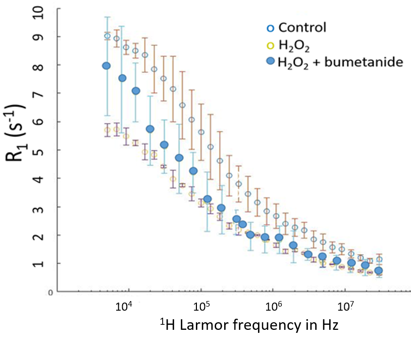
relaxation rate to bumetanide administration at ultra-low field