1291
Simultaneous two-voxel functional magnetic resonance spectroscopy of the motor cortex at 7T1Department of Radiology and Nuclear Medicine, Amsterdam University Medical Center, University of Amsterdam, Amsterdam, Netherlands, 2Sir Peter Mansfield Imaging Centre, School of Physics and Astronomy, University of Nottingham, Nottingham, United Kingdom
Synopsis
Keywords: Spectroscopy, Spectroscopy, functional MRS
Motivation: Functional magnetic resonance spectroscopy (fMRS) shows promise in studying task-related metabolite changes but has been largely confined to single-voxel.
Goal(s): To evaluate two-voxel fMRS at 7 T to measure simultaneous bilateral metabolite changes during a unilateral motor task.
Approach: A modified Hadamard-encoded MRS scheme with dynamic fMRS spectral-temporal fitting for analysis was employed.
Results: Distinct patterns of BOLD activation in contra- and ipsilateral VOIs were detected with significant increases in Glutamate (Glu) in either VOI during a unilateral task
Impact: We demonstrate the feasibility of simultaneous two-voxel MRS to detect bilateral glutamate changes in response to a unilateral motor task. This approach holds promise to increase our understanding of the neurochemical underpinnings of fMRI signals across interconnected brain regions.
Introduction
Functional MRS (fMRS) is a powerful technique to measure changes in metabolite concentrations during a stimulus or task. In the motor cortex, fMRS studies have revealed increases in glutamate (Glu) in regions of activation [1,2]. However, fMRS of single voxels neglects possible metabolite fluctuations occurring in functionally interconnected brain regions. Two-voxel MRS, using multi-band excitation with spatial encoding, e.g. PRIAM [3], vGRAPPA [4] or Hadamard [5], enables simultaneous measurement from two regions with high data quality. Yet, simultaneous two-voxel MRS has not been widely applied to functional studies. Our aim was to implement and assess the feasibility of two-voxel Hadamard-encoded fMRS at 7 T to measure bilateral metabolite changes during a unilateral motor task. We hypothesized an increase in Glu in the contralateral hemisphere to the task, while Glu changes were expected to be smaller or even negative in the ipsilateral hemisphere [6,7].Methods
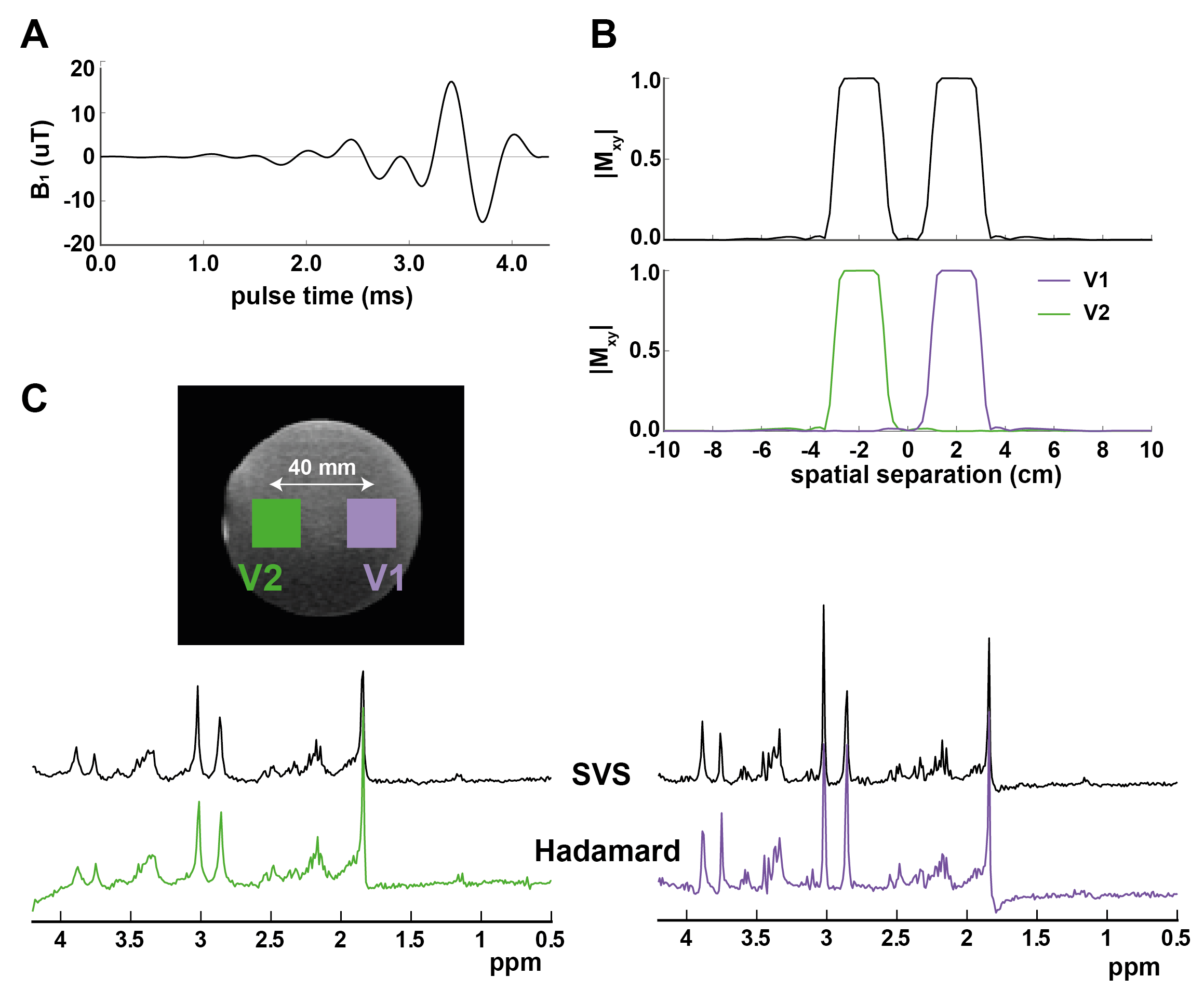
Two-voxel fMRSTwo-voxel localisation was achieved using a Hadamard-encoded semi-LASER (TE/TR=30ms/6.1s) scheme with cosine-modulated excitation pulse (Fig. 1A; dur=4.4ms, BW=1.5kHz). Interleaved VAPOR+OVS [8] was adapted to suppress lipids surrounding each voxel (OVS: HS1, R=40, gap/width=10/60mm). The sequence was validated in a phantom (Fig. 1C). B0-shimming (second-order) was performed on B0 maps (GRE, 2mm isotropic, dTE/TR=1ms/20ms) over two non-contiguous ROIs defined by each volume-of-interest (VOI) [9].
Protocol
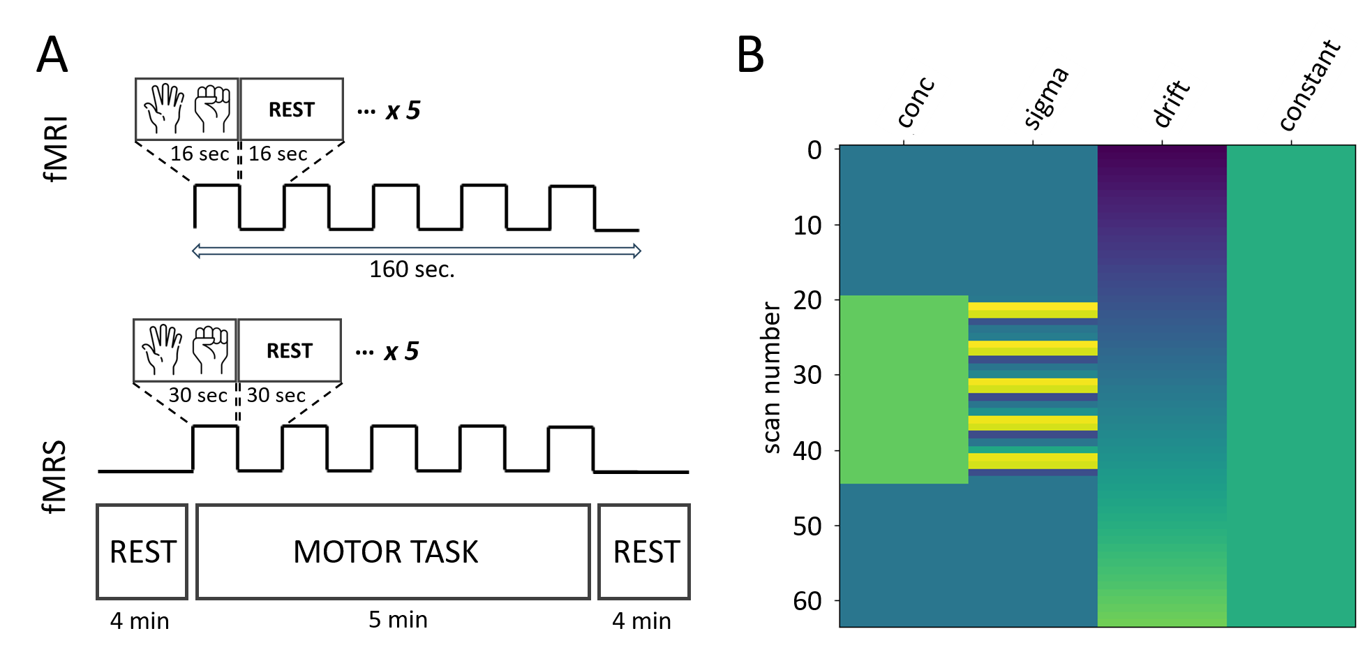
Data were acquired from seven right-handed healthy volunteers on a 7T Philips Achieva. Whole-brain fMRI data (TE/TR=17/2000ms, EPI factor=29, FOV=218x218x65mm3, voxel size=1.24x1.24x2.5mm3, 78 dynamics) and fMRS data (BW=6kHz, 2048 points, VOI size = 20x20x20mm3, NT=126, TA=13min) were acquired from left (L; contralateral) and right (R, ipsilateral) primary motor cortex (M1) during right-handed motor task (Fig. 2A). VOI separation was varied according to individual anatomy.
Analysis
fMRI: Preprocessing included motion correction, high-pass temporal filtering (0.01Hz) and spatial smoothing (4mm FWHM), coregistration to anatomical T1-weighted scans. First-level general linear model (GLM) analyses were carried out using FSL-FEAT [10] to assess the task>rest contrast.
fMRS: Data were fit dynamically using FSL-MRS [11,12]. Spectral fitting was performed using a simulated basis with measured macromolecules (MMs) [13]. Phase, frequency, Lorentzian lineshape- and baseline parameters were fixed. Metabolite concentrations and Gaussian lineshape parameters (sigma) were fit using a temporal model (Fig. 2B) (separately for MMs).
Statistics
fMRI time courses and z-stats were extracted from within the MRS VOIs. Group level fMRS statistics were calculated with second-level GLMs using FSL’s flameo. Significance threshold was p<0.05.
Results
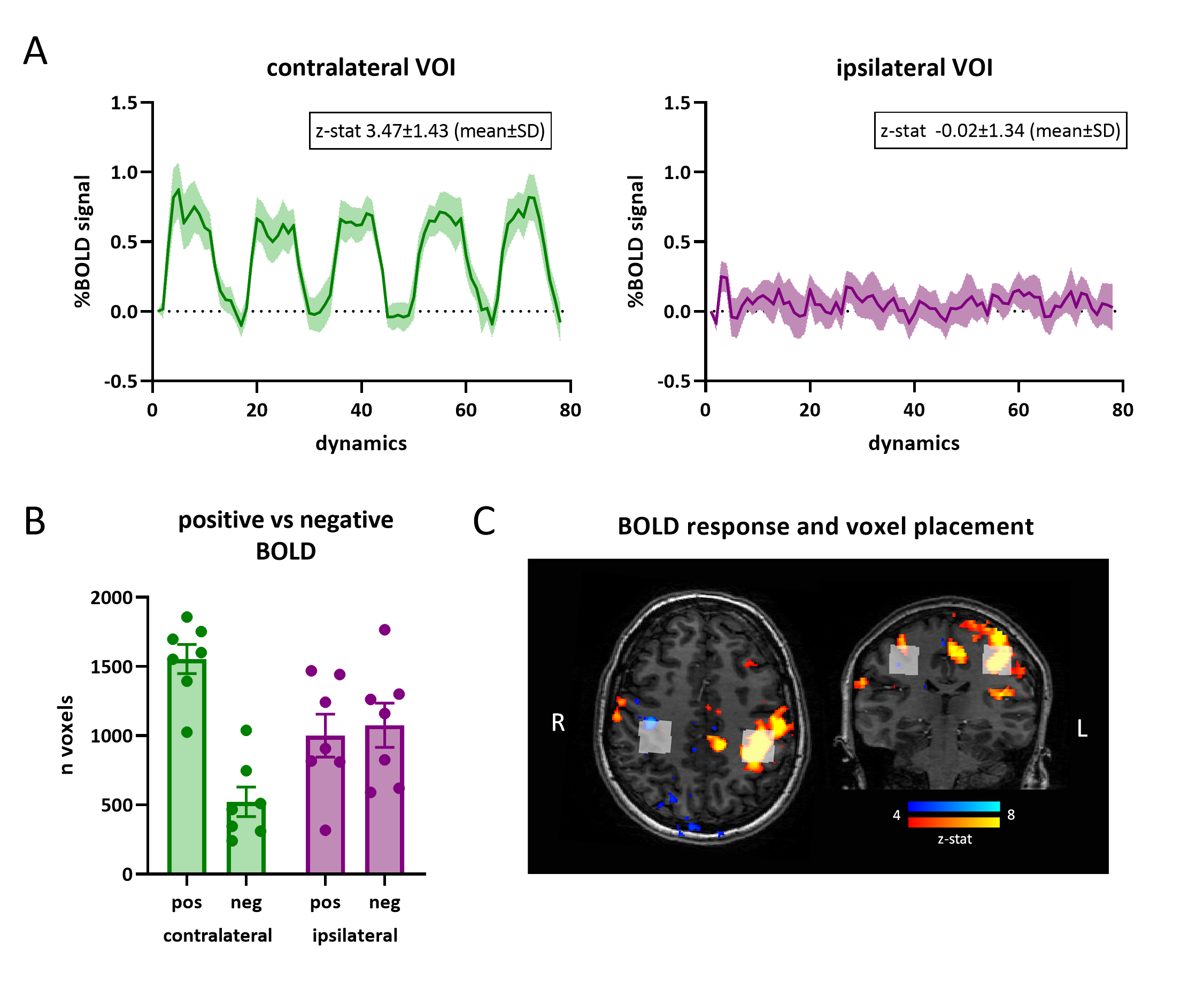
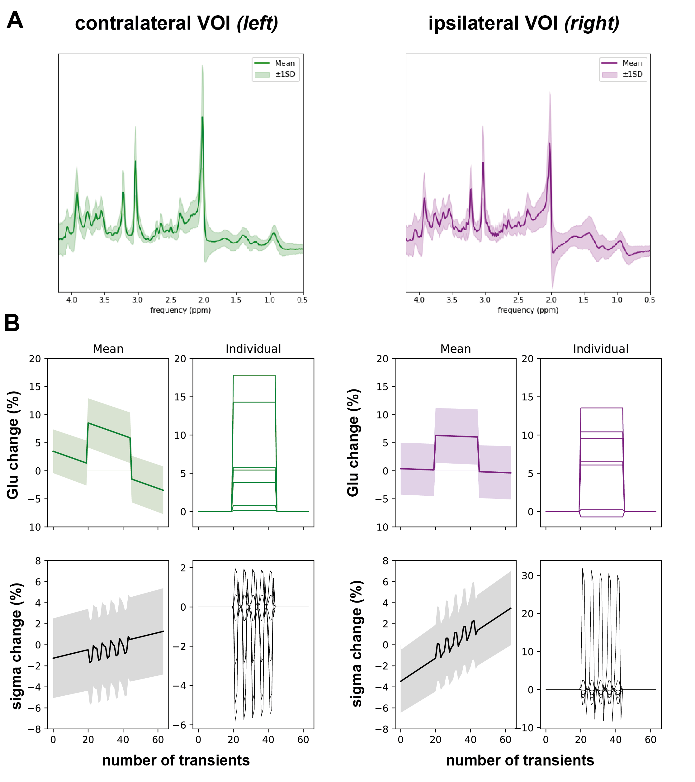
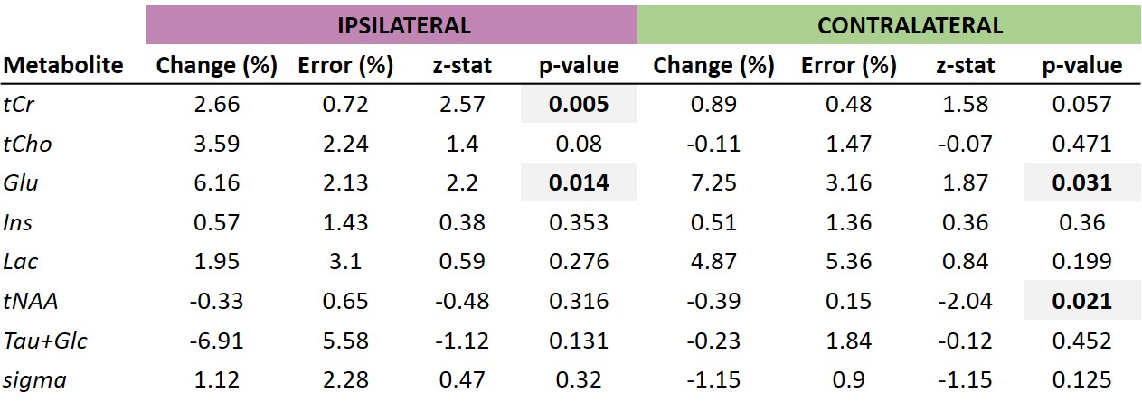
fMRI: Across participants, a strong consistent positive BOLD response was observed in contralateral VOI (z-stat=3.5±1.4), while in ipsilateral VOI, there was a smaller mean response (z-stat=-0.02±1.3), which in four participants was negative (Fig. 3A). In general, there was a large variability in the polarity of BOLD response in the ipsilateral VOI (Fig. 3B).fMRS: The averaged spectrum across participants in both VOIs is displayed in Fig. 4A. Spectra were of high quality with excellent average linewidths in contralateral [(10.9±1.7)Hz] and ipsilateral [(10.9±1.1)Hz] VOIs. In group-level analyses, we observed a significant Glu increase in contralateral and ipsilateral VOIs of (7.3±3.2)% (z=1.9, p=0.03) and (6.2±2.1)% (z=2.2, p=0.014), respectively (Fig. 4B). The change in linewidth (sigma) trended negative in contralateral VOI [(-1.2±0.9)%, z=1.2, p=0.12], yet no trend was observed in ipsilateral VOI [(+1.1±2.3)%, z=0.47, p=0.3] with a large inter-subject variability (Fig. 4B). Group-level changes in other metabolites are shown in Fig. 5.
Discussion and conclusion
We have demonstrated the feasibility of simultaneous 7 T two-voxel fMRS in bilateral motor cortices during a unilateral hand-clenching task. Two-voxel spectra were of high quality (linewidths ~11Hz). As expected, we observed Glu increase in contralateral VOI (+7%). Yet, contrary to our hypothesis, we also measured a significant mean Glu increase in ipsilateral VOI (+6%). Our task elicited strong BOLD-activation in contralateral VOI, yet a mixed response (activation/deactivation) in ipsilateral VOI. Our findings may reflect neuronal activation across the connected motor cortex during planning/execution of the unimanual task [14], yet future refinement is needed using interleaved fMRI-fMRS to directly relate concurrent BOLD signals to metabolite changes. Additionally, the Hadamard approach is SNR-efficient yet leads to an effective time-resolution of 12s for dynamic fMRS. Alternative approaches [7] may allow faster sampling of neurochemical signals.Acknowledgements
We would like to acknowledge Daphne Boucherie for her help with data acquisitionReferences
[1] Schaller B, Xin L, O'Brien K, Magill AW, Gruetter R. Are glutamate and lactate increases ubiquitous to physiological activation? A (1)H functional MR spectroscopy study during motor activation in human brain at 7Tesla. Neuroimage. 2014; 93 Pt 1:138-45. doi: 10.1016/j.neuroimage.2014.02.016.
[2] Chen, C., Sigurdsson, H. P., Pépés, S. E., Auer, D. P., Morris, P. G., Morgan, P. S., Gowland, P. A., & Jackson, S. R. Activation induced changes in GABA: Functional MRS at 7 T with MEGA-sLASER. NeuroImage. 2017 156(May), 207–213. https://doi.org/10.1016/j.neuroimage.2017.05.044
[3] Boer VO, Klomp DW, Laterra J, Barker PB. Parallel reconstruction in accelerated multivoxel MR spectroscopy. Magn Reson Med. 2015; 74(3):599-606. doi: 10.1002/mrm.25718.
[4] Riemann LT, Aigner CS, Mekle R, Speck O, Rose G, Ittermann B, Schmitter S, Fillmer A. Fourier-based decomposition for simultaneous 2-voxel MRS acquisition with 2SPECIAL. Magn Reson Med. 2022; 88(5):1978-1993. doi: 10.1002/mrm.29369.
[5] Dehghani M, Edden RAE, Near J. Hadamard-encoded dual-voxel SPECIAL: Short-TE MRS acquired in two brain regions simultaneously using Hadamard encoding. Magn Reson Med. 2022; 87(4):1649-1660. doi: 10.1002/mrm.29129.
[6] Boillat Y, Xin L, van der Zwaag W, Gruetter R. Metabolite concentration changes associated with positive and negative BOLD responses in the human visual cortex: A functional MRS study at 7 Tesla. J Cereb Blood Flow Metab. 2020 Mar;40(3):488-500. doi: 10.1177/0271678X19831022.
[7] Koush Y, de Graaf RA, Kupers R, Dricot L, Ptito M, Behar KL, Rothman DL, Hyder F. Metabolic underpinnings of activated and deactivated cortical areas in human brain. J Cereb Blood Flow Metab. 2021 May;41(5):986-1000. doi: 10.1177/0271678X21989186.
[8] Tkáć, I., & Gruetter, R. Methodology of 1H NMR spectroscopy of the human brain at very high magnetic fields. Applied Magnetic Resonance. 2005 29(1), 139–157. https://doi.org/10.1007/BF03166960
[9] Boer VO, Andersen M, Lind A, Lee NG, Marsman A, Petersen ET. MR spectroscopy using static higher order shimming with dynamic linear terms (HOS-DLT) for improved water suppression, interleaved MRS-fMRI, and navigator-based motion correction at 7T. Magn Reson Med. 2020; 84(3):1101-1112. doi: 10.1002/mrm.28202.
[10] Woolrich MW, Ripley BD, Brady M, Smith SM. Temporal autocorrelation in univariate linear modeling of FMRI data. Neuroimage. 2001; 14(6):1370-86. doi: 10.1006/nimg.2001.0931.
[11] Clarke WT, Stagg CJ, Jbabdi S. FSL-MRS: An end-to-end spectroscopy analysis package. Magn Reson Med. 2021; 85(6):2950-2964. doi: 10.1002/mrm.28630.
[12] Clarke, L., Ligneul, C., Cottaar, M., Ip, I. B., & Jbabdi, S. Simultaneous fitting of spectral and dynamic models to 2D MRS. 2023 BioRxiv. https://doi.org/10.1101/2023.06.15.544935
[13] Tkác I, Oz G, Adriany G, Uğurbil K, Gruetter R. In vivo 1H NMR spectroscopy of the human brain at high magnetic fields: metabolite quantification at 4T vs. 7T. Magn Reson Med. 2009; 62(4):868-79. doi: 10.1002/mrm.22086.
[14] Horenstein C, Lowe MJ, Koenig KA, Phillips MD. Comparison of unilateral and bilateral complex finger tapping-related activation in premotor and primary motor cortex. Human Brain Mapping. 2009; 30(4): 1397–1412. https://doi.org/10.1002/hbm.20610
[15] Glover GH. Deconvolution of impulse response in event-related BOLD fMRI. NeuroImage 1999; 9:416-429.
Figures