1289
EPTIMA: Echo Planar Time-resolved Imaging derived Millisecond-scale temporal resolution Acquisition1Athinoula A. Martinos Center for Biomedical Imaging, Massachusetts General Hospital, Charlestown, MA, United States, 2Department of Radiology, Harvard Medical School, Boston, MA, United States, 3Harvard-MIT Division of Health Sciences and Technology, Massachusetts Institute of Technology, Cambridge, MA, United States
Synopsis
Keywords: fMRI Acquisition, Data Acquisition, fMRI
Motivation: Achieving millisecond-scale temporal resolution MRI has the potential to provide exciting insights into fast functional/physiological processes of the brain.
Goal(s): Develop a new acquisition method, EPTIMA, that can achieve millisecond-scale temporal resolution, while improving efficiency by acquiring a time-series trial of 2D-images in a single excitation for high robustness to physiological-noise/motion.
Approach: EPTIMA captures fast temporal dynamics occurring within the readout by measuring the rate at which the baseline signal evolution is changing, and employs spatiotemporal encodings to acquire a complete time-series trial in a single-excitation.
Results: EPTIMA can image rapid electric current changes in a phantom and resolve stable phase/magnitude changes in-vivo.
Impact: A new acquisition, EPTIMA, was developed to achieve millisecond-scale temporal resolution and to image ultra-fast dynamic processes of human brain. It improves efficiency by acquiring a time-series trial of 2D-images in a single excitation with high robustness to motion/physiological noises.
Introduction
Achieving ultra-high temporal-resolution MRI can enable the investigation of fast functional/physiological processes of the human brain. With the advancement of many acquisition technologies1-7, fast temporal resolutions of <100ms can be achieved to map fast BOLD fMRI8-9. Further increasing the temporal resolution, for example, to a millisecond scale, has the potential to provide exciting insights into even faster dynamic processes such as direct neuronal activities10-16, but presents technical challenges due to the long RF pulses, readout, and spatial encoding. Several acquisition techniques have been developed to push the boundaries of temporal resolution down to the millisecond range17-23. Among them, SPEEDI17-19 uses an FID/gradient-echo train to measure electric-current changes in phantoms and to capture aortic valve opening/closing. The recent DIANA20-22 work achieves a 5-ms temporal resolution using a line-by-line sampling in FLASH sequence towards investigating fast direct neuronal activity. However, most of the existing methods require repeated sampling of the time-series for different spatial-encodings (e.g.,phase-encodings), often resulting in extended scan times even with the use of parallel imaging/compressed sensing19. This significantly limits its spatial coverage. In addition, the spatial encoding is usually acquired in different trials/repetitions. Combining these temporally-separate spatially-encoded time-series to generate a 2D-image time-series can make the data more susceptible to physiological noise/motion. This can severely compromise the detection sensitivity, especially when measuring inherently subtle signals.Here, we described a new method, EPTIMA, that can achieve millisecond-scale temporal resolution for both magnitude and phase changes, while significantly improving the acquisition efficiency by sampling a time-series trial of 2D images in a single excitation. For example, in EPTIMA, a time-series trial sampled at millisecond-scale temporal resolution (e.g.,~0.53ms) with a duration of ~120ms can be acquired in just 150ms instead of several seconds (e.g., 7s due to repeated multi-excitations for phase-encodings). This can significantly increase the robustness to physiological noise/motion and improve detection sensitivity and can also enable much more flexible paradigm designs (e.g.,resting-state). The proof-of-concept results demonstrated that the technique was able to image rapid electric current changes at milliseconds-scale in a phantom and resolve stable phase and magnitude changes in a preliminary in-vivo experiment.
Methods
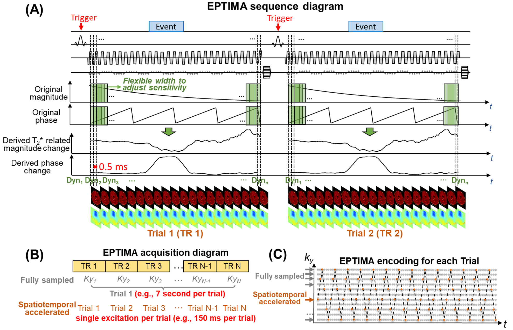
Different from our previous EPTI24-27 where multi-echo images with slow baseline signal evolution are resolved, EPTIMA aims to capture the fast temporal dynamics occurring within the readout by measuring the rate at which the baseline evolution is changing. As shown in Fig.1, in EPTIMA, a time series of gradient echo images will be acquired in each TR. The slow baseline T2* decay and B0-phase evolution of these images will be removed by deriving the magnitude/phase changes that represent the rate at which the baseline evolution is changing as a function of time within the readout. Specifically, using a sliding window that advances at a millisecond temporal-interval (~0.5ms), the phase change is calculated by taking the derivative/phase difference between the adjacent echoes; and the T2*-related magnitude change (or T2* itself) by dividing the magnitude of adjacent echoes. The window size can be flexibly adjusted: a longer window increases the sensitivity for measuring phase and magnitude changes (e.g., 8ms), while a shorter window prioritizes high temporal frequency information (e.g.,1ms). For each TR (e.g.,150ms), a time-series of 2D magnitude/phase images will be obtained (a trial) at millisecond/sub-millisecond temporal intervals. For spatial encoding, multiple excitations are usually needed to acquire different ky-lines to generate 2D-images of the time-series (e.g., 7s for a 150-ms trial using fully-sampled acquisition). In contrast, EPTIMA acquires the same time-series of 2D-images within a single excitation (e.g.,150ms), so more trials (e.g.,48x) can be acquired in the same amount of time with high robustness to physiological noises/motion. This is achieved by employing single-shot EPTI trajectory27 that encodes both space and time (Fig.1C) in each TR/trial and recovering the fully-sampled k-t space by applying GRAPPA-like-kernels24.Results
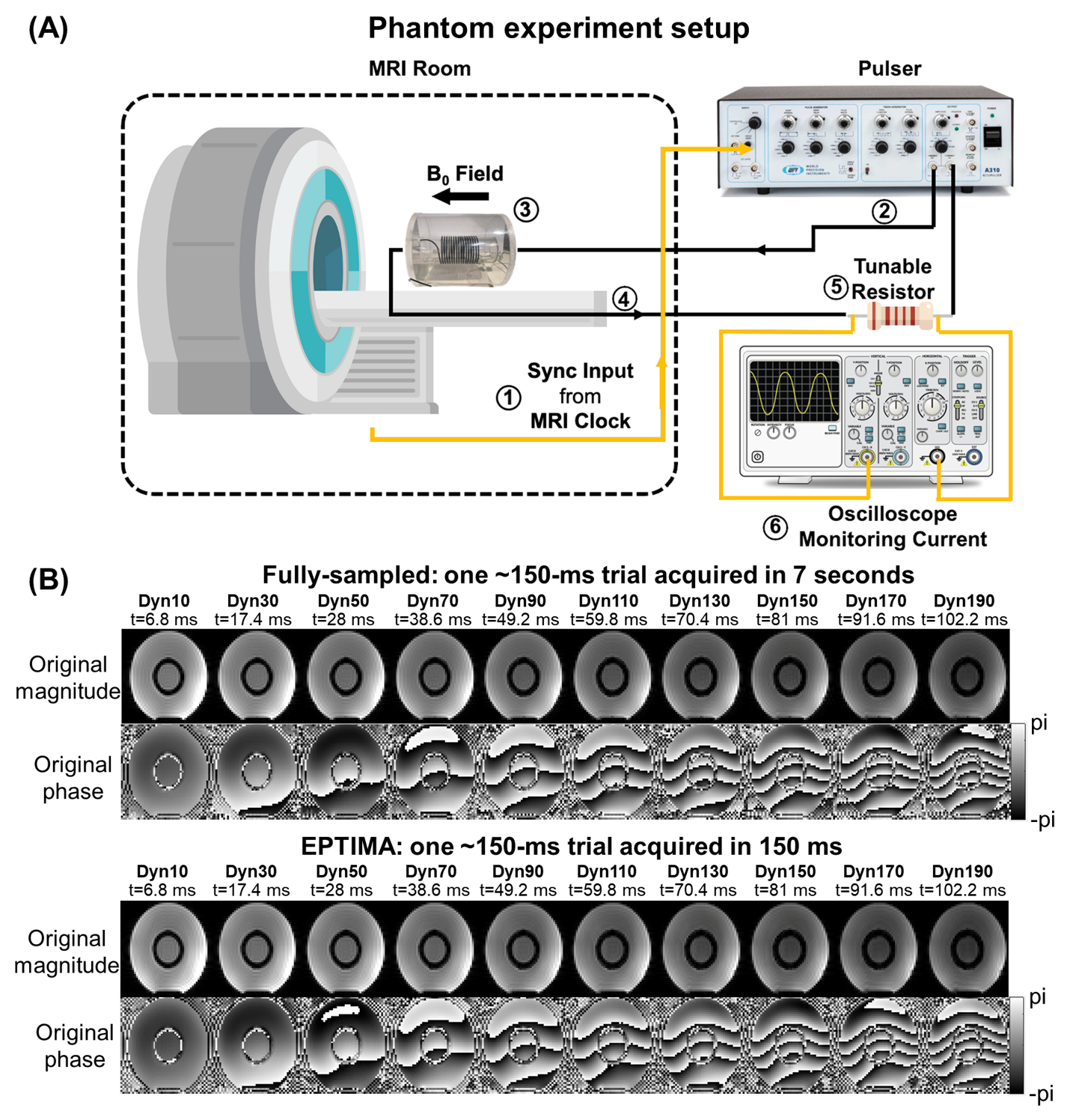
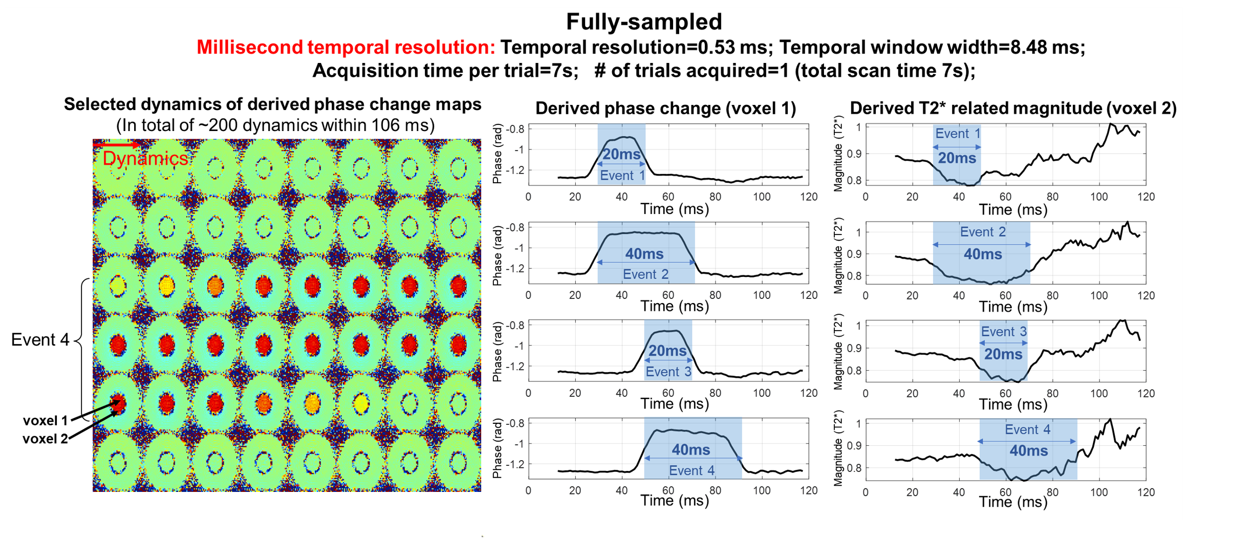
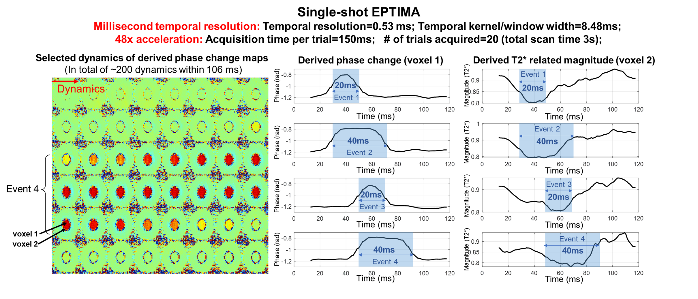
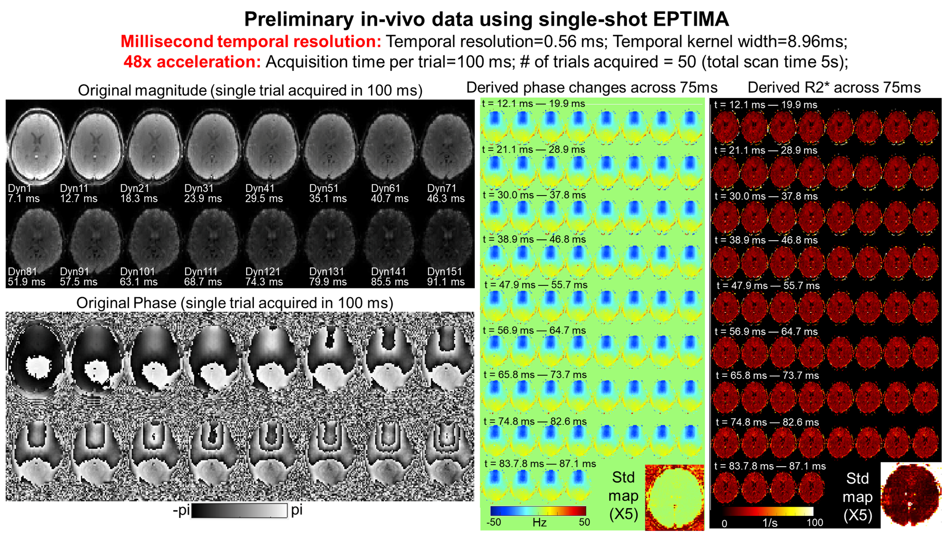
The time-series images for a single 150-ms trial acquired by EPTIMA (48x accelerated) in 150ms resulted in good image quality compared to the lengthy 7s fully sampled acquisition (Fig.2). EPTIMA was able to capture the magnetic field changes accompanying the rapid electric current changes with different durations (20ms vs. 40ms) and start times (30ms vs. 50ms) in both phase and magnitude (Fig.3 vs. Fig.4). Fig.5 demonstrates the feasibility of EPTIMA to obtain good in-vivo image quality for each trial, and to derive phase and magnitude changes with high stability within the readout during resting state.Conclusion
We developed a novel acquisition method EPTIMA that can achieve ultra-fast imaging at millisecond-scale temporal resolution, while providing robustness to physiological noise/motion. Future work will explore the use of EPTIMA for mapping fast brain functional/physiological dynamics.Acknowledgements
This work was supported by the NIH BRAIN Initiative (U24NS129893, 1R01NS112183) and NIH (K99AG083056, R01-EB019437, P41EB030006), and the instrumentation Grants (S10OD023637).References
[1] Moeller S, Yacoub E, Olman CA, Auerbach E, Strupp J, Harel N, Uğurbil K. Multiband multislice GE‐EPI at 7 tesla, with 16‐fold acceleration using partial parallel imaging with application to high spatial and temporal whole‐brain fMRI. Magnetic resonance in medicine. 2010 May;63(5):1144-53.
[2] Feinberg DA, Moeller S, Smith SM, Auerbach E, Ramanna S, Glasser MF, Miller KL, Ugurbil K, Yacoub E. Multiplexed echo planar imaging for sub-second whole brain FMRI and fast diffusion imaging. PloS one. 2010 Dec 20;5(12):e15710.
[3] Setsompop K, Gagoski BA, Polimeni JR, Witzel T, Wedeen VJ, Wald LL. Blipped‐controlled aliasing in parallel imaging for simultaneous multislice echo planar imaging with reduced g‐factor penalty. Magnetic resonance in medicine. 2012 May;67(5):1210-24.
[4] Hennig J, Zhong K, Speck O. MR-Encephalography: Fast multi-channel monitoring of brain physiology with magnetic resonance. Neuroimage. 2007 Jan 1;34(1):212-9.
[5] Zahneisen B, Grotz T, Lee KJ, Ohlendorf S, Reisert M, Zaitsev M, Hennig J. Three‐dimensional MR‐encephalography: fast volumetric brain imaging using rosette trajectories. Magnetic resonance in medicine. 2011 May;65(5):1260-8.
[6] Lin FH, Witzel T, Mandeville JB, Polimeni JR, Zeffiro TA, Greve DN, Wiggins G, Wald LL, Belliveau JW. Event-related single-shot volumetric functional magnetic resonance inverse imaging of visual processing. Neuroimage. 2008 Aug 1;42(1):230-47.
[7] Lin FH, Tsai KW, Chu YH, Witzel T, Nummenmaa A, Raij T, Ahveninen J, Kuo WJ, Belliveau JW. Ultrafast inverse imaging techniques for fMRI. Neuroimage. 2012 Aug 15;62(2):699-705.
[8] Chen JE, Glover GH. BOLD fractional contribution to resting-state functional connectivity above 0.1 Hz. Neuroimage. 2015 Feb 15;107:207-18.
[9] Lewis LD, Setsompop K, Rosen BR, Polimeni JR. Fast fMRI can detect oscillatory neural activity in humans. Proceedings of the national academy of sciences. 2016 Oct 25;113(43):E6679-85
[10] Bandettini PA, Petridou N, Bodurka J. Direct detection of neuronal activity with MRI: fantasy, possibility, or reality?. Applied Magnetic Resonance. 2005 Mar;29(1):65-88.
[11] Petridou N, Plenz D, Silva AC, Loew M, Bodurka J, Bandettini PA. Direct magnetic resonance detection of neuronal electrical activity. Proceedings of the National Academy of Sciences. 2006 Oct 24;103(43):16015-20.
[12] Bodurka J, Bandettini PA. Toward direct mapping of neuronal activity: MRI detection of ultraweak, transient magnetic field changes. Magnetic Resonance in Medicine. 2002 Jun;47(6):1052-8.
[13] Witzel T, Lin FH, Rosen BR, Wald LL. Stimulus-induced Rotary Saturation (SIRS): a potential method for the detection of neuronal currents with MRI. Neuroimage. 2008 Oct 1;42(4):1357-65.
[14] Sundaram P, Nummenmaa A, Wells W, Orbach D, Orringer D, Mulkern R, Okada Y. Direct neural current imaging in an intact cerebellum with magnetic resonance imaging. Neuroimage. 2016 May 15;132:477-90.
[15] Truong TK, Roberts KC, Woldorff MG, Song AW. Toward direct MRI of neuro‐electro‐magnetic oscillations in the human brain. Magnetic resonance in medicine. 2019 Jun;81(6):3462-75.
[16] Patz S, Fovargue D, Schregel K, Nazari N, Palotai M, Barbone PE, Fabry B, Hammers A, Holm S, Kozerke S, Nordsletten D. Imaging localized neuronal activity at fast time scales through biomechanics. Science advances. 2019 Apr 17;5(4):eaav3816.
[17] Zhong Z, Sun K, Karaman MM, Zhou XJ. Magnetic resonance imaging with submillisecond temporal resolution. Magn Reson Med. 2021;85:2434-2444.
[18] Zhong Z, Sun K, Dan G, et al. Visualization of human aortic valve dynamics using magnetic resonance imaging with sub-millisecond temporal resolution. J Magn Reson Imaging. 2021;54:1246-1254.
[19] Luo Q, Zhong Z, Sun K, Scotti A, Zhou XJ. Gradient‐echo‐train‐based sub‐millisecond periodic event encoded dynamic imaging with random (k, t)‐space undersampling: k‐t get‐SPEEDI. Magnetic Resonance in Medicine. 2022 Oct;88(4):1690-701.
[20] Toi PT, Jang HJ, Min K, Kim SP, Lee SK, Lee J, Kwag J, Park JY. In vivo direct imaging of neuronal activity at high temporospatial resolution. Science. 2022 Oct 14;378(6616):160-8.
[21] Hodono S, Rideaux R, van Kerkoerle T, Cloos MA. Initial experiences with Direct Imaging of Neuronal Activity (DIANA) in humans. arXiv preprint arXiv:2303.00161. 2023 Mar 1.
[22] Choi SH, Im GH, Choi S, Yu X, Bandettini PA, Menon RS, Kim SG. No Replication of Direct Neuronal Activity-related (DIANA) fMRI in Anesthetized Mice. bioRxiv. 2023:2023-05.
[23] Zhang Y, Sun K, Ren J, Hu Q, Wang Y, Li S, Chen T, Xu N, Guo N, Fu X, Liu X, Cao Z, Gao J, Liu H. High-resolution Dynamic Human Brain Neural Activity Recording Using 3T MRI. bioRxiv. 2023:2023-05.
[24] Wang F, Dong Z, Reese TG, Bilgic B, Katherine Manhard M, Chen J, Polimeni JR, Wald LL, Setsompop K. Echo planar time‐resolved imaging (EPTI). Magnetic resonance in medicine. 2019 Jun;81(6):3599-615.
[25] Dong Z, Wang F, Reese TG, Bilgic B, Setsompop K. Echo planar time‐resolved imaging with subspace reconstruction and optimized spatiotemporal encoding. Magnetic resonance in medicine. 2020 Nov;84(5):2442-55.
[26] Wang F, Dong Z, Wald LL, Polimeni JR, Setsompop K. Simultaneous pure T2 and varying T2′-weighted BOLD fMRI using Echo Planar Time-resolved Imaging for mapping cortical-depth dependent responses. Neuroimage. 2021 Dec 15;245:118641.
[27] Wang F, Dong Z, Chen J, Setsompop K, Polimeni J, Wald LL. Improving fMRI acquisition using single-shot EPTI with distortion-free high-SNR high-CNR multi-echo imaging. In Proceedings of the 30th Annual Meeting of ISMRM, 2022, p. 3330.
Figures