1281
Evaluation of Magnetic Resonance Mediated Radiofrequency Ablation in Bovine Liver Tissue Using Textile Antenna-Enhanced MR Thermometry.1Seoul Institute of Technology, School of Electrical and Biomedical Engineering, Hanyang University, Seoul, Korea, Republic of, 2Bio-chemical Analysis Team, Korea Basic Science Institute, Cheongju, 28119, South Korea, Cheongju, Korea, Republic of, 3Department of Biomedical Engineering and Department of Electronic Engineering, Hanyang University, Seoul, Korea, Republic of
Synopsis
Keywords: Non-Array RF Coils, Antennas & Waveguides, Thermometry, Magnetic resonance-mediated radiofrequency ablation, antenna
Motivation: Magnetic resonance-mediated radiofrequency ablation (MR-RFA) combines diagnostic and therapeutic functions within MRI scanners, and its significance has grown rapidly, particularly in tumor treatment diagnosis.
Goal(s): In this study, a 3T Philips MRI scanner is channeled toward the ablation site in the bovine liver by means of an antenna and a needle, with the objective of generating RF heating at the tumor location.
Approach: MR thermometry was used to evaluate the MR-RFA procedure and predict local specific absorption rate (SAR) escalation and temperature increase.
Results: Our research demonstrated that temperature maps with a 73 oC peak value were observed at the needle tip.
Impact: In contrast to existing ablation designs, this design provides enhanced patient comfort, localized heating, minimal skin burns, and avoids the use of external RF power sources, all while ensuring there is no distortion in MR images.
Introduction
Currently, ultrasound, computed tomography guidance, and conventional RFA are commonly employed for percutaneous ablation procedures [1]. However, these techniques have several limitations and drawbacks, including the potential for local recurrences, enlarging ablation zones, skin burning, and patient safety [2], [3]. To achieve successful ablation, it is essential to use imaging guidance to accurately position the RFA needle. Other issues exist regarding ground pad handling in MRI scanners, damage to healthy tissues, and patient discomfort [4]. A novel MR-RFA technique has been introduced, involving direct placement of the RF element on the patient's skin to transmit RF energy to the tumor. This approach enhances precision and accuracy, enabling more targeted tumor treatment while minimizing harm to adjacent healthy tissues. The proposed MR-RFA boasts advantages in terms of cost, effectiveness, ease of use, and suitability for diverse patient populations compared to traditional RFA methods.Methodology
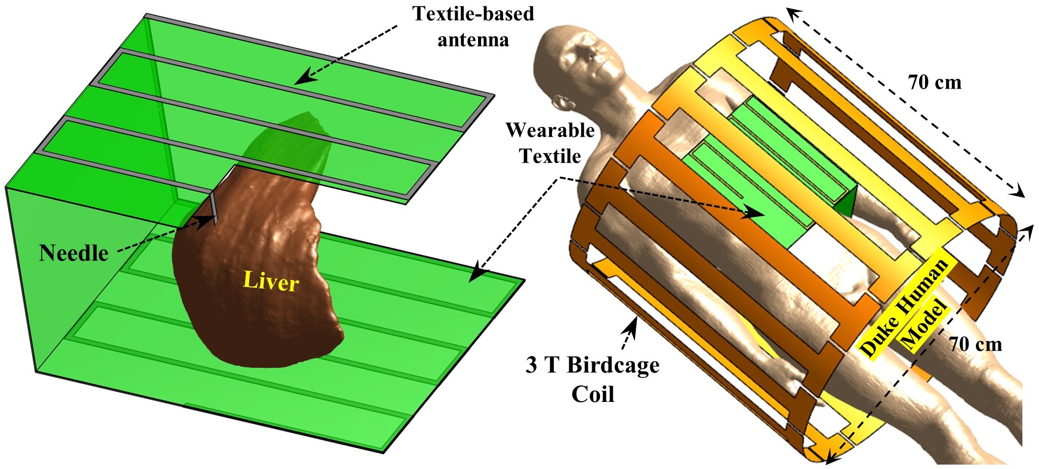
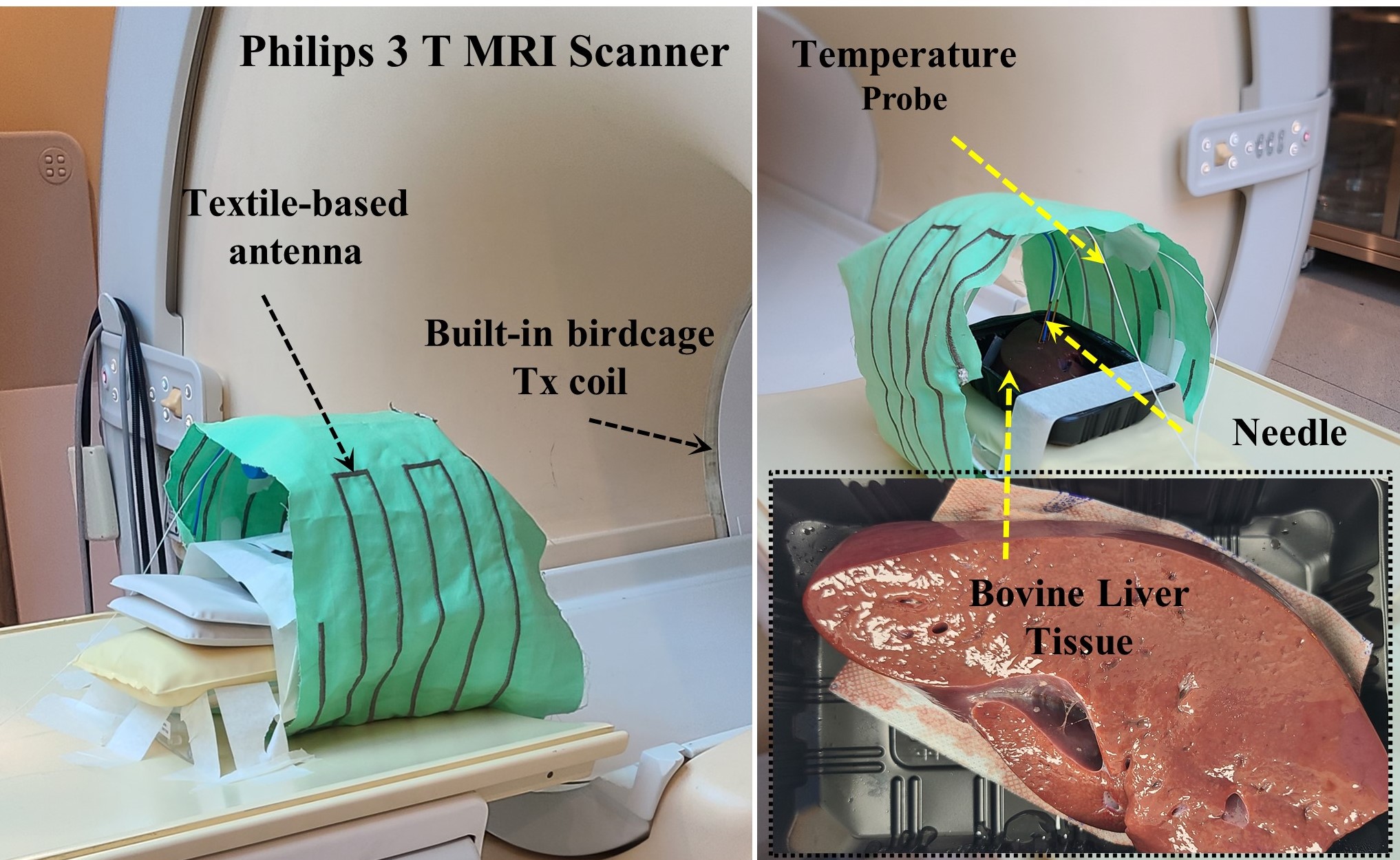
Fig. 1 depicts the simulated setup of the Birdcage coil, liver phantom, and pickup antenna element. The setup includes the RF birdcage coil as an external energy source, with the liver phantom inside the pickup antenna. To evaluate the magnetic field and SAR of the interconnected coil, electromagnetic simulations using Sim4Life by ZMT [5] were performed, while the MR thermometry experiments were conducted on the 3-T MRI scanner (Philips Achieva 3.0T TX, Best, Netherlands) to generate heat for ablating abnormal tissues, as shown in Fig. 2. The experimental setup was carried out under the guidance of a skilled radiologist. Magnetic resonance thermometry scans were conducted by capturing phase images using a gradient-echo (GRE) pulse sequence, with the following scan parameters: a 100-millisecond repetition time (TR), an 8-millisecond echo time (TE), and a 15-degree flip angle (FA). The Proton Resonance Frequency Shift (PRFS) technique was utilized to acquire images showing temperature elevation near the needle, thereby confirming the viability of magnetic resonance thermometry. Temperature variations were determined by assessing the phase disparity between the pre-procedure and post-procedure images.Results and discussion
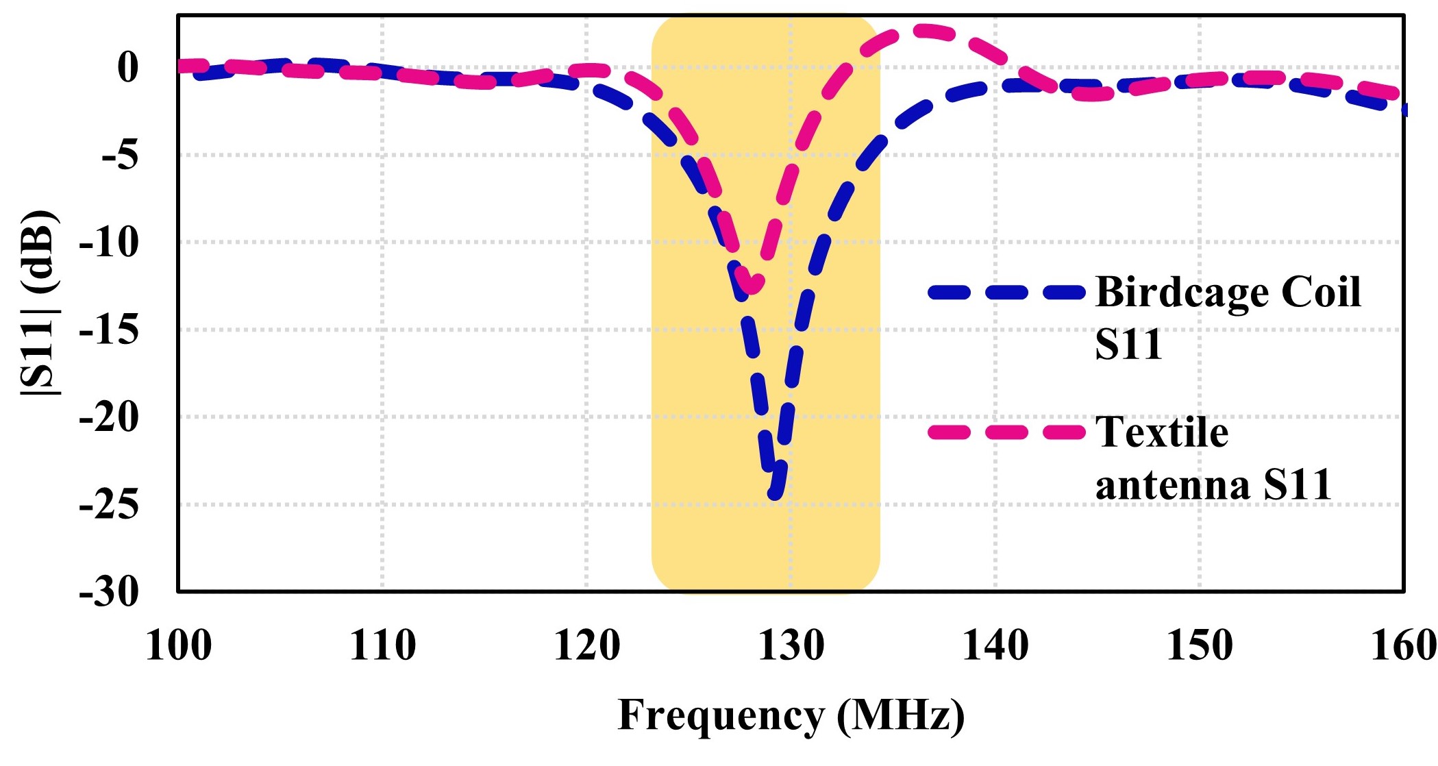
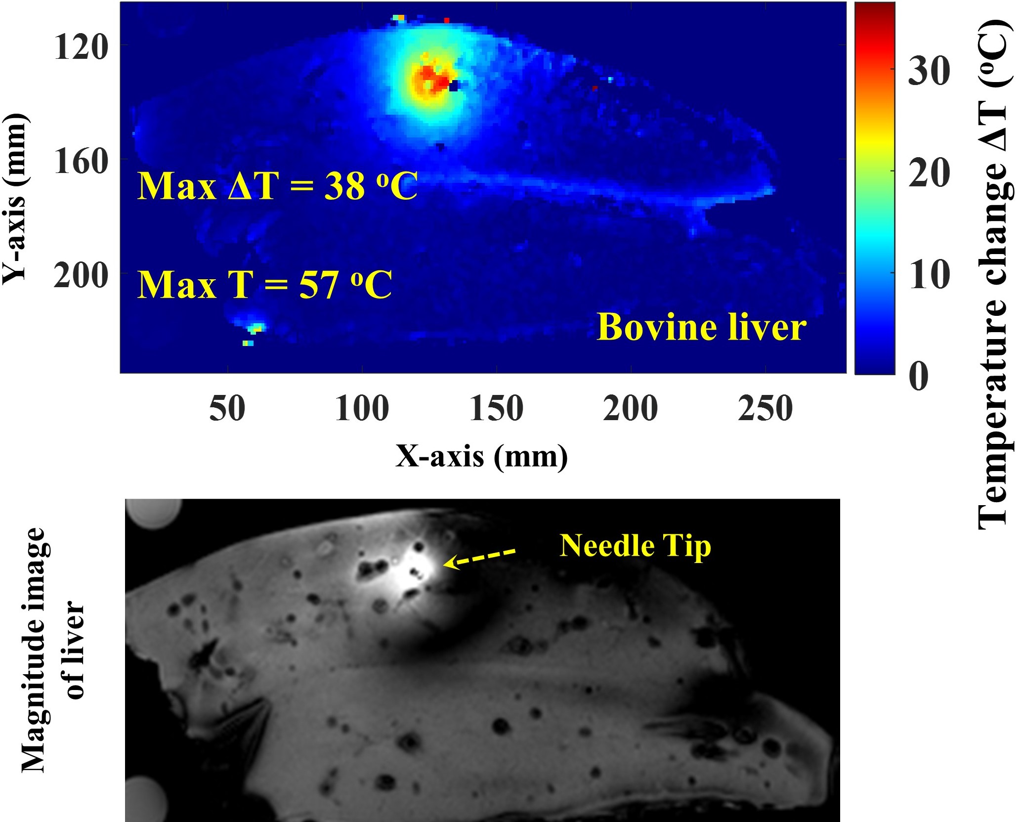
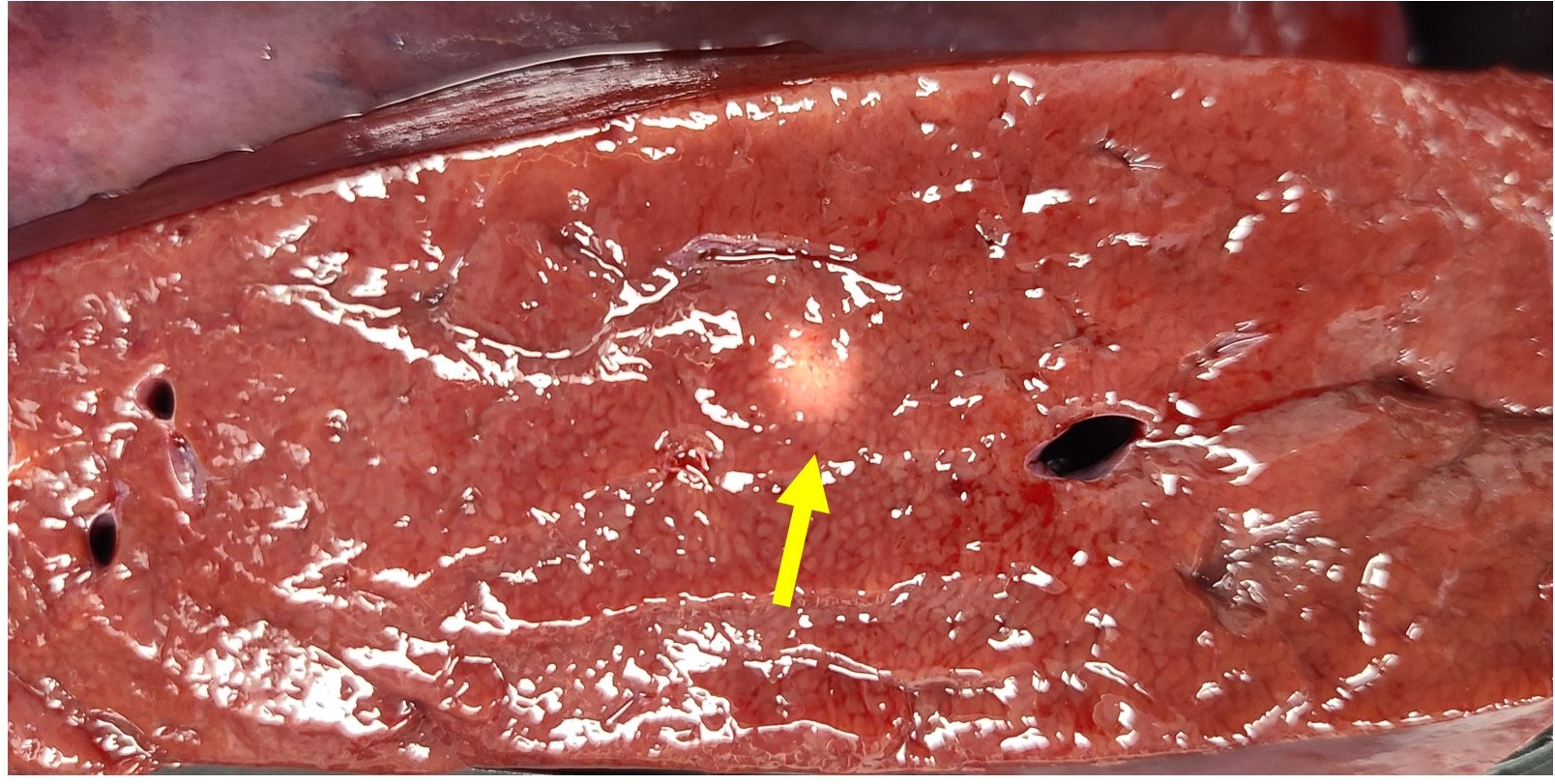
Both the RF birdcage coil and the antenna were adjusted to operate at the resonance frequency of 3 Tesla (corresponding to 298 MHz). The coil exhibited a return loss value that was lower than -13 dB, as shown in Fig. 3. The temperature results show that the antenna captured the power from the scanner and coupled it to the target location in the bovine liver. The temperature rise in the target location is due to the lossy nature of the bovine liver tissue, which can be used for RF ablation tumor therapy with MRI scanning. The temperature map reveals a peak-induced temperature of 57 degrees C (oC) at the needle tip in the bovine liver, as shown in Fig. 4. The temperature at the tip increases from 19 oC to 57 oC, indicating a change of 38 oC. It can also be seen in the figure that the temperature rise is confined to the tip location while the surrounding temperature map is more uniform, which indicates the effectiveness of the proposed antenna for ablation. The figure illustrates a magnitude image revealing notable hyperintense artifacts within a segment of bovine liver tissue. Moreover, the MR-RFA system induces heating in the bovine liver tissue at the tip, resulting in an observable ablation lesion at that specific location, as shown in Fig. 5.Conclusion
All MR thermometry results showed that the proposed pickup antenna can induce a 57-degree rise in the bovine liver tissue. This study concluded that with the inclusion of a potentially temporary ablation device, MR-RFA may enhance the usefulness of MRI. In this study, it was shown that the energy extracted from the MRI scanner was adequate for the effective ablation of abnormal tissues. Through the use of RF pulses, MR thermometry can precisely control thermal heating.Acknowledgements
This work was supported in part by the Institute for Information & Communications Technology Promotion (IITP) Grant funded by the Korean Government [Ministry of Science, ICT and Future Planning (MSIP)] (No. 2021-0-00490), and in part by the Development of Precision Analysis and Imaging Technology for Biological Radio Waves.References
1. F. Fischbach, K. Lohfink et al., “Magnetic resonance–guided freehand radiofrequency ablation of malignant liver lesions: a new simplified and time-efficient approach using an interactive open magnetic resonance scan platform and hepatocyte-specific contrast agent,” Investigative radiology, vol. 48, no. 6, pp. 422–428, 2013.
2. E. K. Abdalla, J.-N. Vauthey et al., “Recurrence and outcomes following hepatic resection, radiofrequency ablation, and combined resection/ablation for colorectal liver metastases,” Annals of surgery, vol. 239, no. 6, p. 818, 2004.
3. I. Sucandy, S. Cheek et al., “Longterm survival outcomes of patients undergoing treatment with radiofrequency ablation for hepatocellular carcinoma and metastatic colorectal cancer liver tumors,” Hpb, vol. 18, no. 9, pp. 756–763, 2016.
4. S. Huffman, N. Huffman, R. J. Lewandowski, and D. B. Brown, “Radiofrequency ablation complicated by skin burn,” in Seminars in interventional radiology, vol. 28, no. 02. © Thieme Medical Publishers, 2011, pp. 179–182.
5. Sim4Life by ZMT, https://www.zmt.swiss.
Figures