1277
Dynamic 3D Thermometry in Moving Tissue using Accelerated Stack-of-Radial MRI and an Image-Navigated Multi-Baseline PRF Method1Radiology, University of California, Los Aneles, Los Angeles, CA, United States, 2Mechanical And Aerospace Engineering, University of California, Los Aneles, Los Angeles, CA, United States
Synopsis
Keywords: Thermometry/Thermotherapy, MR-Guided Interventions, Liver, Motion Correction, Radial MRI, Focused Ultrasound
Motivation: MRI thermometry faces challenges in moving tissues: intra- and inter-scan motion, limited spatio-temporal resolution, and constrained spatial coverage. These obstacles result in temperature mis-calculations, compromising treatment safety and efficacy.
Goal(s): To develop an image-navigated 3D thermometry method to simultaneously track respiratory motion and temperature in moving tissue.
Approach: A stack-of-radial sequence was combined with compressed sensing reconstruction to obtain dynamic 3D images. An image-navigated multi-baseline proton resonance frequency shift (PRF) method was developed to generate motion-resolved temperature maps with tissue tracking.
Results: The proposed method achieved 24-30 slice coverage with a temporal resolution <1 second/volume and mean absolute error <2 degrees during motion.
Impact: The proposed method could improve the safety and efficacy of MRI-guided thermal therapies through reliable temperature monitoring in moving tissues. The capability to simultaneously track motion and temperature evolution enables feedback control, including focused ultrasound beam steering in moving organs.
Introduction
Proton resonance frequency shift-based (PRF) MRI thermometry1 faces multiple challenges when monitoring thermal therapies in moving organs. First, intra-scan motion may lead to ghosting or blurring artifacts. 3D stack-of-radial acquisition with compressed sensing (CS) reconstruction has shown promising results2-4 but has not yet been tested for PRF thermometry. Second, inter-scan motion will cause severe temperature errors due to baseline mis-registration. Multi-baseline (MB) PRF method5,6 can reduce these errors. However, the volumetric coverage was limited to maintain spatio-temporal resolution. Moreover, in MRI-guided high-intensity focused ultrasound (HIFU) procedures, the ultrasound beam needs to be controlled with respect to physiological motion7. Therefore, a stable and accurate motion-resolved and motion-tracked temperature measurement is crucial.In this work, we proposed a dynamic 3D thermometry method for moving tissue using accelerated stack-of-radial MRI acquisition with CS reconstruction and an image-navigated multi-baseline (iNAV-MB) PRF technique. We evaluated our method in an in-vivo swine subject without heating and an ex-vivo tissue motion phantom under HIFU heating.
Methods
Acquisition and Reconstruction:Multi-channel k-space data was acquired using a 3D stack-of-radial gradient echo sequence. Images were reconstructed using CS by solving $$ m = argmin \left\| F \cdot S\cdot \hat{m}-d \right\|^{2}_{2} + \lambda\left\| T\cdot \hat{m} \right\|_{1}$$where $$$F$$$ is the NUFFT operator, $$$S$$$ is the coil sensitivity map, $$$\hat{m}$$$ is the image to be reconstructed, $$$d$$$ is the multi-coil k-space data, $$$\lambda$$$ is the regularization parameter, and $$$T$$$ is a temporal sparsifying transformation. The dynamic reconstruction pipeline is shown in Figures 1B and 1C. Coil-combination8 and phase-unwrapping9 were performed. The first $$$N_b$$$ non-heating frames containing multiple respiratory cycles were used to establish the multi-baseline library, and the rest of the frames were used for temperature mapping.
Image-Navigated Multi-Baseline (iNAV-MB) PRF Thermometry:
The iNAV-MB workflow is demonstrated in Figure 1D. The motion signals $$$\Delta{\overrightarrow{r}}$$$ was first extracted by comparing a dynamic frame to a reference frame (e.g. at end-of-expiration) using a template matching algorithm10.
For standard multi-baseline PRF calculation, the measured temperature $$$T(t,{\overrightarrow{r}})$$$ at voxel $$$ \overrightarrow{r} = (x,y,z)$$$ and time point $$$t$$$ is given by: $$T(t,\overrightarrow{r})=\frac{\phi_d(t,\overrightarrow{r} )–\phi_{ref}(t,\overrightarrow{r})}{\gamma\times\alpha\times TE\times B_0}$$with$$\phi_{ref}(t,\overrightarrow{r}) =\sum_{b=1}^{N_b}\phi_{b}(\overrightarrow{r})w(b,t)$$ where $$$\phi_d(t,\overrightarrow r) $$$ is a dynamic phase image, $$$\phi_{ref}(t,\overrightarrow r) $$$ is the phase reference image after baseline selection, $$$w$$$ is calculated from the structural similarity between each $$$\phi_b$$$ and $$$\phi_d(t) $$$, $$$\gamma$$$ and $$$\alpha$$$ are constants.
Instead of measuring the temperature at a fixed voxel, our proposed iNAV-MB technique tracked a moving voxel $$$ \overrightarrow{r}_{cor}(t) = (x+\Delta{x(t)},y+\Delta{y(t)},z+\Delta{z(t)})$$$: $$T_{cor}(t,\overrightarrow{r}_{cor}(t)) =\frac{\phi(t,\overrightarrow{r}_{cor}(t))- \phi_{ref}(t,\overrightarrow{r}_{cor}(t))}{\gamma\times\alpha\times TE\times B_0}$$
In Vivo Swine and Ex Vivo HIFU Tissue Motion Phantom Experiments:
In an animal research committee-approved study, we acquired MRI scans from one 33-kg female Yorkshire swine at 3T (Prisma, Siemens, Germany). We conducted MRI-guided HIFU ablation 3T using a research HIFU system (Image Guided Therapy, Bordeaux, France). An ex-vivo tissue sample was placed on a programmable motion stage11. The temperature stability and accuracy were assessed by mean absolute error (MAE) in selected ROIs. The acquisition and reconstruction parameters are shown in Figure 2.
Restuls
For the in-vivo and ex-vivo experiments, 8 radial angles with a temporal resolution of 0.72 seconds and 0.96 seconds were used to reconstruct the images, and a balance between image quality and temporal fidelity was found with $$$\lambda = 0.01$$$ and $$$0.1$$$, respectively. The CS reconstruction pipeline can suppress undersampling artifacts and resolve motion (Figure 3A-B, 3F-G). The proposed method can resolve motion in the temporal profiles of both magnitude and phase (Figure 3C-3E, 3H-3J). Figure 4 and 5 compare the PRF thermometry performance among single-baseline (SB), MB, and iNAV-MB.Discussion
Our proposed achieved an in-plane resolution of 1.37x1.37mm2 or 1.56x1.56mm2 resolution with volumetric coverage of 24 or 30 slices for in-vivo and ex-vivo experiments, respectively. MAE was <2 degrees for both in-vivo and ex-vivo experiments using iNAV-MB.Our iNAV-MB method extends the existing multi-baseline method by producing both motion-resolved PRF temperature maps and accurate motion measurements. This enables accurate motion-corrected temperature and thermal dose measurements during MRI-guided thermal procedures. Furthermore, the simultaneous motion-temperature information could help to enable temperature and/or motion feedback control in HIFU beam steering.
There are several directions to improve: 1) The current reconstruction time is 5 min/volume. Accelerated reconstruction strategies12 could be explored to reduce computation time and latency. 2) Non-rigid motion compensation can be further investigated to reduce residual temperature errors. 3) More subjects need to be evaluated.
Conclusion
A new dynamic 3D MRI thermometry method was developed to track both respiratory motion and temperature evolution in moving tissues. This method can potentially improve temperature measurement in moving organs during MRI-guided thermal therapies.Acknowledgements
This work was supported in part by the NIH/NIBIB (R01 EB031934) and the Department of Radiological Sciences at UCLA.References
1. Rieke, Viola, and Kim Butts Pauly. "MR thermometry." Journal of magnetic resonance imaging: JMRI 27.2 (2008): 376-390.
2. Usman, Muhammad, et al. "Motion corrected compressed sensing for free‐breathing dynamic cardiac MRI." Magnetic resonance in medicine 70.2 (2013): 504-516.
3. Feng, Li, et al. "Golden‐angle radial sparse parallel MRI: combination of compressed sensing, parallel imaging, and golden‐angle radial sampling for fast and flexible dynamic volumetric MRI." Magnetic resonance in medicine 72.3 (2014): 707-717.
4. Chen, Lihua, et al. "Free‐breathing dynamic contrast‐enhanced MRI for assessment of pulmonary lesions using golden‐angle radial sparse parallel imaging." Journal of Magnetic Resonance Imaging 48.2 (2018): 459-468.
5. Vigen, Karl K., et al. "Triggered, navigated, multi‐baseline method for proton resonance frequency temperature mapping with respiratory motion." Magnetic Resonance in Medicine: An Official Journal of the International Society for Magnetic Resonance in Medicine 50.5 (2003): 1003-1010.
6. Grissom, William A., et al. "Hybrid referenceless and multibaseline subtraction MR thermometry for monitoring thermal therapies in moving organs." Medical physics 37.9 (2010): 5014-5026.
7. Auboiroux, Vincent, et al. "An MR-compliant phased-array HIFU transducer with augmented steering range, dedicated to abdominal thermotherapy." Physics in Medicine & Biology 56.12 (2011): 3563.
8. Walsh, David O., Arthur F. Gmitro, and Michael W. Marcellin. "Adaptive reconstruction of phased array MR imagery." Magnetic Resonance in Medicine: An Official Journal of the International Society for Magnetic Resonance in Medicine 43.5 (2000): 682-690.
9. Maier, Florian, et al. "Robust phase unwrapping for MR temperature imaging using a magnitude‐sorted list, multi‐clustering algorithm." Magnetic resonance in medicine 73.4 (2015): 1662-1668.
10. Wu, Holden H., et al. "Free‐breathing multiphase whole‐heart coronary MR angiography using image‐based navigators and three‐dimensional cones imaging." Magnetic resonance in medicine 69.4 (2013): 1083-1093.
11. Simonelli, James, et al. "Hydrostatic actuation for remote operations in MR environment." IEEE/ASME Transactions on Mechatronics 25.2 (2019): 894-905.
12. Gao, Chang, et al. "Undersampling artifact reduction for free-breathing 3D stack-of-radial MRI based on a deep adversarial learning network." Magnetic Resonance Imaging 95 (2023): 70-79.
Figures
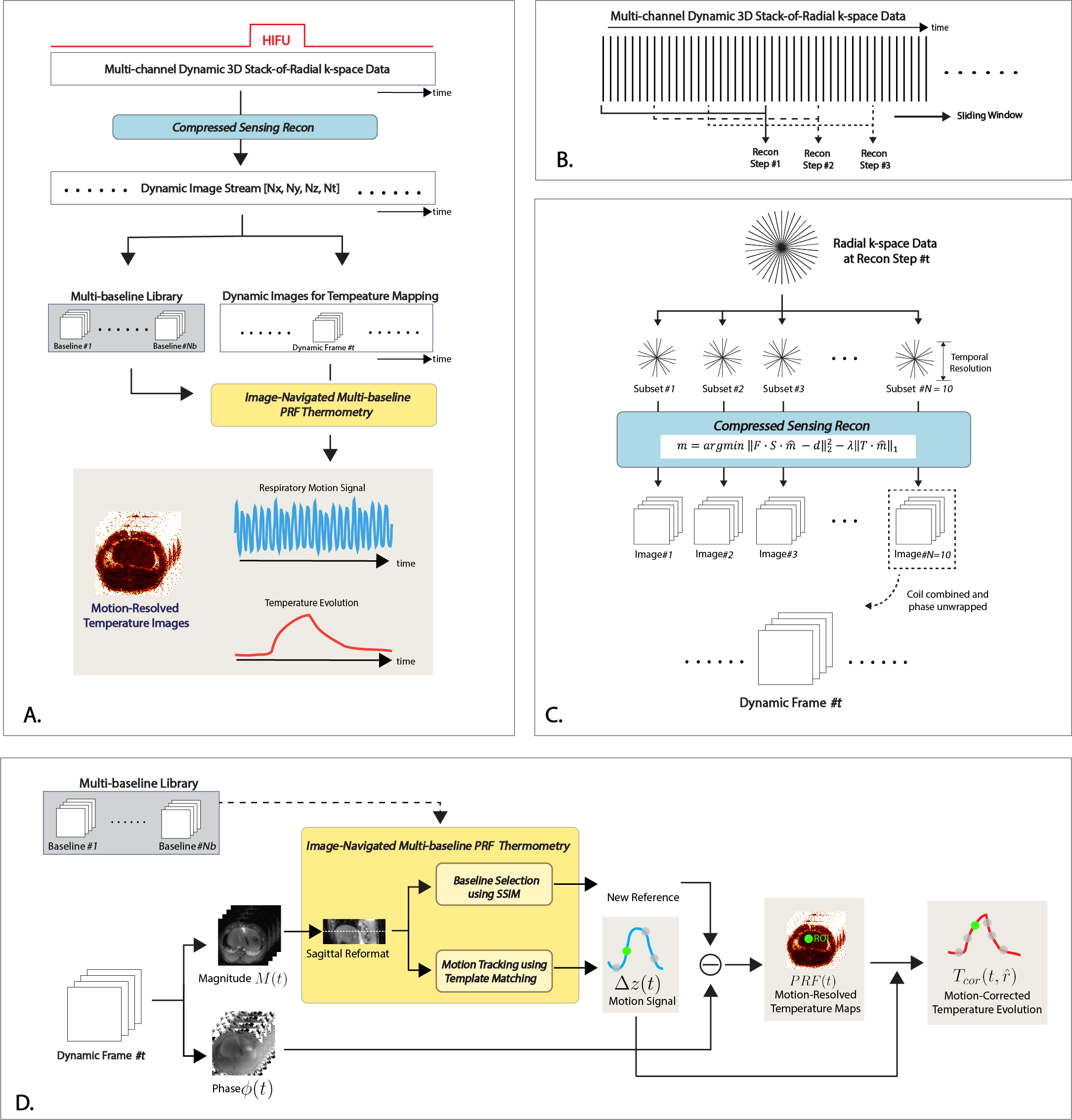
Figure 1: (A) An overview of the iNAV-MB pipeline. (B) Dynamic image reconstruction using a sliding-window approach. (C) At each reconstruction step, the stack-of-radial data was split into subsets along temporal dimensions for CS reconstruction. The time needed to acquire each subset was defined as the temporal resolution. The image reconstructed from the last subset was selected as a dynamic image frame. (D) iNAV-MB PRF temperature calculation. Only motion in the S/I direction Δz was tracked.
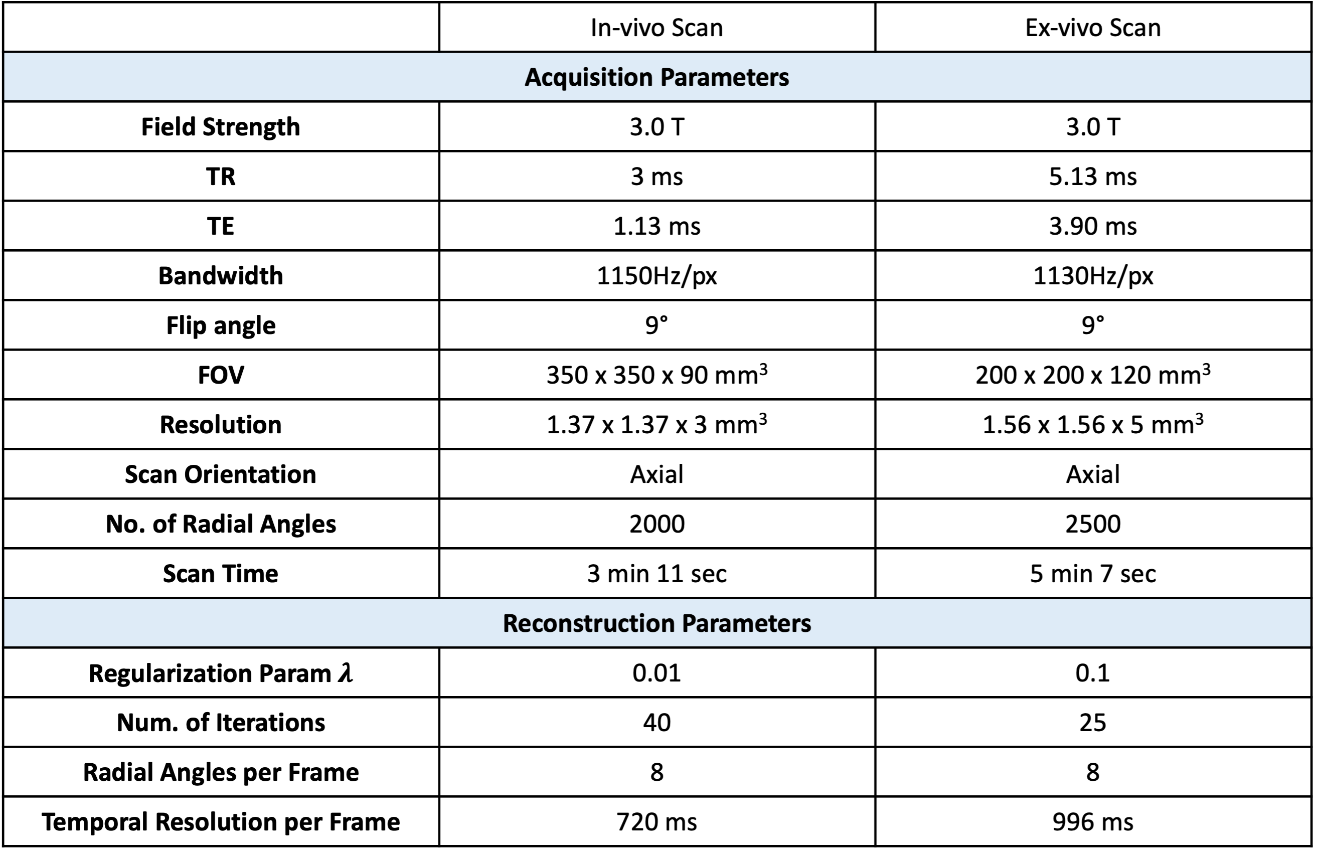
Figure 2: Acquisition and Reconstruction Parameters for the In Vivo and Ex Vivo Experiments. An vivo experiment was performed on one pig without heating. An ex vivo experiment was performed in a tissue motion phantom with HIFU heating.
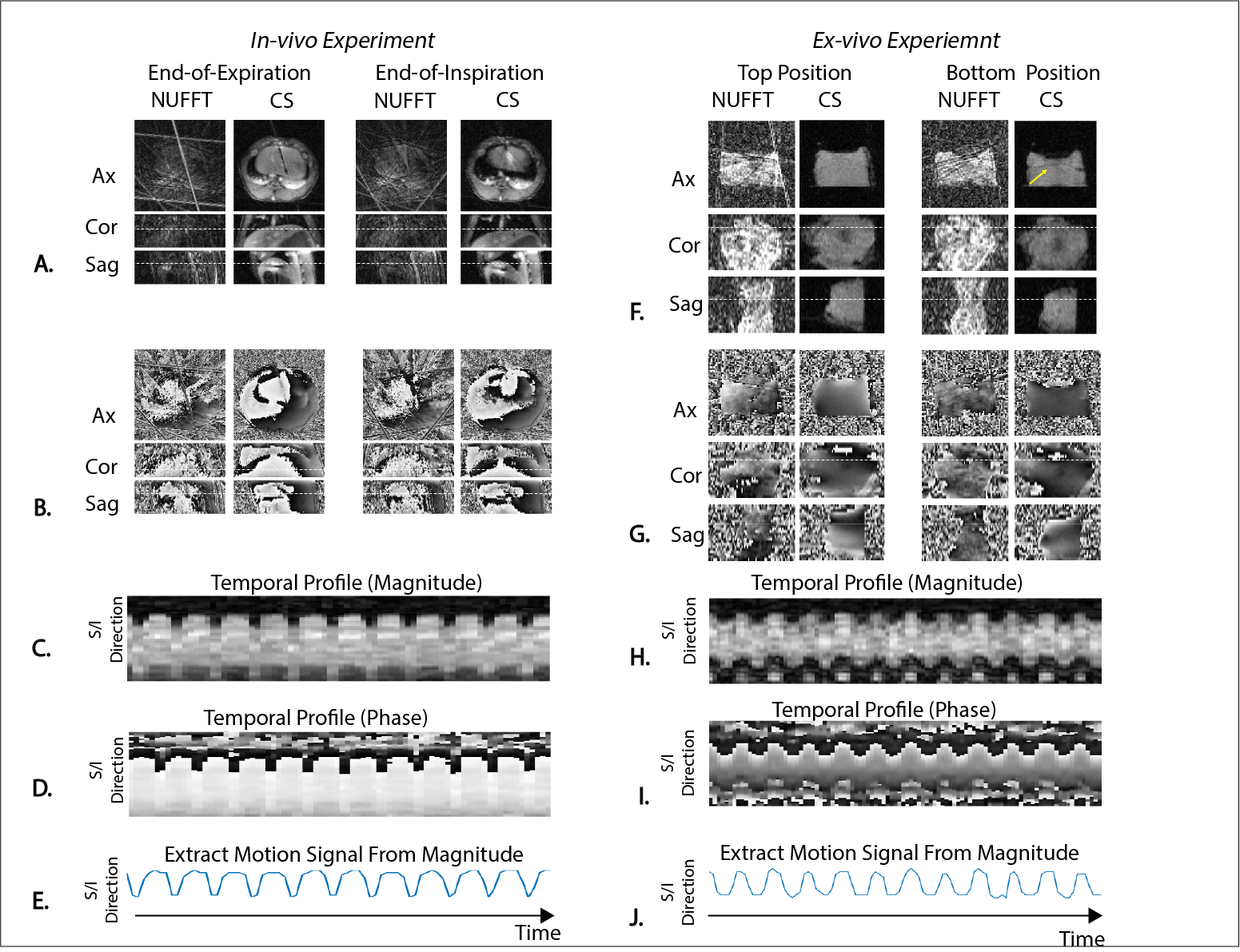
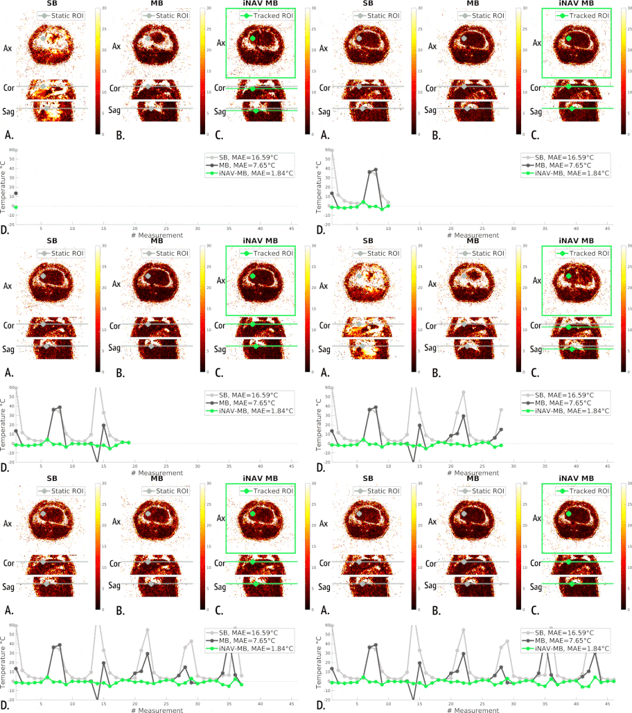
Figure 4: Dynamic 3D PRF Mapping from an In Vivo Swine Subject without Heating. (A-C) PRF maps from single baseline (SB), multi-baseline (MB), and image-navigated multi-baseline (iNAV-MB) methods. For iNAV-MB, the displayed axial slice location is tracked to follow a representative ROI (green) during motion. (D) Temperature evolution over time from a tracked tissue ROI from C. Note that in this non-heating experiment, the temperature should remain at body temperature throughout. MAE: mean absolute error.
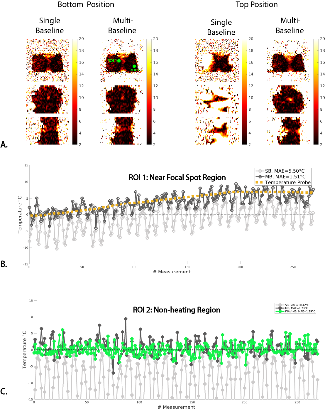
Figure 5: Dynamic 3D PRF Mapping in an Ex Vivo Tissue Motion Phantom During HIFU. (A) Dynamic 3D PRF maps at the top and the bottom positions of the motion stage. (B) Temperature evolution over time at a region near the HIFU focal spot vs. temperature probe (see Figure 3F and 5A). MAE is calculated with respect to temperature probe reading. (C) Temperature evolution over time around a non-heating region. MAE is calculated with respect to zero-degree.