1275
DTI at four directions: an application at ultra-low field1School of Psychology, Cardiff University, Cardiff, United Kingdom, 2University of Valladolid, Valladolid, Spain, 3Hyperfine Inc., Guilford, CT, United States, 4School of Physics and Astronomy, Cardiff University, Cardiff, United Kingdom
Synopsis
Keywords: DWI/DTI/DKI, Low-Field MRI, Tetrahedral Encoding
Motivation: The project aimed to tackle extended DTI scan durations, worsened by low SNR at low fields, striving to boost efficiency while preserving results' accuracy at lower SNR levels.
Goal(s): The study sought to create an ML-based approach to shorten DT-MRI scans while ensuring reliable tensor estimation despite low SNR challenges at ULF.
Approach: ML models, trained on synthetic data, predicted diffusivities and principal eigenvectors from four diffusion-weighted images, factoring in simulated noise and gradient rotations for noise and motion.
Results: The models estimated diffusivities and fibre orientations with fewer data, showing promise for ULF tractography. Suggesting shorter DTI scans are possible at ULF.
Impact: Our results are relevant to clinicians and researchers using low-field MRI, potentially enabling faster DT-MRI scans, opening avenues for efficient DTI in challenging settings, and making fibre mapping more accessible with reduced acquisition scan times.
INTRODUCTION
Estimating the full tensor in diffusion tensor MRI (DT-MRI) requires at least six diffusion-weighted images (DWIs)[1] . If the b value used is around 1/D, where D is the diffusivity, the resulting signal decays by 1/e. This means the DWI SNR is at least 60% lower than the non-diffusion-weighted image. In cases where the SNR is inherently low (e.g., at ultra-high resolution and/or with low field (LF), i.e., < 0.5T)), this low SNR can be compensated by multiple averages, at the expense of increased scan times, which are challenging for non-compliant participants. We recently reported DT-MRI at 64 mT[2], for which the acquisition time was almost 1 hour. Here, with the aim of reducing scan duration, we revisit previously-reported tetrahedral encoding and tensor estimation strategies, highlight their shortcomings, and propose a machine-learning (ML) based approach to provide more robust DT-MRI results in a shorter scan-time than previously reported.METHODS
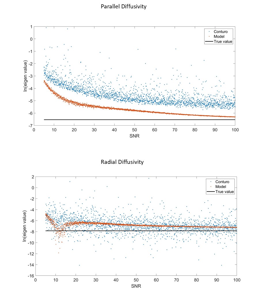
Tetrahedral encoding[3] under the assumption of cylindrical symmetry of the tensor, uses just four DWIs to estimate the axial (l//) and radial (l^) diffusivities, and principal eigenvector (q,f). However, it can be shown this method yields undefined or non-physiological values when the fibre is aligned with the scanner’s ordinal axes. Monte Carlo simulations further reveal that diffusivity estimates are highly sensitive to noise at low SNR, which is problematic at LF (figure 1). Additionally, previously-published formulae[3] become invalid if the gradient directions are not exactly as prescribed, e.g., due to gradient non-uniformity or participant motion between the application of successive diffusion-encodings.We propose a new machine learning (ML) approach to estimate l//, l^, q and f. Our model is trained using data synthetically generated via an algorithm that produces randomly generated diffusion tensors, DWI signal values for given b-values between 500 s/mm2 and 5000 s/mm2, and Rician noise at an SNR of 5 (in the b=0 image) to foster a model adept at low SNR predictions. Rotations were also applied to the gradient directions so the model’s predictions are not limited to one gradient configuration. This approach circumvents issues related to tissue hallucinations and scanner-specific configurations. Consequently, tensor estimation is reframed as a regression problem, with the model generating individual predictions for each voxel.
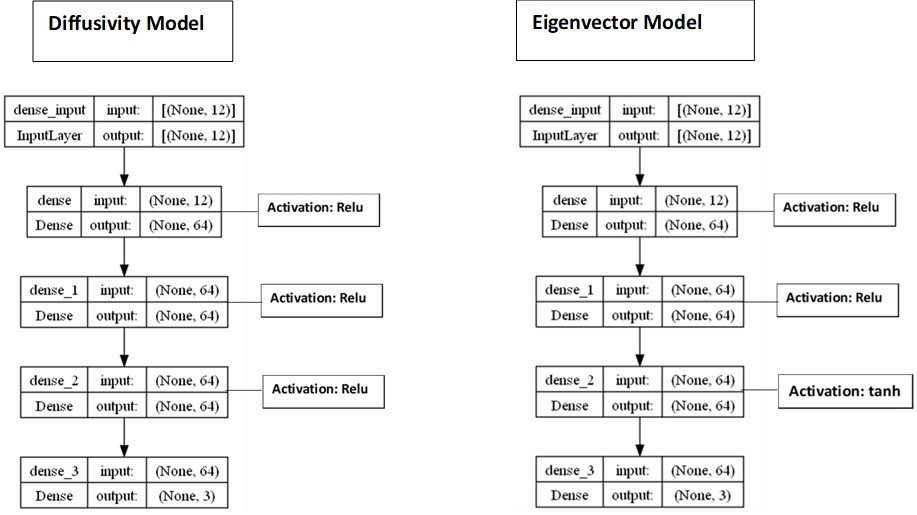
Two models were developed: one fine-tuned for (l//, l^) estimates , hence fractional anisotropy, using a mean squared error loss; and another for the principal eigenvector, using a cosine distance loss, accommodating the orientational nature of the predictions, defined as
1 - |a.b|
where a represents the actual eigenvector and b, the eigenvector predicted by the model(figure 2).
We compared results from our ML-based approach with the original analytical approach[3] using:
(i) digital phantom data[4] (a 128-direction data set, from which we synthesised 4 tetrahedral-encoded signals);
(ii) data acquired on a 64 mT Hyperfine Swoop system, using the scheme previously-reported[2]
RESULTS
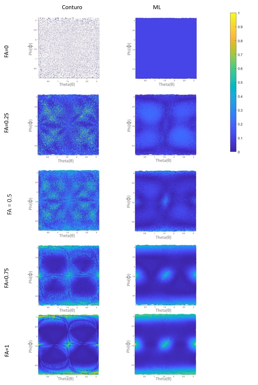
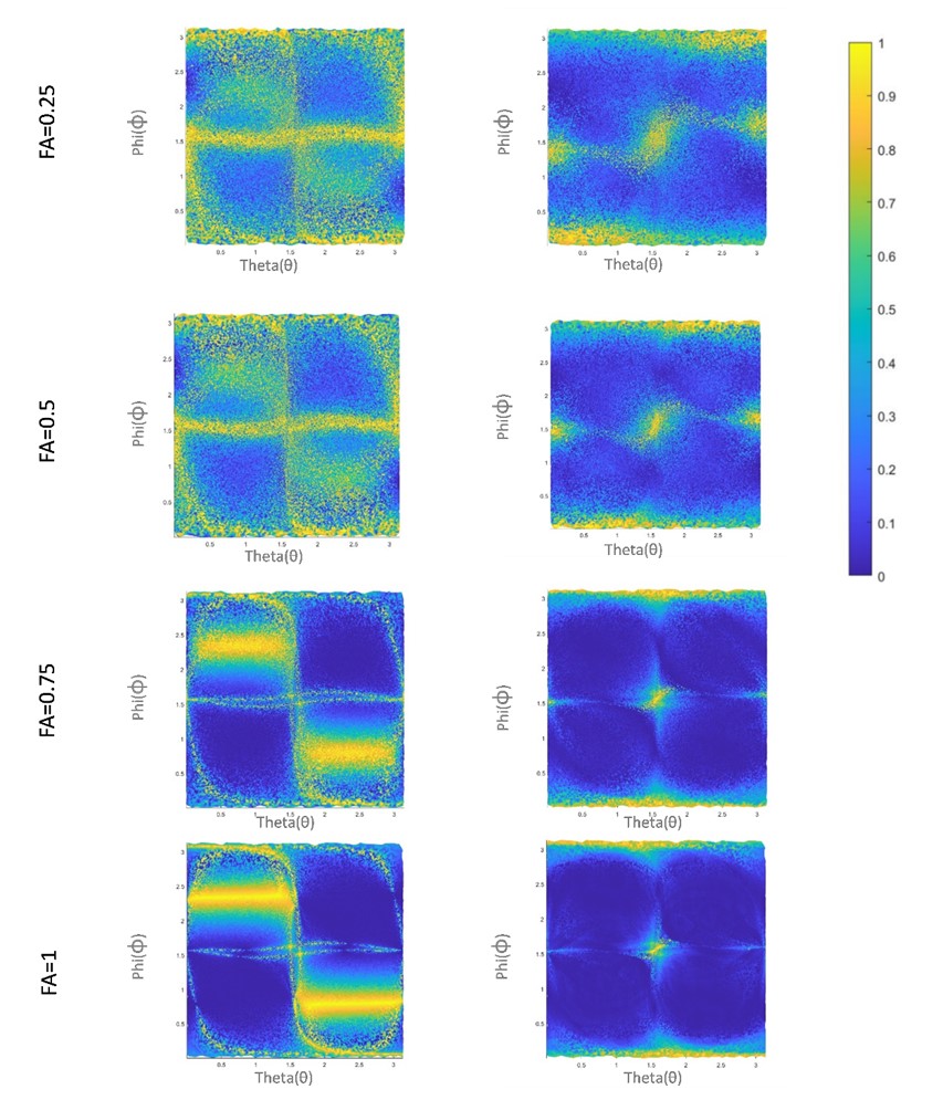
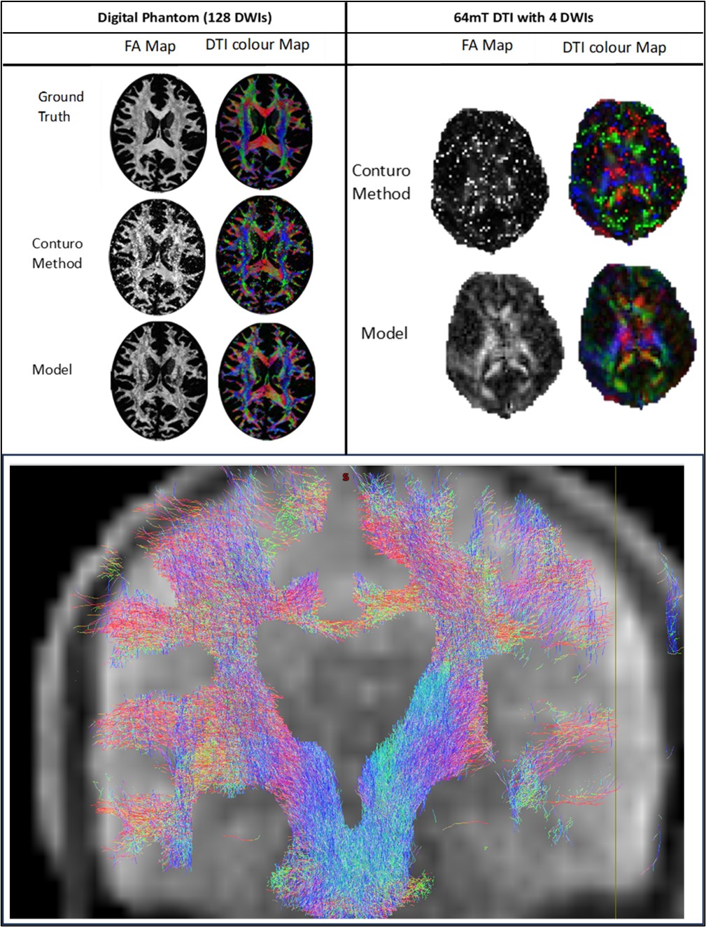
Our ML-models successfully predicted diffusivities, fractional anisotropy and principal eigenvectors with four-direction DWI data. They were also robust to rotation of gradients due to gradient non-uniformity or participant motion. Testing on the 4-direction digital phantom revealed that the model could generate broadly similar colour-encoded FA maps to those from the full 128-direction set, although significant errors were observable when fibre orientations were aligned with the scanner’s ordinal axes (figure 3) (figure 4). These errors stem from the mathematically indeterminate nature of the problem. When tested with ULF data from the Hyperfine Swoop scanner, the models showed potential for tractography applications.DISCUSSION
Our study demonstrates the feasibility of obtaining reliable diffusivity and fractional anisotropy maps using four-direction data, even at low field (figure 5). Compared to the Conturo method, our method showed more realistic FA colour maps at ULF. Nevertheless, significant errors in principal eigenvector estimates are evident, particularly with fibres aligned with the scanner's ordinal axes (figure 4), attributable to the insufficient directionality information to resolve degeneracy in the estimation with only four DWI directions. Exploring methods to incorporate additional directional data could enhance the outcomes.CONCLUSION
Our study shows the possibility of conducting DT-MRI with only four directions, while avoiding the numerical instability of previous methods, and without excessive computational demands, albeit with a margin of error in fibre orientation (figure 4). We demonstrate that even with the reduced number of directions and reduced SNR, there is the potential for tractography with this method at ULF. This advancement holds promise for substantially reducing DTI scan times, especially beneficial for ULF applications.Acknowledgements
This work was made possible by generous support from the Bill and Melinda Gates Foundation through the UNITY projectReferences
1. Basser PJ, Mattiello J, LeBihan D. MR diffusion tensor spectroscopy and imaging. Biophysical journal 1994;66(1):259-267.
2. Plumley A, Padormo F, Cercignani M, O'Halloran R, Teixeira R, Planchuelo-Gómez Á, Legouhy A, Luo T, Jones D. Tensors and Tracts at 64 mT. ISMRM and ISMRT Annual Meeting and Exhibition. Toronto2023.
3. Conturo TE, McKinstry RC, Akbudak E, Robinson BH. Encoding of anisotropic diffusion with tetrahedral gradients: a general mathematical diffusion formalism and experimental results. Magnetic resonance in medicine 1996;35(3):399-412.
4. Esteban O, Caruyer E, Daducci A, Bach-Cuadra M, Ledesma-Carbayo MJ, Santos A. Diffantom: whole-Brain diffusion MRI Phantoms derived from real datasets of the human connectome project. Frontiers in neuroinformatics 2016;10:4.
Figures