1274
High-resolution whole brain multi-shell diffusion MRI at 5.0 Tesla1Center for Biomedical Imaging Research, Department of Biomedical Engineering, School of Medicine, Tsinghua University, Beijing, China, 2Center for Nano and Micro Mechanics, Department of Engineering Mechanics, Tsinghua University, Beijing, China
Synopsis
Keywords: DWI/DTI/DKI, Diffusion Tensor Imaging
Motivation: In neuroscientific researches, 3T DWI provides limited resolution while 7T DWI suffers from challenges such as shorter relaxation time and increased field inhomogeneity.
Goal(s): To evaluate the performance of 5T in high-resolution whole brain multi-shell DWI.
Approach: 1.1 mm-isotropic whole brain DWI with 2 shells was acquired using multi-band single-shot 2D EPI on 5T. DTI metrics and MSMT-CSD model were computed. The nearly identical acquisition parameters and data processing were applied to the same subject on 3T.
Results: 5T DWI resolved brain structural connectivity more accurately than 3T. Better FA contrast was observed at 5T, demonstrating higher SNR achieved at 5T.
Impact: The feasibility of simultaneously achieving high spatial resolution and adequate q-space sampling in practical acquisition time at 5T demonstrated its potential as a new tool in DWI-based neuroscientific studies.
Introduction
Simultaneously achieving high spatial resolution and adequate q-space sampling in practical acquisition time is crucial for accurately resolving the complex tissue microstructure throughout the entire brain1,2, such as crossing/kissing fibers. Compared to 3T, 7T can provide increased SNR, which enables higher spatial resolution, thus reducing partial volume effects leading to gyral bias in tractography3. Given this, 7T DWI has been applied to large-scale population study projects such as HCP3-5. However, 7T DWI suffers from severe B0 and B1+ inhomogeneity, shorter T2 and T2*, as well as higher SAR, all of which weaken its intrinsic SNR advantage6. In this study, we aim to explore the performance of 5T in high-resolution whole-brain multi-shell dMRI.Method
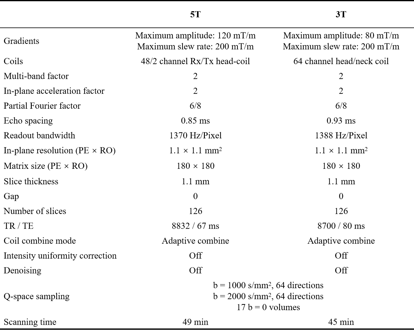
Data Acquisition: Data were acquired on a 5.0T MR system (uMR Jupiter, United Imaging Healthcare, Shanghai, China) equipped with 120mT/m gradients and a 48/2 channel Rx/Tx head-coil. The dMRI data were acquired from a healthy volunteer. Two shells (b = 1000 and 2000 s/mm2) were sampled in q-space, with 64 non-collinear gradient directions uniformly distributed on each shell. DWI volumes and additional 17 b=0 volumes with two phase-encoding directions (AP/PA) were acquired, resulting in 290 volumes and a scanning time of 49 minutes. 3D-MPRAGE T1-weighted images with 0.7 mm-isotropic voxel size were also obtained. For comparison, the same volunteer was scanned using a nearly identical protocol on a 3.0T MR system (MAGNETOM Prisma, Siemens Healthcare, Erlangen, Germany). Detailed DWI sequence parameters are shown in Table 1. Images were reconstructed online by the vendors directly. Note that denoising, intensity uniformity correction, and other image filters were turned off on both scanners.Data Processing: The processing pipeline was identical between 3T and 5T data. QSIPrep7 was used in preprocessing to avoid blurriness induced by multiple interpolations and imprecise spatial resampling. Specifically, MP-PCA8 denoising was implemented with a 5-voxel window. FSL’s eddy9,10 was used for head motion correction and eddy current correction. Fieldmaps were estimated from the b=0 image pairs from data acquired with reversed phase-encoding directions using a TOPUP-style method11 to correct susceptibility-induced distortion, after which the two phase-encoding groups were averaged. All DWI images were co-registered with the T1W images. After preprocessing, DTI metrics were computed using b=0 and b=1000 images. All volumes sampled in q-space were fed into multi-shell multi-tissue CSD (MSMT-CSD) method12 to estimate white matter fiber orientation distribution (WM FODs).
Results and Discussion
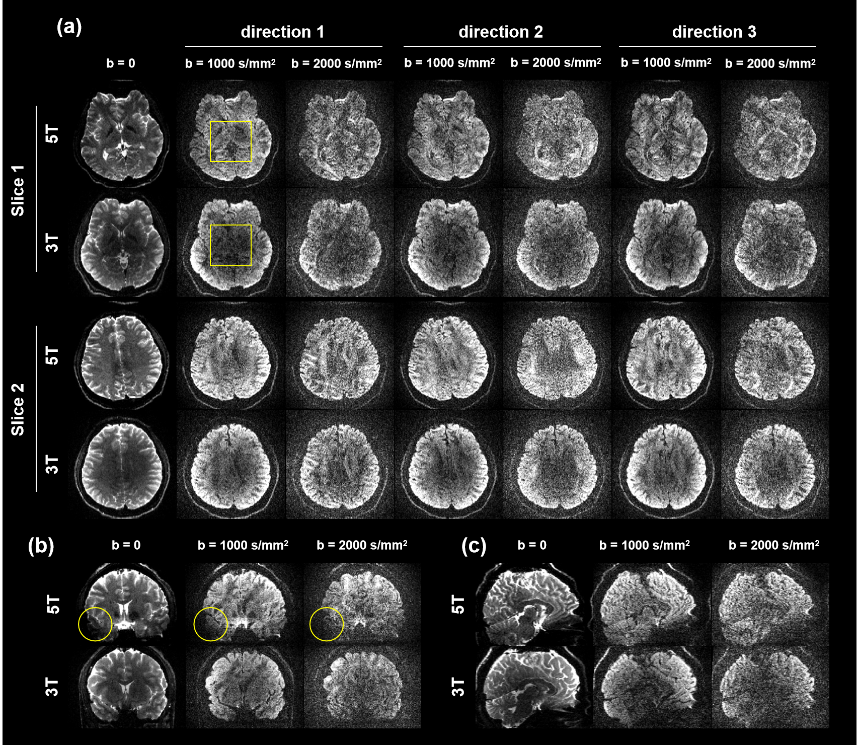
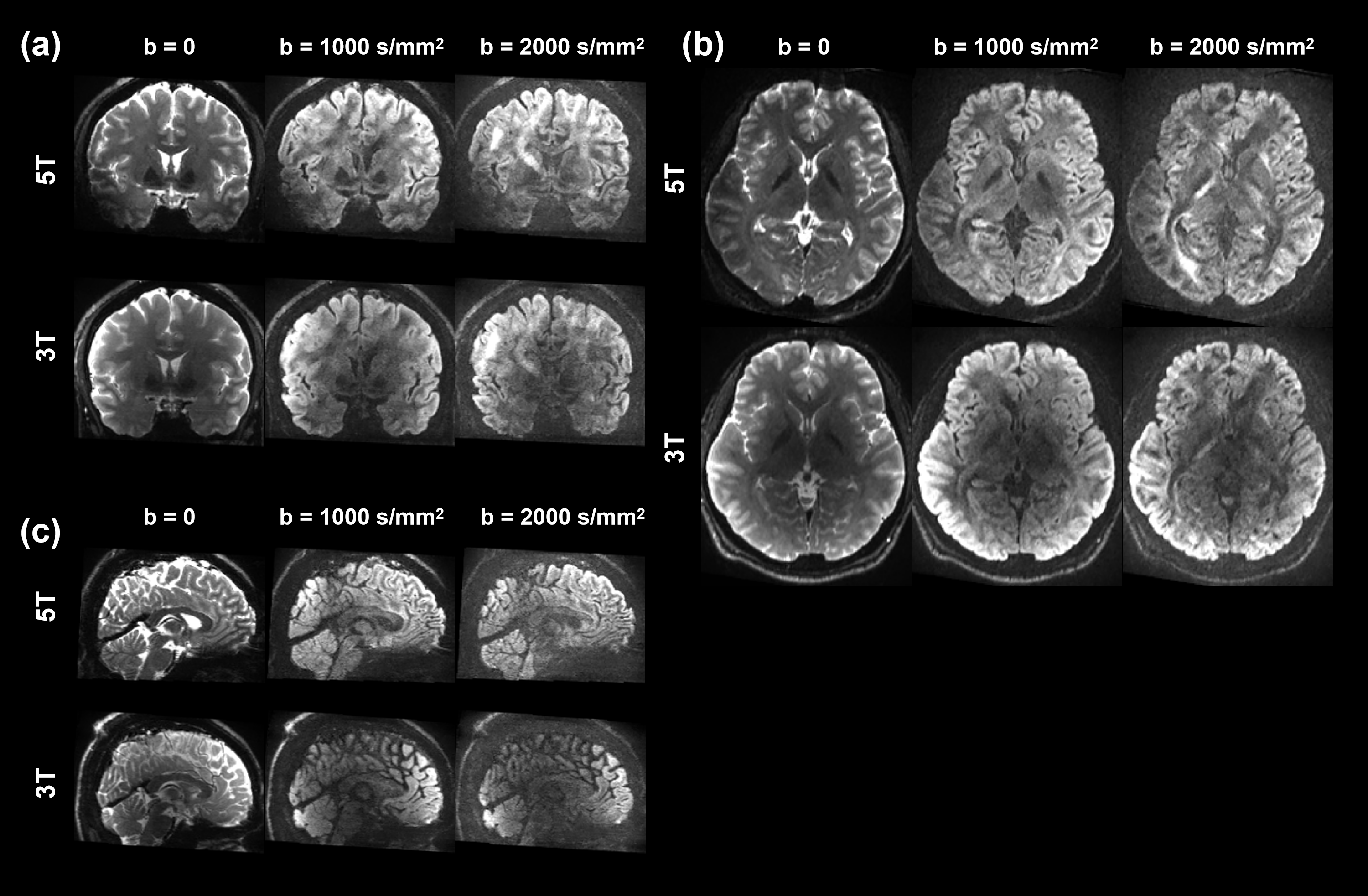
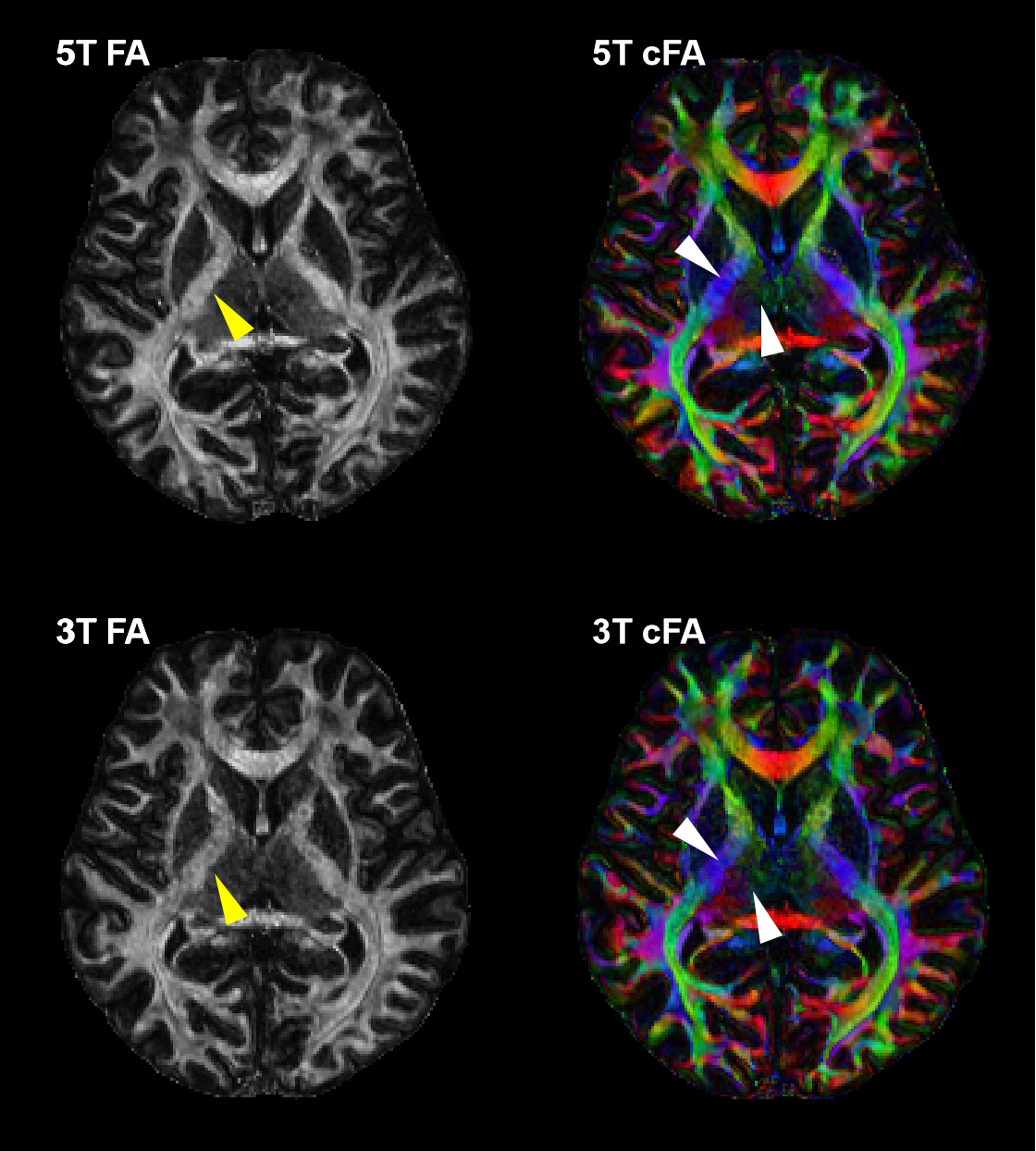
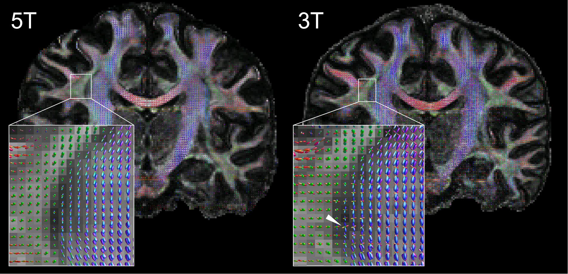
Figure 1 shows DWI images acquired at 5T and 3T before preprocessing. Lower noise levels are observed at 5T DWI despite of shorter T2/T2* values, especially in the center of the brain. Additionally, 5T DWI showed better intensity uniformity in the central brain than that of 3T, as highlighted by yellow rectangles in Figure 1a. This might result from two reasons: (1) B1+ variation exists (B1+ shimming was used by the scanner on 5T). (2) a larger receive coil unit size at 5T could achieve deeper penetration. However, signal loss was observed at the temporal lobe of 5T DWI (Figure 1b, yellow circles) due to B1+ inhomogeneity. Also, spatial distortion at 5T DWI was more severe due to increased B0 inhomogeneity. Figure 2 shows images after preprocessing. The spatial distortion and noise were properly handled at both 5T and 3T while contrast was well preserved.As shown in Figure 3, FA contrast was stronger at 5T, as highlighted with yellow arrowheads, where the border between post internal capsules and thalamus was sharper at 5T. Additionally, the details of the structural connectivity of post-internal capsules and thalamus were clearer at 5T, as shown in color FA maps in Figure 3 with white arrowheads. Figure 4 indicated that 5T DWI could resolve kissing fibers more accurately, while 3T DWI might incorrectly connect the superior longitudinal fasciculus and cortico-spinal tract, as indicated by white arrowheads. Furthermore, sharper FODs in the cortico-spinal tract could be computed from 5T DWI.
For a fair comparison, we set uniform sequence parameters to the fullest extent, but some of them still have different values depending on the hardware. Thus, a comprehensive comparison using gradient systems with identical configurations will be done in the future. Also, data corruption due to severe physiological motion during DWI scanning can be better controlled by using either data rejection13 or cardiac triggering. More comprehensive investigations are needed to compare the diffusion images acquired at different platforms by scanning more subjects.
Conclusion
Preliminary results show high-resolution whole brain multi-shell diffusion MRI at 5T provided higher SNR than 3T, thus exhibiting finer brain microstructural details. This may indicate the potential of 5T as a new tool in neuroscientific studies.Acknowledgements
No acknowledgement found.References
1. Descoteaux M. High angular resolution diffusion imaging (HARDI). Wiley encyclopedia of electrical and electronics engineering 1999:1-25.
2. Zhan L, Leow AD, Aganj I, et al. Differential information content in staggered multiple shell HARDI measured by the tensor distribution function. 2011 IEEE International Symposium on Biomedical Imaging: From Nano to Macro, 2011. p. 305-309.
3. Vu AT, Auerbach E, Lenglet C, et al. High resolution whole brain diffusion imaging at 7 T for the Human Connectome Project. Neuroimage 2015;122:318-331.
4. Van Essen DC, Smith SM, Barch DM, et al. The WU-Minn human connectome project: an overview. Neuroimage 2013;80:62-79.
5. Uğurbil K, Xu J, Auerbach EJ, et al. Pushing spatial and temporal resolution for functional and diffusion MRI in the Human Connectome Project. Neuroimage 2013;80:80-104.
6. Gallichan D. Diffusion MRI of the human brain at ultra-high field (UHF): A review. NeuroImage 2018;168:172-180.
7. Cieslak M, Cook PA, He X, et al. QSIPrep: an integrative platform for preprocessing and reconstructing diffusion MRI data. Nature methods 2021;18(7):775-778.
8. Veraart J, Novikov DS, Christiaens D, Ades-Aron B, Sijbers J, Fieremans E. Denoising of diffusion MRI using random matrix theory. Neuroimage 2016;142:394-406.
9. Woolrich MW, Jbabdi S, Patenaude B, et al. Bayesian analysis of neuroimaging data in FSL. Neuroimage 2009;45(1):S173-S186.
10. Andersson JL, Graham MS, Zsoldos E, Sotiropoulos SN. Incorporating outlier detection and replacement into a non-parametric framework for movement and distortion correction of diffusion MR images. Neuroimage 2016;141:556-572.
11. Andersson JL, Sotiropoulos SN. An integrated approach to correction for off-resonance effects and subject movement in diffusion MR imaging. Neuroimage 2016;125:1063-1078.
12. Tournier J-D, Yeh C-H, Calamante F, Cho K-H, Connelly A, Lin C-P. Resolving crossing fibres using constrained spherical deconvolution: validation using diffusion-weighted imaging phantom data. Neuroimage 2008;42(2):617-625.
13. Zhang Z, Huang F, Ma X, Xie S, Guo H. Self-feeding MUSE: a robust method for high resolution diffusion imaging using interleaved EPI. NeuroImage 2015;105:552-560.
Figures