1272
Dealing with Gradient Nonlinearities for High-Performance Gradient Diffusion MRI - Application to the Human Connectome Project (HCP) Dataset1QMI/NIBIB, NIBIB/NIH, Bethesda, MD, United States, 2NatBrainLab, KCL, London, United Kingdom, 3Systems Neuroscience and Psychopathology Lab, CAHBIR, Rutgers University, New Jersey, NJ, United States
Synopsis
Keywords: Diffusion Analysis & Visualization, Diffusion/other diffusion imaging techniques, Gradient nonlinearity correction
Motivation: Correction of gradient nonlinearity effects on diffusion-sensitization is important. However, not all software packages and/or diffusion models can incorporate spatially-varying bvals/bvecs information.
Goal(s): Determine how significant nonlinearities are in the Human Connectome Project(HCP) dataset and investigate the feasibility of a spherical harmonics (SH)-based signal regeneration technique to directly incorporate nonlinearity effects and eliminate the need for additional information sharing.
Approach: FA, MD and angular error maps from 200 subjects were compared using different formats of correction.
Results: Nonlinearities are significant on HCP-dMRI data. The proposed SH-based signal regeneration approach allows the use of spatially invariant diffusion gradient tables with substantially reduced residual error.
Impact: Diffusion models and software not designed to incorporate spatially-varying bvals/bvecs can now take advantage of gradient nonlinearity correction. A reprocessed version of HCP dMRI dataset will be made publicly available in this format, along with the conventional gradient deviation tensors.
Introduction
Gradient nonlinearities (GNL) lead to image distortions1 and spatially-varying deviations of b-values and diffusion gradient directions from those prescribed2-5. These deviations are typically ignored on clinical scanners; however, they have been shown to affect the accuracy of diffusion-derived metrics3. With high performance but highly nonlinear gradients being more and more available, the dissemination of GNL information to users and its inclusion in dMRI model-fitting is becoming increasingly important.GNL information can be distributed to users in different ways: Field-describing spherical harmonics coefficients are generally inaccessible. Alternatively, gradient deviation tensor images5 (GDEV), or voxelwise B-matrices6,7,8 (VBMAT), (which also consider subject motion), can be distributed to provide spatial diffusion encoding information. To our knowledge, only one software package7 can take advantage of VBMAT, which also increases data size significantly (6x DWI size). Only a handful of software can employ GDEV mainly because for deconvolution-based models, its use is impractical due to the need to recompute response functions voxelwise. In this work, we investigate the effects of GNL on the Human Connectome Project (HCP) dataset and provide a simplified alternative to gradient nonlinearly correction, (gradient-deviation first-order approximation (GDFA)). This approach directly integrates GNL into DWI signal and generates spatially-invariant gradient tables that can be used by any dMRI software.
Materials & Methods
DMRI data from 200 HCP9 subjects were fully reprocessed with TORTOISEV4 following novel and advanced pre-processing techniques7,10. Manufacturer-provided coefficients were used during pre-processing to output the analytically computed GNL information6 in GDEV format, in subject anatomical image space.The GDFA was defined in the following way: First, all diffusion signals were voxelwise recomputed for nominal b-value by computing the correct ADC, based on the effective and nominal b-values assuming mono-exponential decay. Second, a voxelwise spherical harmonic (SH) decomposition (l=10) was performed on each shell using the effective gradient directions. The final corrected signal was regenerated using these SH coefficients and the prescribed, spatially invariant gradient orientations. This regenerated dataset can directly be used by any diffusion model or software as no additional GNL information need to be considered.
To analyze the effects of GNL and to validate the accuracy of GDFA, FA and MD maps were computed from data with no GNL, GDEV correction and GDFA correction. Additionally, primary eigenvector angular difference maps were also computed between GDEV-NOCORR and GDEV-GDFA.
A diffeomorphic, diffusion tensor-based atlas was created11,12 from all 200 subjects, and the difference images were warped onto this population template and averaged, to determine the GNL effects and GDFA accuracy at population level.
DTI-Tractography was performed on two pathways to evaluate the impact on the accuracy of the final anatomical reconstructions.
Results
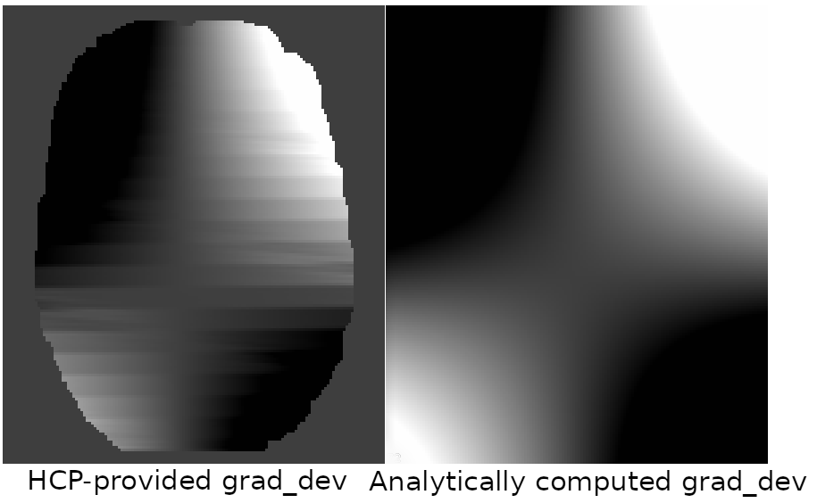
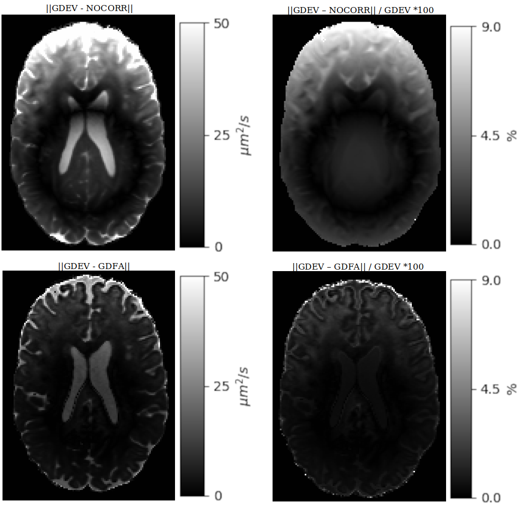
Figure1 displays the Lxy component of the GDEV image, for both the current and original HCP processing. The analytically computed Lxy image does not suffer from the striation artifacts originating from numerical differentiation and interpolation.Figure2 displays the absolute and percentage errors in MD for a single subject for GDEV/NOCORR and GDEV/GDFA. The effects of nonlinearities on diffusivity can be as high as 50 µm2/s (9%) on the displayed slice and up to 80 µm2/s (14%) at the top of the brain (not shown). Even though differences could still be observed, GDFA significantly reduced this systematic error, with a maximum residual of 2%.
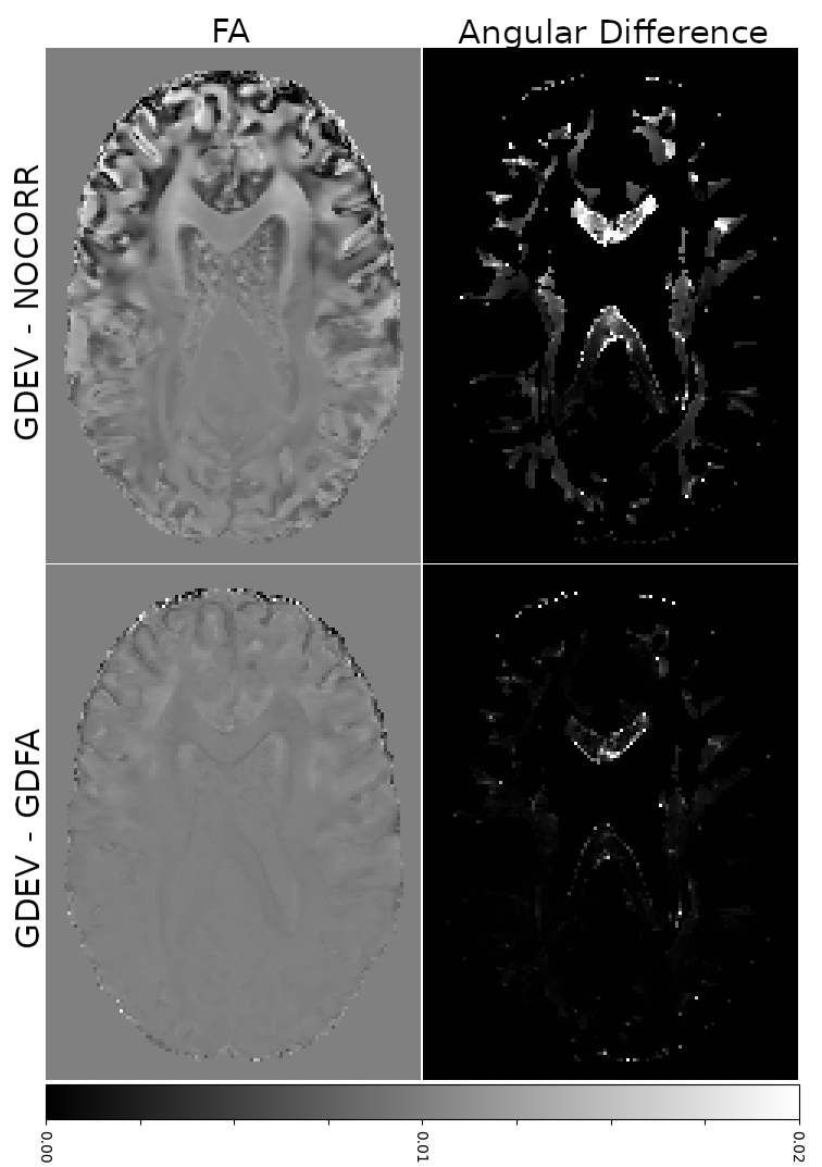
Figure3 displays the FA errors and angular differences in primary eigenvectors. The effect of GNL is still evident, with a maximum FA difference of 0.07 between GDEV and NOCORR and an angle difference of 4 degrees. GDFA was only negligibly different from GDEV.
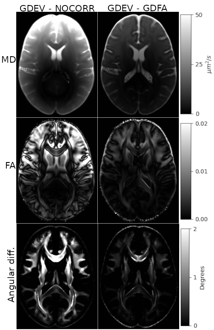
Figure4 displays the difference maps at the population level. Patterns at the population level are similar: GNL have significant effects on the entire population (up to 90 µm2/s for MD, 0.01 for FA and 2.4o for the angular difference). GDFA was able to substantially reduce nonlinearity effects (up to 28µm2 /s for MD, 0.001 for FA and 0.8o for the angular difference).
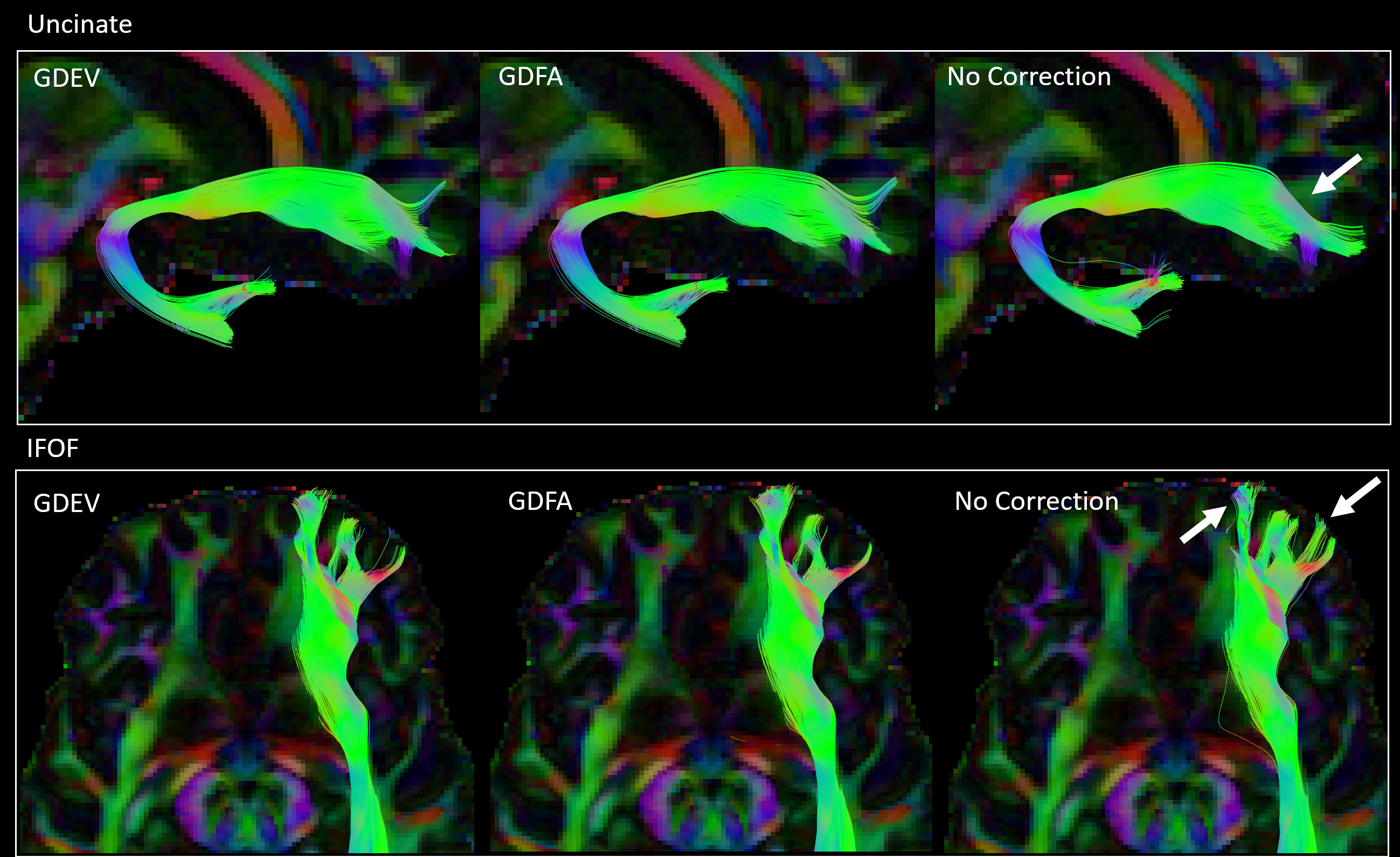
DTI tractography in Figure5 shows clear anatomical differences in the cortical termination in frontal regions between the uncorrected data and both GDEV and GDFA corrections. GDFA and GDEV were able to generate very similar reconstructions with negligible anatomical differences.
Conclusions & Discussion
Compared to the full correction offered by GDEV, GDFA correction provided very close quantitative results, and negligible anatomical differences in tractography reconstructions. By regenerating the DWI signal with spatially invariant gradient tables, we have enabled users to take advantage of GNL correction when using any diffusion models or software packages. The entire HCP1200 dataset, including the newly processed DWIs with GDEV10 and GDFA together, with revised anatomical templates, will be made publicly available to researchers.Acknowledgements
This research was supported by the Intramural Research Program of the National Institute of Biomedical Imaging and Bioengineering (MOI, AT, CP) and King's College London (AB, FD). The contents of this work do not necessarily reflect the position or the policy of the government, and no official endorsement should be inferred.References
1. Glover GH and Pelc NJ. "Method for correcting image distortion due to gradient nonuniformity". US Patent # 4,591,789, May 27, 1986.
2. Dariya I. Malyarenko, Brian D. Ross, and Thomas L. Chenevert, "Analysis and correction of gradient nonlinearity bias in apparent diffusion coefficient measurements". Magn Reson Med. 2014;71(3):1312-23.
3. Malyarenko D, Galbán CJ, Londy FJ, Meyer CR, Johnson TD, Rehemtulla A, Ross BD, Chenevert TL, "Multi-system repeatability and reproducibility of apparent diffusion coefficient measurement using an ice-water phantom", J Magn Reson Imaging. 2013 May;37(5):1238-46.
4. Tan ET, Marinelli L, Slavens ZW, King KF, Hardy CJ., "Improved correction for gradient nonlinearity effects in diffusion-weighted imaging", J Magn Reson Imaging. 2013 Aug;38(2):448-53.
5. Bammer R, Mark M, Barnett A, Acar B, Alley MT, Pelc NJ, Glover GH, Moseley ME. "Analysis and generalized correction of the effect of spatial gradient field distortions in diffusion-weighted imaging". Magn Reson Med. 2003 Sep;50(3):560-9.
6. Barnett, Alan Seth, Irfanoglu, M. Okan, Landman, Bennett, Rogers, Baxter, Pierpaoli, Carlo. Mapping gradient nonlinearity and miscalibration using diffusion-weighted MR images of a uniform isotropic phantom. Magn Reson Med. 2021; 86: 3259–3273. https://doi.org/10.1002/mrm.28890
7. M. O Irfanoglu, A. nayak, P. Taylor, C. Pierpaoli, "TORTOISEV4: ReImagining the NIH Diffusion MRI Processing Pipeline", ISMRM 2023, p-80.
8. Rudrapatna, U., Parker, G.D., Roberts, J., Jones, D.K., 2021. A comparative study of gradient nonlinearity correction strategies for processing diffusion data obtained with ultra-strong gradient MRI scanners. Magn Reson Med 85 (2), 1104–1113. doi:10.1002/mrm.28464.
9. Sotiropoulos SN, Jbabdi S, Xu J, Andersson JL, Moeller S, Auerbach EJ, Glasser MF, Hernandez M, Sapiro G, Jenkinson M, Feinberg DA, Yacoub E, Lenglet C, Van Essen DC, Ugurbil K, Behrens TE; WU-Minn HCP Consortium. Advances in diffusion MRI acquisition and processing in the Human Connectome Project. Neuroimage. 2013 Oct 15;80:125-43. doi: 10.1016/j.neuroimage.2013.05.057. Epub 2013 May 20. PMID: 23702418; PMCID: PMC3720790.
10. M. Okan Irfanoglu, Ahmad Beyh, Flavio Dell’Acqua, Marco Catani, Carlo Pierpaoli, "ReImagining the Young Adult Human Connectome Project (HCP) Diffusion MRI Dataset", ISMRM2022, p.4305
11.Irfanoglu MO, Nayak A, Jenkins J, Hutchinson EB, Sadeghi N, Thomas CP, Pierpaoli C. DR-TAMAS: Diffeomorphic Registration for Tensor Accurate Alignment of Anatomical Structures. Neuroimage. 2016 May 15;132:439-454. doi: 10.1016/j.neuroimage.2016.02.066. Epub 2016 Feb 28. PMID: 26931817; PMCID: PMC4851878.
12. M. Okan Irfanoglu, Amritha Nayak, Carlo Pierpaoli,"Diffusion MRI Atlases from the Human Connectome Project Data", ISMRM 2020, p4035.
Figures