1270
Axon diameter mapping in the living human brain with ultra-high gradient diffusion MRI using 500 mT/m gradient strength1Martinos Center for Biomedical Imaging, Charlestown, MA, United States
Synopsis
Keywords: Microstructure, Gradients
Motivation: Noninvasive quantification of axon diameter in the living human brain offers valuable insights into the mesoscopic organization of white matter. Current methods for mapping axon diameter using diffusion MRI are limited by gradient strength.
Goal(s): To evaluate the sensitivity of axon diameter mapping to small diameter axons on the Connectome 2.0 scanner (Gmax=500mT/m) compared to the original Connectome scanner (Gmax=300mT/m).
Approach: The AxCaliber-SMT model was fitted to diffusion MRI data in 10 healthy subjects scanned on Connectome 2.0 and Connectome 1.0.
Results: Median axon diameter in the corticospinal tract was 2.63um on Connectome 2.0 and 4.00um on Connectome 1.0.
Impact: Connectome 2.0 pushes the resolution limit and signal-to-noise ratio of axonal diameter mapping, allowing for greater sensitivity toward small diameter axons at the individual level for a variety of neuroscientific and clinical applications.
Introduction
Axon diameter mapping in the living human brain is important for advancing an understanding of brain connectivity, white matter integrity, and the microstructural changes associated with neurological and psychiatric disorders. Axon diameter and myelin content are key determinants of conduction velocity and delays across regional and whole-brain networks [1]. Previous studies have shown that gradient strengths up to 300 mT/m on the 3T Connectome MRI scanner can sensitize the diffusion MRI signal to intra-axonal water diffusion and enable estimation of axon diameters in the living human brain down to a diffusion resolution limit of 3-4 um [2-5]. However, such measurements of effective axon diameter are weighted by the largest axons in the distribution and remain insensitive to smaller axons (~1 um or less) making up most white matter in the brain [5]. To access smaller diffusion length scales that capture a greater proportion of small diameter axons, simulations and theoretical work [3,5] have shown that stronger and faster gradients are needed.These findings have motivated our efforts to upgrade the original Connectome scanner to reach even higher gradient strengths up to 500 mT/m and slew rates up to 600 T/m/s – the highest ever achieved for in vivo human imaging. This next-generation Connectome MRI scanner (Connectome 2.0) has now been installed and rendered operational at MGH for human use [6,7]. Here, we report the first demonstration of axonal diameter mapping in the living human brain on Connectome 2.0 (C2) and compare the results to axonal diameter mapping on the original Connectome MRI scanner (C1) [8].
Methods
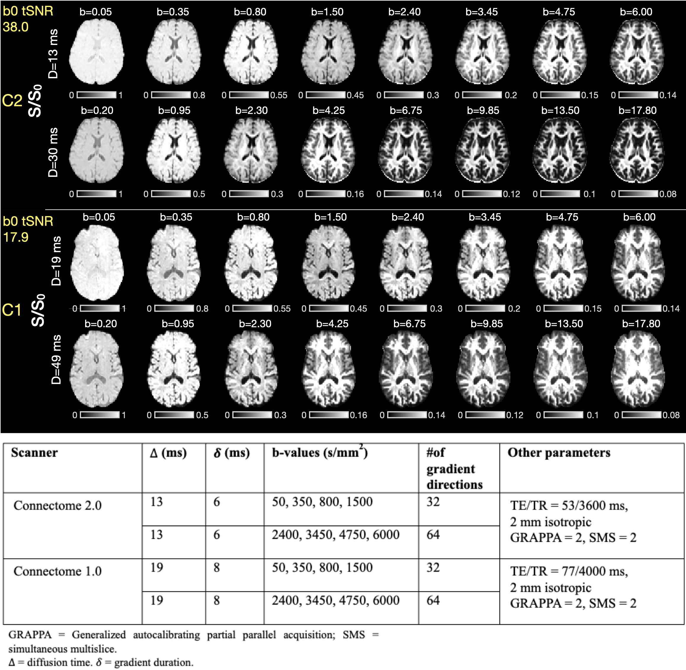
Acquisition10 healthy subjects (34.1±7.2 years, 9 female) were imaged on the Connectome 2.0 scanner (MAGNETOM Connectom.X, Siemens Healthineers, Erlangen, Germany) equipped with Gmax=500mT/m and SRmax=600T/m/s. 10 age- and sex-matched healthy subjects (34.5±6.6 years, 9 female) were scanned on the Connectome 1.0 scanner (MAGNETOM Connectom) equipped with Gmax=300mT/m and SRmax=200T/m/s. Diffusion MRI acquisitions on C2 and C1 were harmonized to the previously published TractCaliber protocol [4] utilizing Gmax=500mT/m and 300mT/m, respectively, with parameters shown in Figure 1.
Diffusion preprocessing
Diffusion-weighted images (DWIs) were preprocessed following a previously established pipeline for correcting susceptibility- and eddy-current-induced distortions in high-gradient diffusion MRI data [9]. The temporal signal-to-noise ratio (tSNR) was estimated through voxel-wise calculation of the mean divided by standard deviation of 10 consecutive b=0 scans and used for Rician noise floor correction.
Model-fitting
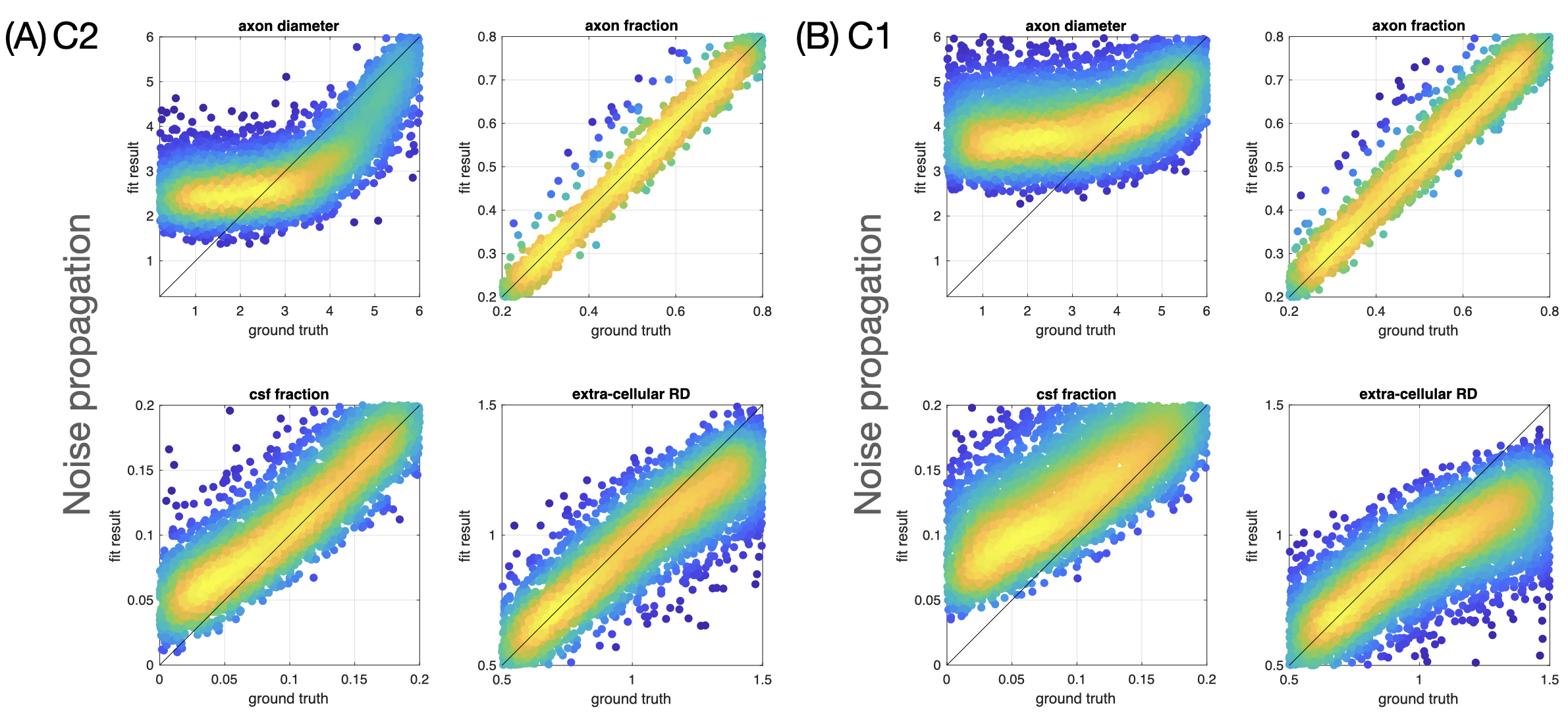
Spherical mean signals from the multi-shell DWIs were used for AxCaliber-SMT model fitting with Markov chain Monte Carlo sampling [8]. Noise propagation was used to check the performance of model fitting; theoretical resolution limits were also calculated using Eq. 40 of [3].
Analysis
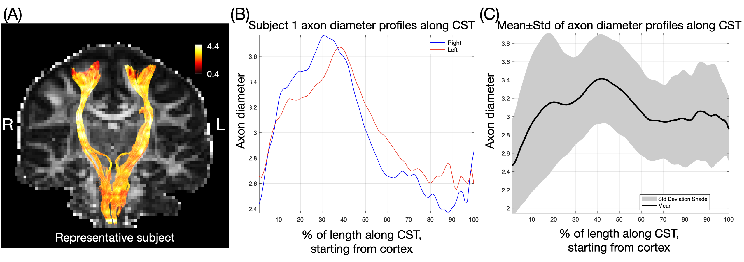
All subjects were transformed to MNI space and averaged within the group for visualization. To probe the variation in axonal diameter along tracts at the single-subject level, the corticospinal tract (CST) was segmented using TractSeg [10], fiber tracking was run using Tensor_Prob algorithms, axon diameters were sampled along the bilateral CST.
Results
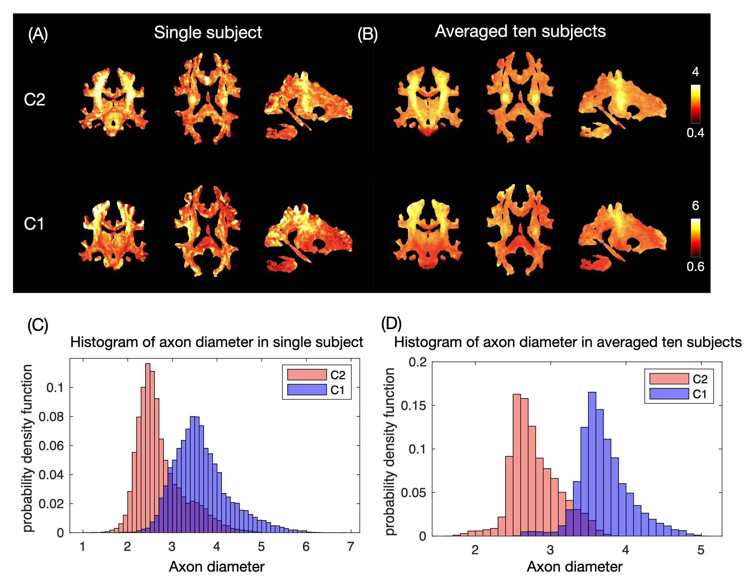
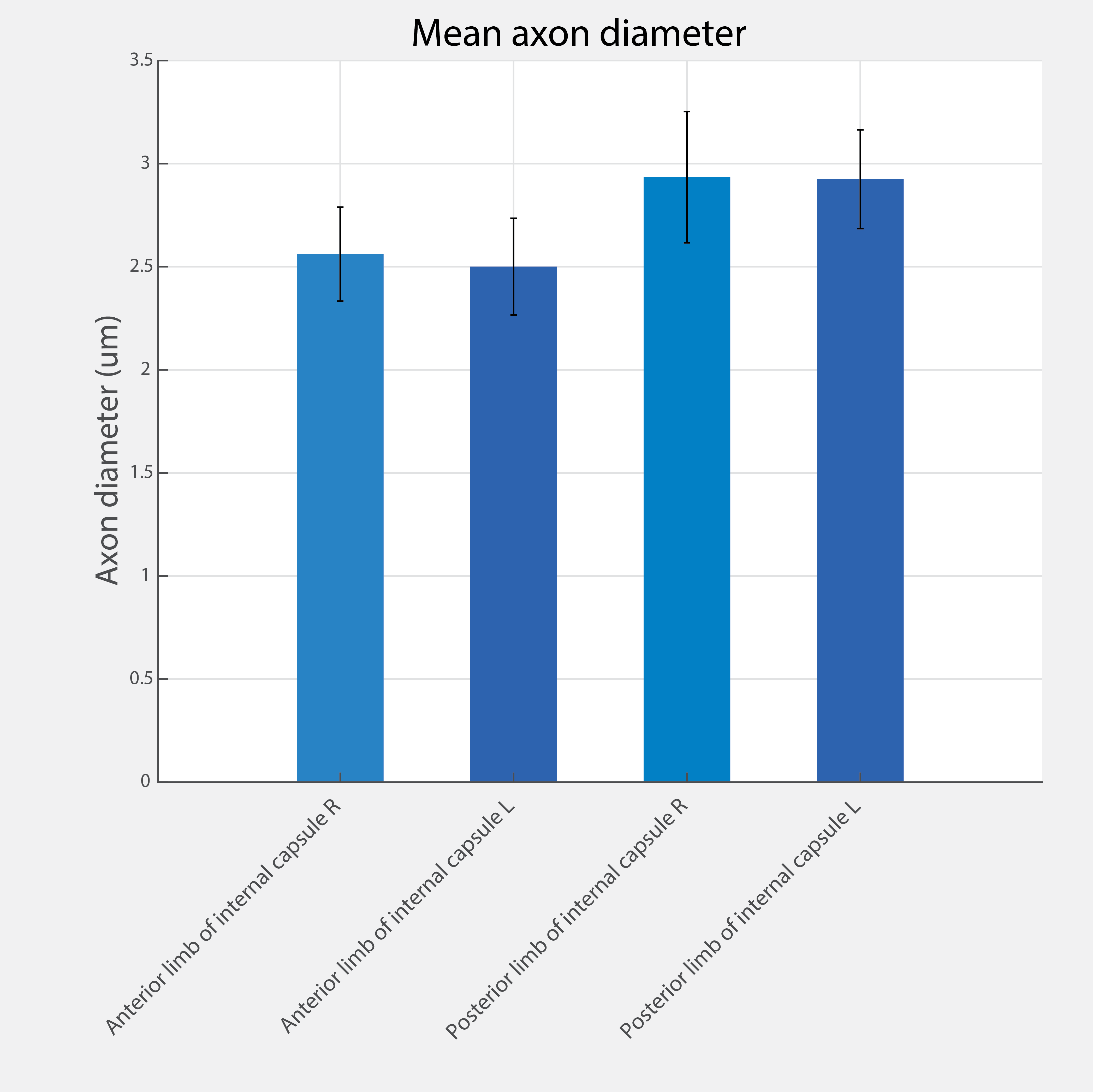
Figure 1 shows representative averaged DWIs for each b-value acquired on C2 and C1. tSNR of white matter on C2 was 38.0 and 17.9 on C1. Figure 2 shows the noise propagation results. The resolution limit for axon diameter of the C2 protocol was lower and demonstrated less bias and variance compared to C1. The theoretical resolution limit for dispersed axons was 1.85 um for C2 and 2.54 um for C1. Figure 3 shows single subject and averaged axonal diameter maps from 10 subjects in each group registered to MNI space. In the center of the brain, where C1 suffers from low SNR, C2 outperforms C1 and shows continuous delineation of the CST bundle. Overall, C2 shows lower estimated axonal diameter, quantified by the histogram and its median (2.63 um on C2 vs 4.00 um on C1). Figure 4 shows the estimated mean axon diameter averaged across the 10 subjects scanned on C2 for the right and left CST and anterior/posterior limbs of the internal capsule, demonstrating generally larger diameter axons posteriorly compared to anteriorly, consistent with trends seen on C1 [4]. Figure 5A shows CST fiber tracts color-coded with relative axonal diameter in a representative subject. Figure 5B shows the axon diameter profile along the CST tracts. Figure 5C shows axon diameter profiles along the CST averaged over all subjects, which replicates the single-subject-level trends of axonal diameter along the CST tracts.Conclusion
Initial experimental results on Connectome 2.0 show that 500mT/m gradient strengths achieve estimation of smaller diameter axons compared to 300mT/m, in line with trends predicted from theory and simulations.Acknowledgements
The research reported in this abstract was supported by the National Institute of Biomedical Imaging and Bioengineering of the National Institutes of Health under award number U01EB026996 and P41EB030006.References
[1] Waxman SG, Kocsis JD, Stys PK. The Axon: Structure, Function and Pathophysiology. New York: Oxford University Press; 1995.
[2] Huang SY, Nummenmaa A, Witzel T, Duval T, Cohen-Adad J, Wald LL, McNab JA. The impact of gradient strength on in vivo diffusion MRI estimates of axon diameter. Neuroimage. 2015 Feb 1:106:464-72.
[3] Nilsson M, Lasič S, Drobnjak I, Topgaard D, Westin CF. Resolution limit of cylinder diameter estimation by diffusion MRI: The impact of gradient waveform and orientation dispersion. NMR Biomed. 2017;30(7):e3711.
[4] Huang SY, Tian Q, Fan Q, Witzel T, Wichtmann B, McNab JA, Daniel Bireley J, Machado N, Klawiter EC, Mekkaoui C, Wald LL, Nummenmaa A. High-gradient diffusion MRI reveals distinct estimates of axon diameter index within different white matter tracts in the in vivo human brain. Brain Struct Funct. 2020;225(4):1277-1291.
[5] Veraart J, Nunes D, Rudrapatna U, Fieremans E, Jones DK, Novikov DS, Shemesh N. Nonivasive quantification of axon radii using diffusion MRI. Elife. 2020 Feb 12;9:e49855.
[6] Ramos-Llorden G et al. Connectome 2.0: Performance evaluation and initial in vivo human brain diffusion MRI results. Proc ISMRM. 2024 (submitted).
[7] Huang SY et al. "Connectome 2.0: Ultra-high gradient strength MRI for connectome studies." Neuroimage. 2021;243:118530.
[8] Fan Q, Nummenmaa A, Witzel T, Ohringer N, Tian Q, Setsompop K, Klawiter EC, Rosen BR, Wald LL, Huang SY. Axon diameter index estimation independent of fiber orientation distribution using high-gradient diffusion MRI. Neuroimage. 2020 Nov 15;222:117197.
[9] Tian Q et al. Comprehensive diffusion MRI dataset for in vivo human brain microstructure mapping using 300 mT/m gradients. Sci Data. 2022 Jan 18;9(1):7.
[10] Wasserthal J et al. "TractSeg: Fast white matter tract segmentation." NeuroImage. 2018;183:239-253.
Figures