1261
Sub-space Low-rank Imaging for mapping of blood-brain barrier Water Exchange Rate (SLIWER) using multi-PLD diffusion-weighted pCASL1Laboratory of FMRI Technology (LOFT), Mark & Mary Stevens Neuroimaging and Informatics Institute, Keck School of Medicine, University of Southern California, Arcadia, CA, United States, 2Laboratory of FMRI Technology (LOFT), Mark & Mary Stevens Neuroimaging and Informatics Institute, Keck School of Medicine, University of Southern California, Los Angeles, CA, United States, 3Siemens Medical Solutions USA, Inc., Urbana, IL, United States, 4Beckman Institute for Advanced Science and Technology, University of Illinois at Urbana-Champaign, Urbana, IL, United States, 5Department of Electrical and Computer Engineering, University of Illinois at Urbana-Champaign, Urbana, IL, United States, 6Lawson Health Research Institute, London, ON, Canada, 7Department of Medical Biophysics, Western University, London, ON, Canada
Synopsis
Keywords: Arterial Spin Labelling, Permeability, Blood-brain barrier, water exchange, cerebral small vessel disease
Motivation: Non-invasive MRI mapping of the blood-brain barrier (BBB) function using water as an endogenous tracer can be a valuable tool for early detection of subtle BBB dysfunctions.
Goal(s): To develop an advanced MRI technique and reconstruction methods for reliable BBB water exchange rate (kw) mapping.
Approach: We introduce a Sub-space Low-rank Imaging method for mapping BBB Water Exchange Rate (SLIWER) with an innovative pulse sequence termed motion compensated diffusion weighted pCASL (MCDW-pCASL).
Results: The SLIWER method demonstrated high test-retest reliability, indicating its potential in clinical settings, such as in evaluating cerebral small vessel disease (cSVD).
Impact: We developed a reliable tool for early BBB dysfunction detection, potentially transforming the diagnosis and treatment of neurological disorders such as cerebral small vessel disease.
Background
Non-invasive MRI mapping of blood-brain barrier (BB) function using water as an endogenous tracer is desirable since it can be more sensitive to subtle BBB dysfunction at an earlier stage of disease progression. A diffusion-prepared pseudo-continuous arterial spin labeling (DP-pCASL) technique was developed for mapping BBB water exchange with good test-retest reliability1, however, SNR and spatial resolution were limited. Recently an innovate pulse sequence termed motion compensated diffusion weighted pCASL (MCDW-pCASL) was proposed for high resolution mapping of cerebral blood flow (CBF) while differentiating intra-/extra-vascular ASL signals (b=40 s/mm2) 2. BBB water exchange rate (kw) can be quantified from the MCDW-pCASL signals using single-pass approximation (SPA) model2,8. However, SPA models require high SNR and long scan time, which makes it challenging for clinical studies. In this study we propose an innovative Sub-space Low-rank Imaging method for mapping perfusion and BBB Water Exchange Rate (SLIWER) with enhanced robustness to noise, while a similar framework has been successfully applied in MRSI3-5 and EPTI6. We evaluated test-retest reliability of the method and demonstrated its clinical feasibility in cerebral small vessel disease (cSVD).Method
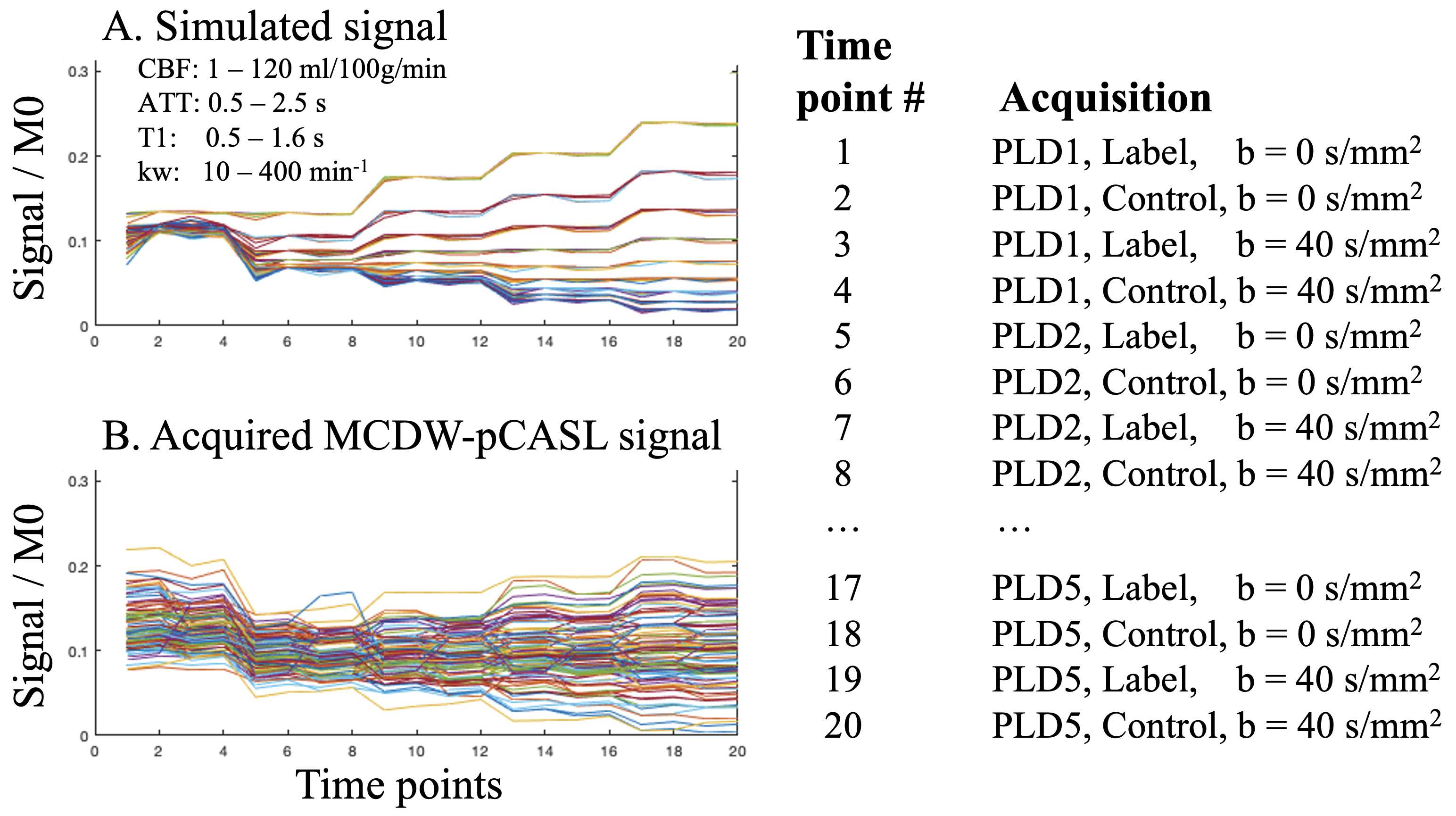
MRI experiments: MRI scans were acquired from a Siemens 3T Prisma system using a 32‐channel head coil. Imaging parameters for MCDW-pCASL were: FOV=224mm, matrix size=64×64, 24 slices (10% oversampling), resolution=3.5×3.5×4mm3, EPI/turbo factor=63/10, 3-fold acceleration along Z direction7, TE = 45.7ms, labeling duration=1.87s. Six label/control pairs were acquired with b=0 or 40.4 sec/mm2 at five PLDs = 1.6, 1.9, 2.2, 2.5 and 2.8s. Corresponding TRs were 4.9 to 6.1 s for 5 PLDs, and total scan time was 11 mins. DP-pCASL scans were also acquired to demonstrate the improvement of SNR and spatial resolution1.We have collected test-retest scans (~1 week apart) from 7 young healthy subjects (4M, 31±5 years) and co-enrolled two elderly subjects (1M 66years, 1F 81years) from MarkVCID study to evaluate the clinical feasibility in cSVD.Sub-space Low-rank method for mapping BBB Water Exchange Rate (SLIWER): The proposed method utilizes singular value deposition (SVD) to extract the major components of the signal evolution across 5 PLDs (A total of 20 time points: 5PLD × 2 L/C × diffusion on/off). In-vivo data were combined with simulated (SPA model2,8) MCDW-pCASL signals incorporating physiological parameters, including T1 (0.5–1.6s), CBF (1–120 ml/100g/min), ATT (0.5–2.5s) and kw (10–400 min-1), to extract the basis of signals, and tissue T1 was incorporated into simulation to account for control signal evolutions induced by background suppression pulses.
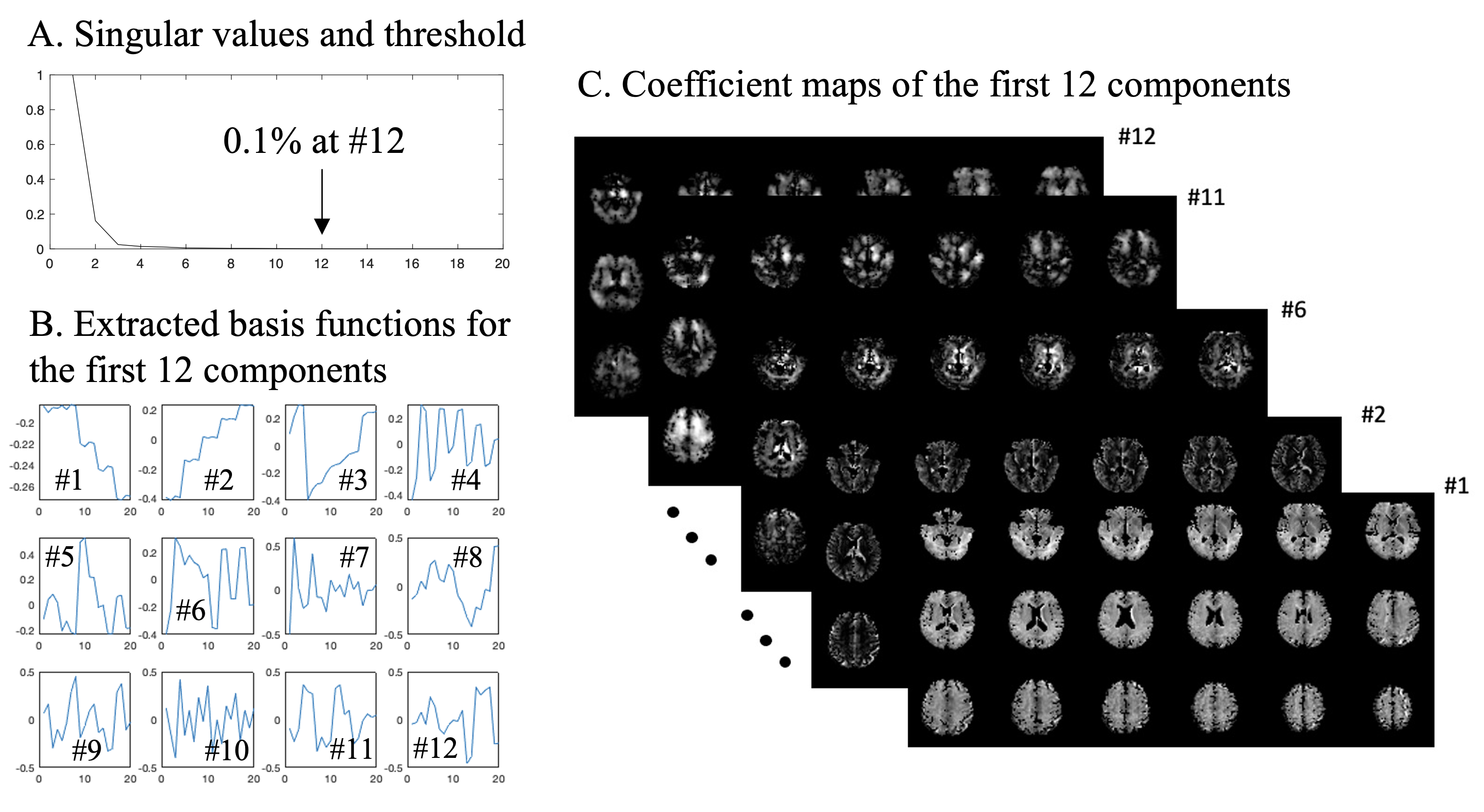
Figure 1A and B show examples of simulated and in-vivo MCDW-pCASL signals across 20 time points. Figure 2A shows singular values obtained by SVD of combined simulated and in-vivo MCDW-pCASL signals across 20 time points. Basis functions with singular value smaller than 0.1% was considered as noise, thus the first 12 components were considered as major components. Extracted basis functions of the first 12 components are shown in Fig.2B and corresponding coefficient maps from one subject are shown in Fig. 2C, respectively.
We expect signals can be sufficiently expressed as a combination of these 12 basis functions4, voxel-wise MCDW-pCASL signals were then projected onto the 12 major basis for denoising and obtaining corresponding CBF and kw using dictionary matching with a slight TV regularization term on kw to improve robustness to noise.
Results and discussion
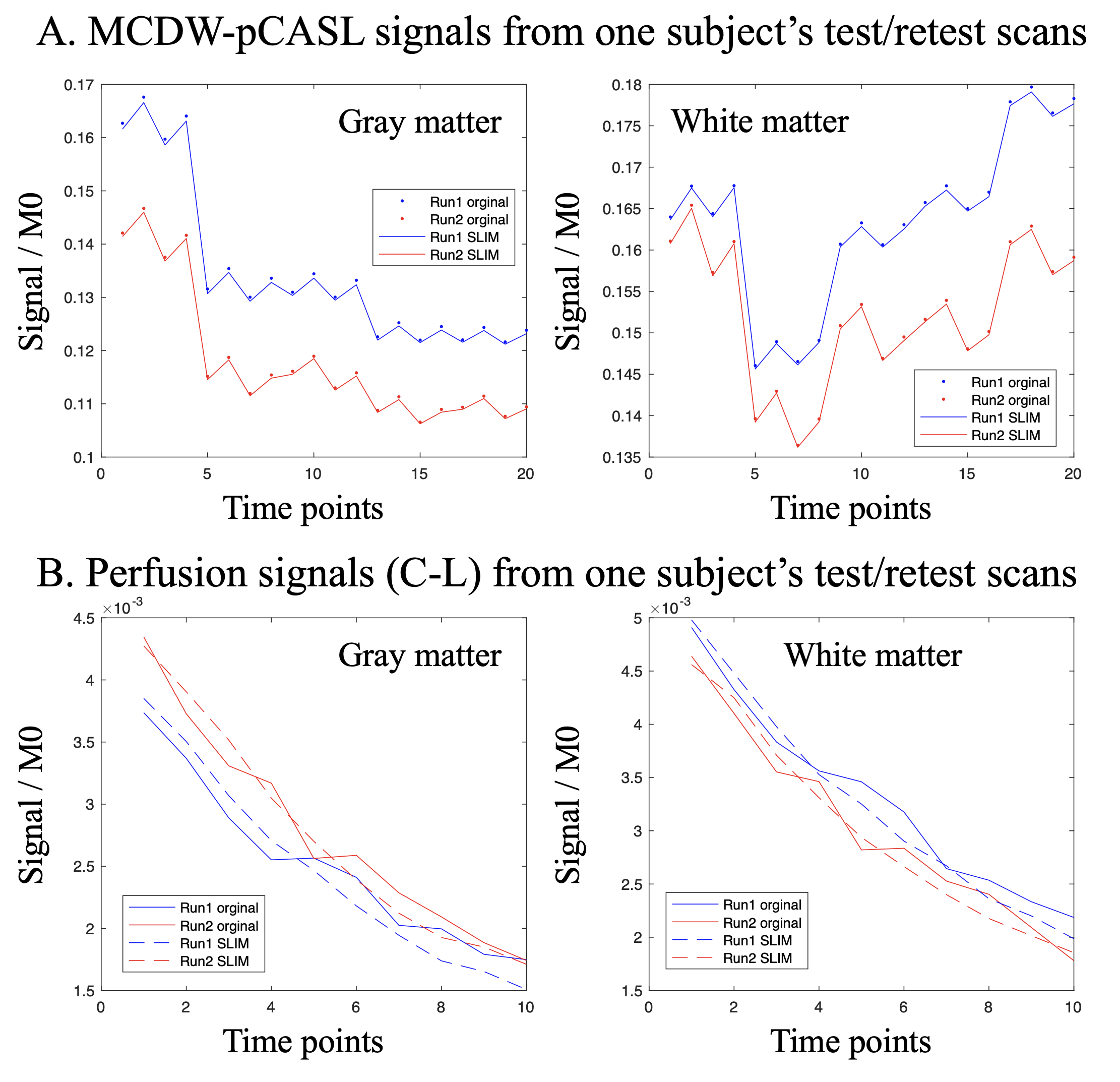
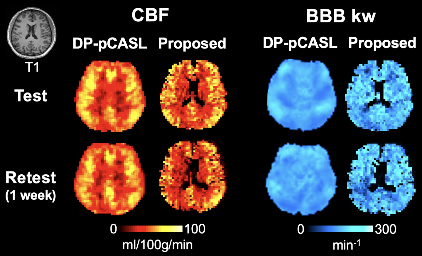
Figure 3A shows original and SLIWER reconstructed MCDW-pCASL signals from one subject’s test and retest scans in gray (left) and white matter (right). Perfusion signals (interleaved diffusion on and off) were subtracted and shown in Figure 3B to demonstrate the improved robustness to noise and higher consistency between test and retest scans. Fluctuations in the perfusion signals were significantly minimized by SLIWER.Figure 4 shows CBF and kw maps acquired from one subject’s test and retest scans using the DP-pCASL (10 min) and proposed MCDW-pCASL (11 min) with SLIWER reconstruction. Improved spatial resolution of CBF and kw maps can be observed using the proposed method. Average test/retest kw values were 140.9±11.8 and 136.7±10.8 min-1, respectively, indicating high reliability of the proposed method (wsCV=10.1%).
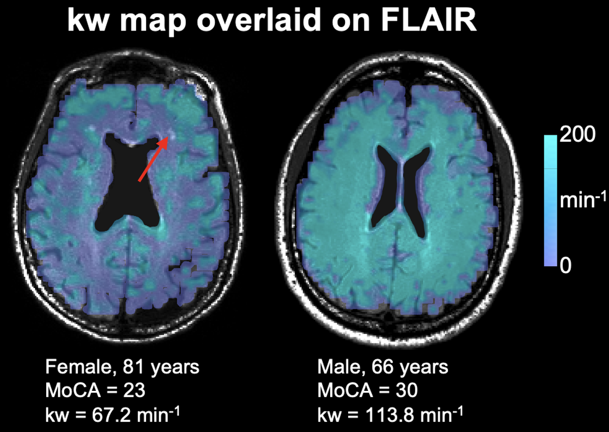
Figure 5 shows kw maps overlaid on FLAIR from the two subjects with MoCA scores of 23 and 30 respectively. The cognitively normal subject (MoCA=30) had overall higher kw, which demonstrates the feasibility of the proposed technique in cSVD. Moreover, the SLIWER improved spatial resolution of kw map so that BBB dysfunction between WM lesion and surrounding penumbra can be potentially distinguished (red arrow).
Conclusion
Our study introduces a novel SLIWER method for reliable BBB kw mapping within 11 mins. This technique is promising for studying BBB dysfunction in clinical settings, such as cerebral small vessel disease cases.Acknowledgements
This work was supported by National Institute of Health (NIH) grant UF1-NS100614, R01-NS114382 and R01-EB028297.References
1. Shao X, Ma SJ, Casey M, D'Orazio L, Ringman JM, Wang DJJ. Mapping water exchange across the blood-brain barrier using 3D diffusion-prepared arterial spin labeled perfusion MRI. Magn Reson Med 2019;81(5):3065-3079.
2. Shao X, Zhao C, Shou Q, St Lawrence KS, Wang DJ. Quantification of blood–brain barrier water exchange and permeability with multidelay diffusion‐weighted pseudo‐continuous arterial spin labeling. Magnetic Resonance in Medicine 2023.
3. Guo R, Zhao Y, Li Y, Li Y, Liang ZP. Simultaneous metabolic and functional imaging of the brain using SPICE. Magnetic resonance in medicine 2019;82(6):1993-2002.
4. Liang Z-P. Spatiotemporal imagingwith partially separable functions. 2007. IEEE. p 988-991.
5.Lam F, Li Y, Guo R, Clifford B, Liang ZP. Ultrafast magnetic resonance spectroscopic imaging using SPICE with learned subspaces. Magnetic resonance in medicine 2020;83(2):377-390.
6. Dong Z, Wang F, Reese TG, Bilgic B, Setsompop K. Echo planar time‐resolved imaging with subspace reconstruction and optimized spatiotemporal encoding. Magnetic resonance in medicine 2020;84(5):2442-2455.
7. Spann SM, Shao X, Wang DJ, Aigner CS, Schloegl M, Bredies K, Stollberger R. Robust single-shot acquisition of high resolution whole brain ASL images by combining time-dependent 2D CAPIRINHA sampling with spatio-temporal TGV reconstruction. NeuroImage 2020;206:116337.
8. St Lawrence KS, Owen D, Wang DJ. A two-stage approach for measuring vascular water exchange and arterial transit time by diffusion-weighted perfusion MRI. Magn Reson Med 2012;67(5):1275-1284.
Figures