1260
Vessel-Encoded Arterial Spin Labeling at 7 Tesla1Institute of Science and Technology for Brain-inspired Intelligence, Fudan University, Shanghai, China, 2University of Oxford, Wellcome Centre for Integrative Neuroimaging, FMRIB Division, Nuffield Department of Clinical Neurosciences, Oxford, United Kingdom, 3Key Laboratory of Computational Neuroscience and Brain-Inspired Intelligence (Fudan University), Ministry of Education, Shanghai, China
Synopsis
Keywords: Arterial Spin Labelling, Arterial spin labelling, ultra-high field
Motivation: Vessel-encoded arterial spin labeling (VEASL) allows the visualization of collateral blood flow and blood supply to lesions, but has limited SNR. At ultra-high field, ASL benefits from significantly improved SNR and longer blood T1 relaxation time. However, B0 field inhomogeneity can reduce labeling efficiency and disrupt encoding patterns.
Goal(s): Implementing VEASL robustly at 7 Tesla.
Approach: Optimized ASL parameters, dynamic B0 shimming and OES-based correction methods were used to mitigate the impact of B0 field inhomogeneity.
Results: Good vascular territory maps, labeling both the neck and above the circle of Willis, were achieved, including at high spatial resolution.
Impact: We demonstrate the first vessel-encoded ASL perfusion maps at ultra-high field, and the vascular territory maps significantly improved after applying B0 correction techniques, with the potential to push for even higher spatial resolution.
Introduction
Vessel-Encoded Arterial Spin Labeling (VEASL) generates a series of spatial labeling patterns, allowing the unique encoding of blood signals arising from each feeding artery. This provides information about the vascular territories and thus non-invasive assessment of collateral circulation. For simple vessel arrangements, VEASL has equivalent SNR efficiency to conventional non-selective ASL1,2, but for more complex vascular geometries (above the circle of Willis, CoW), encoding efficiency may be reduced. Thus far, VEASL has mainly been implemented on 3T scanners. The intrinsically low SNR of ASL combined with the reduced encoding efficiency in scenarios with complex vascular geometries mean the application of VEASL can require several repeated scans over each perfusion weighted image for robust territorial separation. At ultra-high field the SNR of ASL can be greatly improved and the blood T1 relaxation time becomes longer. This may result in a greatly enhanced ability to separate multiple vascular territories even under the pathological conditions of different arterial transit time within relatively short scan times and/or push the spatial resolution to examine small vascular territories with greater fidelity. B0 inhomogeneity is particularly problematic for VEASL and is exacerbated at higher field strengths3, so we implemented VEASL at 7T, using optimized parameters, combined with B0 correction, and achieved good results in labeling both the neck and above the CoW.Materials and methods
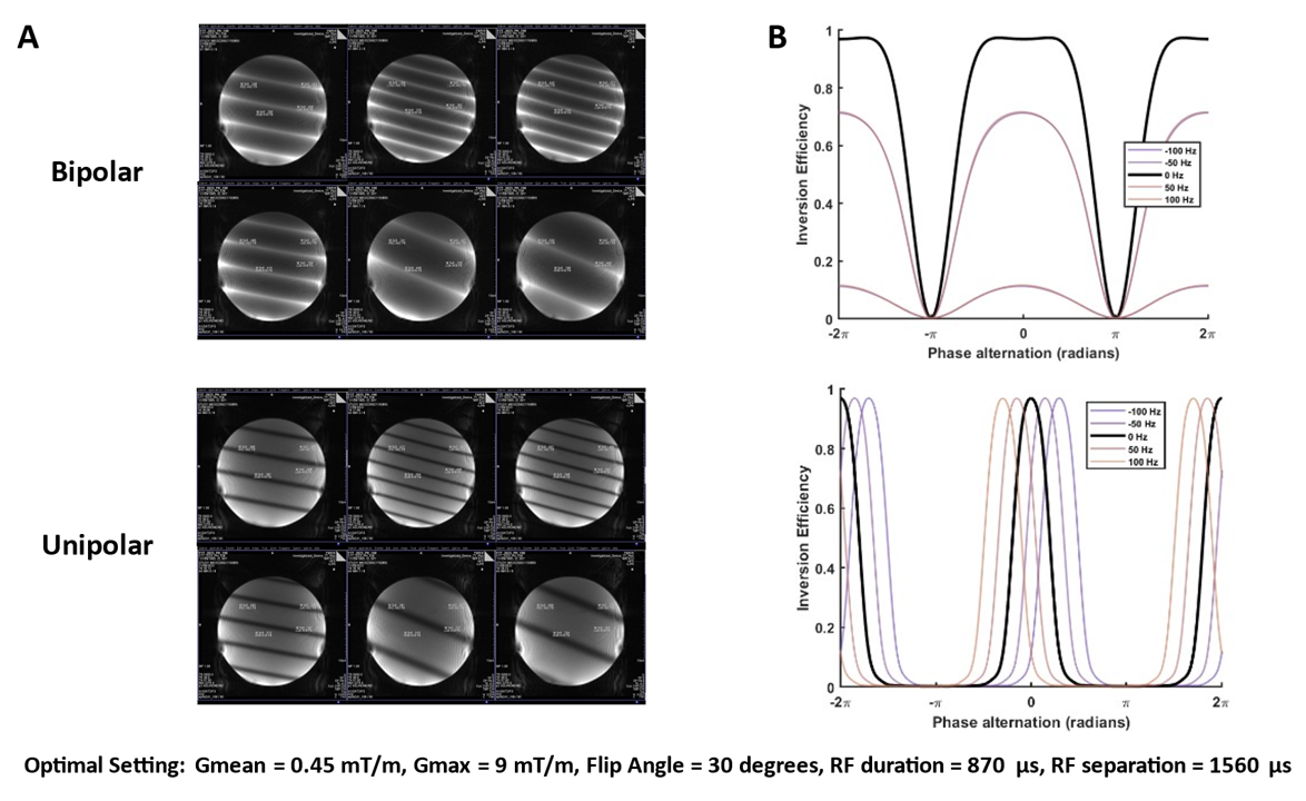
SimulationTo overcome the complex vascular structure and enhance the SNR efficiency of VEASL, we further optimized the original optimized encoding scheme (OES)4. Additionally, we optimized the PCASL parameters to achieve a thin-slice labeling plane, to minimize the effect of vessel tortuosity within the labeling plane, whilst maintaining high labeling efficiency. Using optimal parameters, we simulated the effects of field inhomogeneity on the inversion profiles of two VEASL gradient modes.
Image acquisition
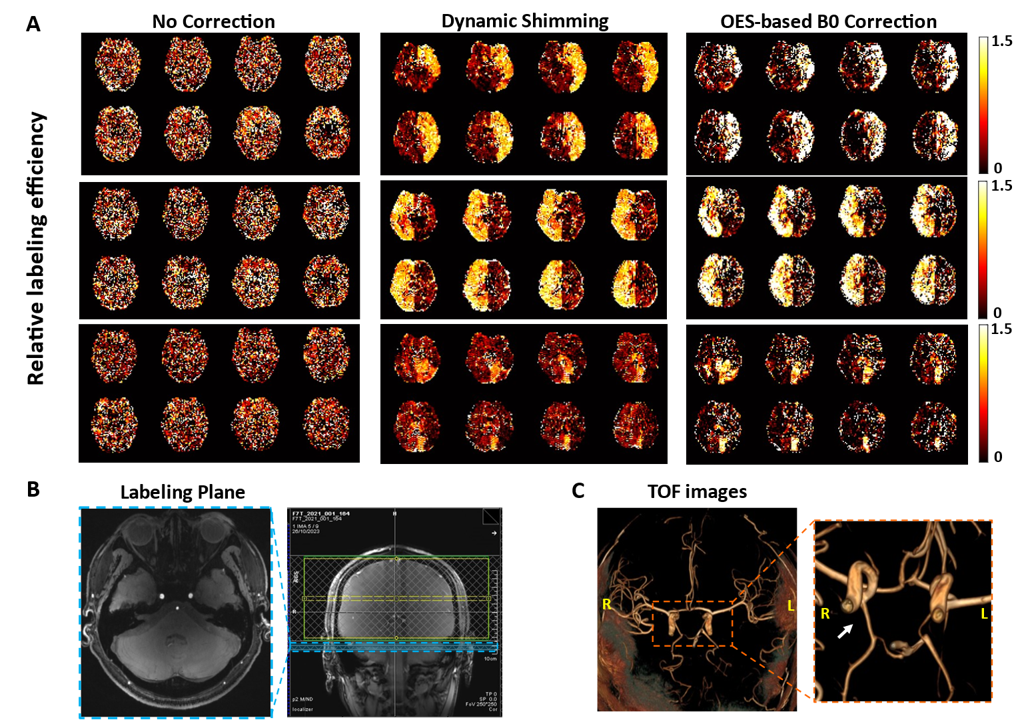
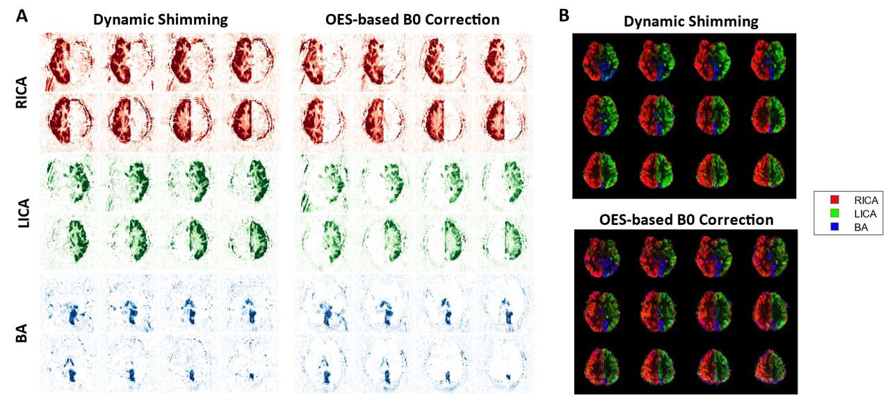
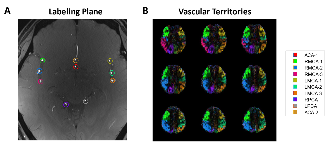
Data was acquired on a 7T Plus Siemens Magnetom scanner using pTx coil. Phantom: The labeling plane was overlapped with the imaging region to visualize the bipolar/unipolar encoding patterns. In-vivo: Three healthy volunteers were scanned using optimized parameters: For the first subject, the labeling plane was positioned just below the CoW with three brain-feeding arteries, and we compared the performance of no B0 correction, dynamic shimming and OES-based correction3. For the second subject, the labeling plane was positioned in the neck, and high-resolution VEASL imaging (2×2×4 mm³) was performed using dynamic shimming. The third subject had the labeling plane placed above the CoW. Benefiting from better field homogeneity, we only used the static B0 shimming for the imaging region and tagging plane. Maximum a posteriori solution to the Bayesian framework was used for VEASL analysis5.
Results
The impact of field inhomogeneity on the encoding pattern differs between bipolar and unipolar approaches. For optimized parameters, unipolar leads to a very narrow label region, which could make achieving an efficient encoding more difficult and make this approach more sensitive to subject motion. In the in-vivo experiments, the bipolar approach was chosen for dynamic shimming, reserving the utilization of unipolar as required for the OES-based B0 correction. In the in-vivo scans the relative labeling efficiency of each artery was high, both for dynamic shimming and OES-based B0 corrections (Figure 2A). Both correction approaches also produced good quality vascular territory maps (Figure 3). In Figure 4, the high SNR achievable at 7T allowed VEASL to be pushed to higher spatial resolutions, we still obtained a clear delineation of vascular territories. For labeling above the CoW, the territories of ACAs did not receive a clear delineation, likely due to significant inhomogeneity in the frontal lobe, which was challenging to address with standard static shimming. However, the six segments of the MCAs and RPCA were still clearly visible, as shown in Figure 5B.Discussion and conclusions
Here we demonstrate the first vessel-encoded ASL at 7T and the application of B0 correction techniques to improve the resulting vascular territory maps. Benefiting from the significantly improved SNR at 7T, we showcased the potential of pushing VEASL to higher resolutions. Above the CoW, good results could be achieved for most arterial branches even without advanced field correction approaches, although the ACA signal was poorly labeled. However, in areas like the neck where B0 issues were more pronounced, field correction became crucial. The optimized PCASL settings increased the RF interval, making it more sensitive to B0 inhomogeneity, making these corrections particular important. Both dynamic shimming and OES were effective methods for VEASL in these situations. In the future, it will be necessary to collect more subjects and perform quantitative analyses to evaluate the effectiveness of different B0 correction methods for VEASL, including their application above the CoW.Acknowledgements
This work was enabled by a Sir Henry Dale Fellowship jointly funded by the Wellcome Trust and the Royal Society (220204/Z/20/Z). The Wellcome Centre for Integrative Neuroimaging is supported by core funding from the Wellcome Trust (203139/Z/16/Z).References
1. Wong, E.C. Vessel-encoded arterial spin-labeling using pseudocontinuous tagging. Magnetic Resonance in Medicine 58, 1086-1091 (2007).
2. Okell, T.W., Chappell, M.A., Kelly, M.E. & Jezzard, P. Cerebral Blood Flow Quantification Using Vessel-Encoded Arterial Spin Labeling. Journal of Cerebral Blood Flow & Metabolism 33, 1716-1724 (2013).
3. Berry, E.S.K., Jezzard, P. & Okell, T.W. Off-resonance correction for pseudo-continuous arterial spin labeling using the optimized encoding scheme. NeuroImage 199, 304-312 (2019).
4. Berry, E.S.K., Jezzard, P. & Okell, T.W. An Optimized Encoding Scheme for Planning Vessel-Encoded Pseudocontinuous Arterial Spin Labeling. Magnetic Resonance in Medicine 74, 1248-1256 (2015).
5. Chappell, M.A., Okell, T.W., Payne, S.J., Jezzard, P. & Woolrich, M.W. A fast analysis method for non-invasive imaging of blood flow in individual cerebral arteries using vessel-encoded arterial spin labelling angiography. Medical Image Analysis 16, 831-839 (2012).
Figures