1256
Field-Correcting GRAPPA (FCG) for improved mitigation of even-odd and field-related artifacts in EPI1Radiology Department, Stanford University, Stanford, CA, United States, 2Electrical Engineering, Stanford University, Stanford, CA, United States, 3Cognitive and Neurobiological Imaging Center, Stanford University, Stanford, CA, United States, 4Athinoula A. Martinos Center for Biomedical Imaging, Massachusetts General Hospital, Charlestown, MA, United States, 5Department of Radiology, Harvard Medical School, Boston, MA, United States, 6Harvard-MIT Division of Health Sciences and Technology, Massachusetts Institute of Technology, Cambridge, MA, United States, 7Functional Magnetic Resonance Facility (FMRIF), National Institutes of Health, Bethesda, MD, United States
Synopsis
Keywords: Artifacts, Artifacts
Motivation: Field perturbation from gradient error and eddy current has been a long-standing issue for EPI, especially with high gradient performance or ramp sampling
Goal(s): To develop a MLP based Field-Correcting GRAPPA (FCG) that accounts for spatial-varying field and produces artifacts-mitigated images
Approach: Calibration data with dual polarity were acquired. A MLP based kernel was trained to take single-polarity data as input and output the clean averaged data. It was applied for both phantom and in vivo undersampled EPI with ramp-sampling.
Results: FCG produced best correction results for ramp-sampling EPI, with potentials for wide applications including fMRI.
Impact: A Field-Correcting GRAPPA (FCG) technique was developed to correct the field perturbation induced image artifacts for EPI, which accounts for spatial varying field and produced promising resulting on ramp-sampling cases, with great potentials for wide applications including fMRI.
Introduction
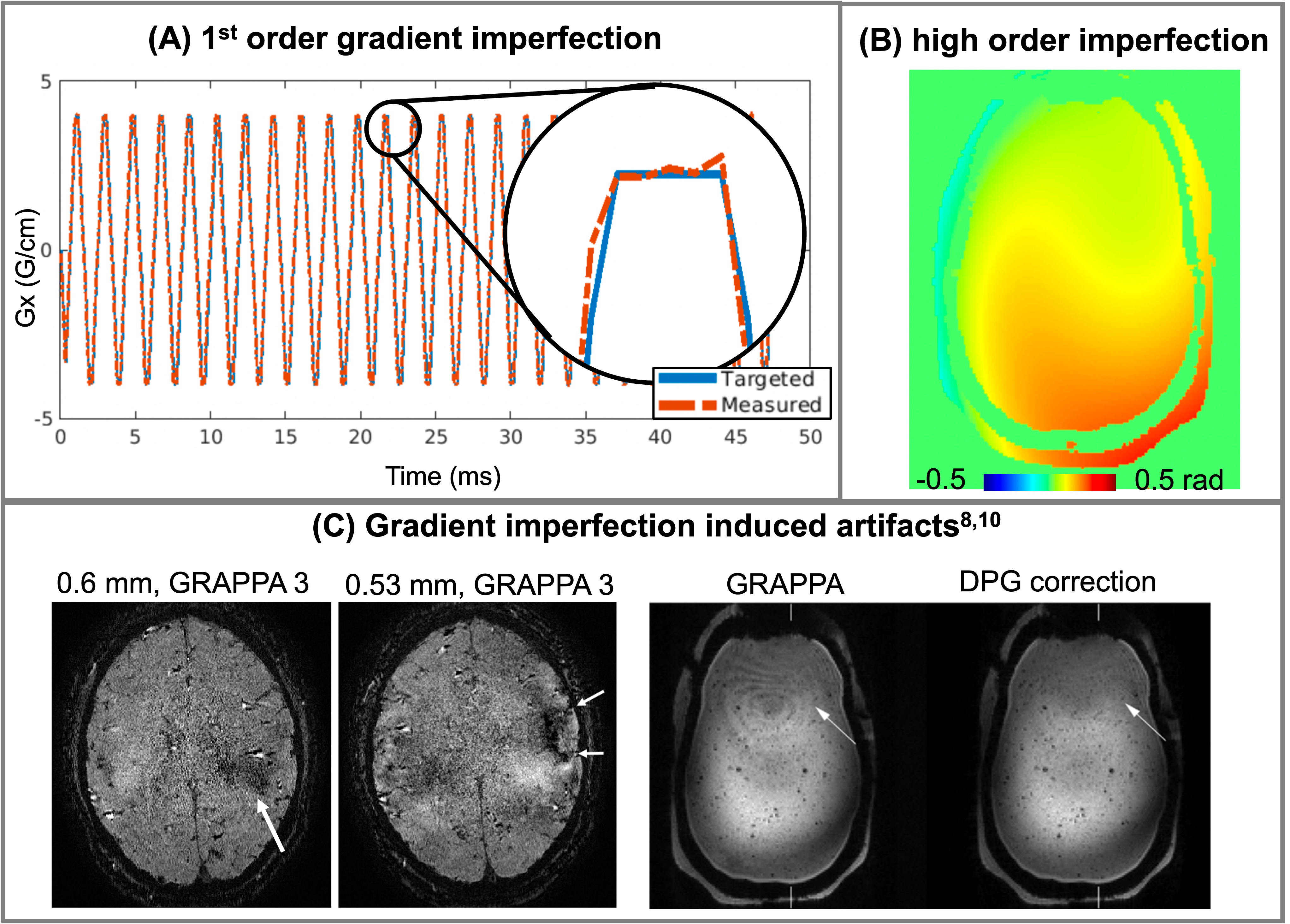
Echo Planar Imaging (EPI) is a rapid imaging method1 that has been extensively used in numerous MRI applications2-5. However, it is subject to field perturbations (Figure 1) from gradient errors and system interaction (e.g., eddy-current)6,7. The associated artifacts significantly degrade image quality especially for high-resolution imaging with more undersampling, stronger gradient system or higher field8. Tremendous efforts have been made to resolve this issue. Field probe based measurements can be used to improved image quality, but relies on an additional external device9. Dual-Polarity-GRAPPA (DPG) showed promising correction in EPI with limited ramp-sampling but produced higher error when adopting to cases with large ramp sampling due to changing field across kx8. Dual polarity average (DPA)10, which directly acquire two datasets with opposite readout polarity sequentially and complex-averaged them, demonstrated encouraging results, but led to twice of the scan time. To address the problem, we developed a Field-Correcting GRAPPA (FCG) technique11, which utilizes machine learning to achieve k-space location-varying correction on the data and can be used for different sampling and reconstruction frameworks without extra scan time.Methods
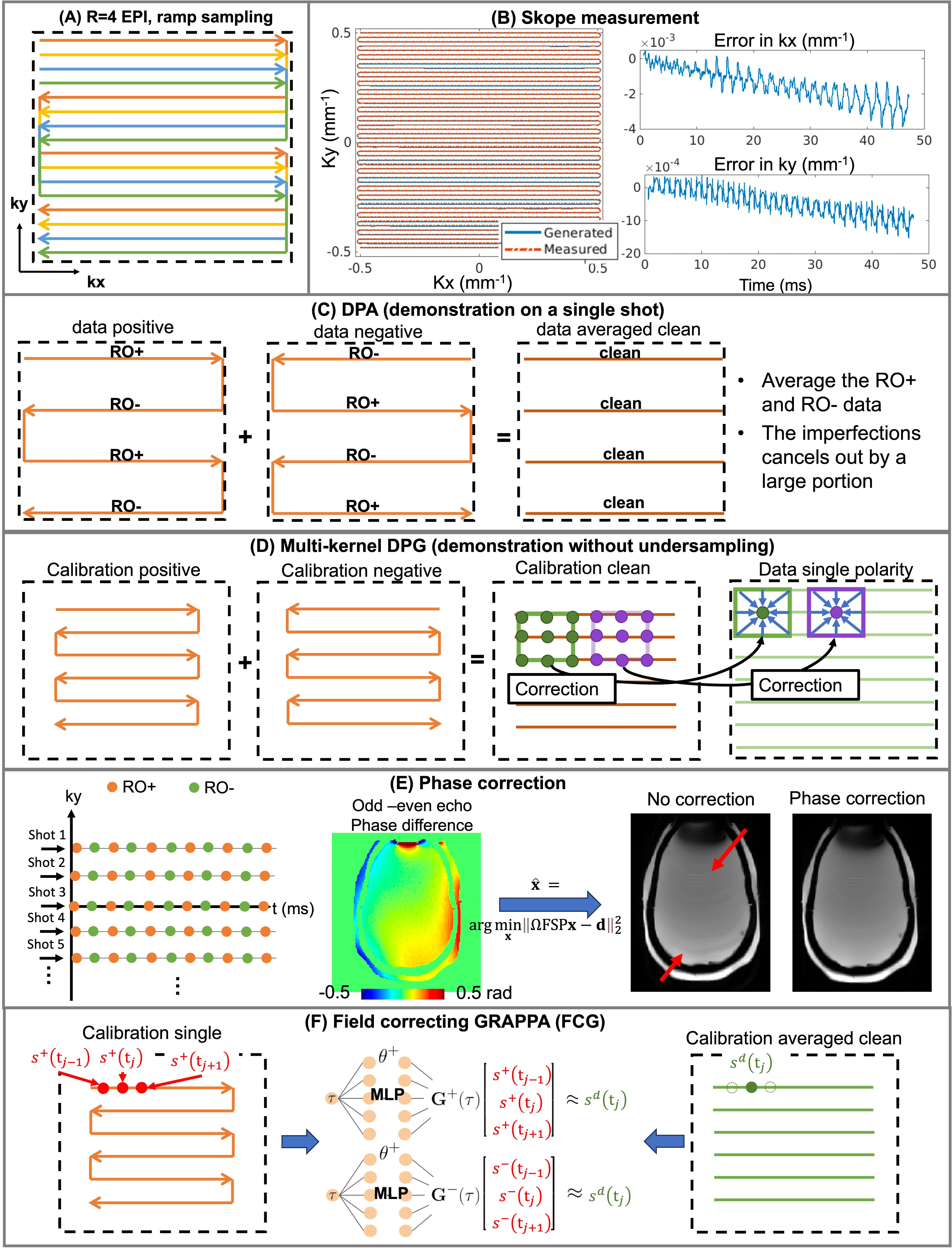
Sequence and sampling design: a 2D gradient echo sequence with EPI sampling were used. The EPI trajectory is R=4, and all the 4 shots were acquired (Figure 2A). The maximum gradient and slew rate were 40mT/m and 12mT/m/ms.Skope measurement: Skope field-probes (Skope, Switzerland) were used to provide the gold-standard field measurement (Figure 2B) as:
$$S(t)=\int_{\boldsymbol{r}}\rho(\boldsymbol{r})e^{i\left(C_0(t)+C_1(t)\boldsymbol{r}+C_2(t)\boldsymbol{r}^2+C_3(t)\boldsymbol{r}^3\right)}d\boldsymbol{r},(1)$$
where $$$S(t)$$$ is the k-space signal at time $$$t$$$, $$$\boldsymbol{r}$$$ is the spatial locations, $$$C_j(t)$$$ is the Skope-measured spherical harmonics coefficient at order $$$j\in[0,3]$$$. The 1st order coefficient $$$C_1(t)$$$ can be used as the trajectory for NUFFT reconstruction with ramp sampling.
DPA: The DPA data was acquired with positive polarity (RO+) and negative polarity (RO-), put to Cartesian grid using 1D-NUFFT, and then complex averaged (Figure 2C) to serve as a “clean” reference.
DPG: For DPG, the calibration was acquired with dual polarity for GRAPPA kernel training to correct the single polarity data. To compensate for the varying field perturbation due to ramp sampling, a multi-kernel DPG was also performed (Figure 2D), with different kernels at different $$$k_x$$$ location.
2D even-odd phase correction12: Phase correction acquires a low-spatial-resolution ky-t space calibration data acquired across multiple echoes using bipolar readout to provide the sensitivity maps, B0 map, and a phase map between odd and even echoes containing the high-order field perturbation. The phase map can be incorporated into the reconstruction of EPI data as
$$\mathbf{\hat{x}}=\arg\min_{\mathbf{x}}\|\Omega\mathrm{FSP}\mathbf{x}-\mathbf{d}\|_2^2,(2)$$
with acquired data $$$\mathbf{d}$$$, image $$$\mathbf{x}$$$, sampling mask $$$\Omega$$$, Fourier transform $$$F$$$, coil-sensitivity $$$S$$$, and phase $$$P$$$ corresponding to each echo (Figure 2E).
Field-Correcting GRAPPA (FCG): Multi-kernel DPG in theory can capture the spatial varying field across kx, but will be a highly ill-posed problem due to limited training data compared to single-kernel DPG. Since the kernel weights across kx are highly correlated, Multilayer Perceptron (MLP)13 can potentially provide a compact representation of the spatial-varying information with limited data. The input for MLP is 3 adjacent kx points from a single polarity and the output is the clean center kx of the 3 (Figure 2F). The training of MLP is performed as
$$\arg\min_\theta\sum_j\left\|\boldsymbol{G}\left(s^{+}\left(\mathrm{t}_{j-1}\right),s^{+}\left(\mathrm{t}_j\right),s^{+}\left(\mathrm{t}_{j+1}\right),\theta\right)-s^d\left(\mathrm{t}_j\right)\right\|,(3)$$
Where $$$\boldsymbol{G}$$$ is the MLP kernel with trainable parameter $$$\theta$$$, $$$s^{+}\left(\mathrm{t}_{j}\right)$$$ is the $$$j$$$th signal acquired with positive polarity, $$$s^d\left(\mathrm{t}_j\right)$$$ is the dual polarity “clean” output.
Experiment design: All phantom and in vivo experiments were performed on 3T system (UHP, GE healthcare) with a 32-channel head coil. Each experiment acquired images in 2D axial orientation including (1) low-resolution calibration data; (2) 4-shot R=4 EPI in both polarity. The EPI sampling parameters are: FOV=220×220mm2, spatial-resolution=1.1×1.1mm2, slice-thickness=5mm, TE=25ms, TR=2000ms, flip-angle=90°, echo-spacing=1.0ms with 70% ramp sampling (Figure 1A).
Results
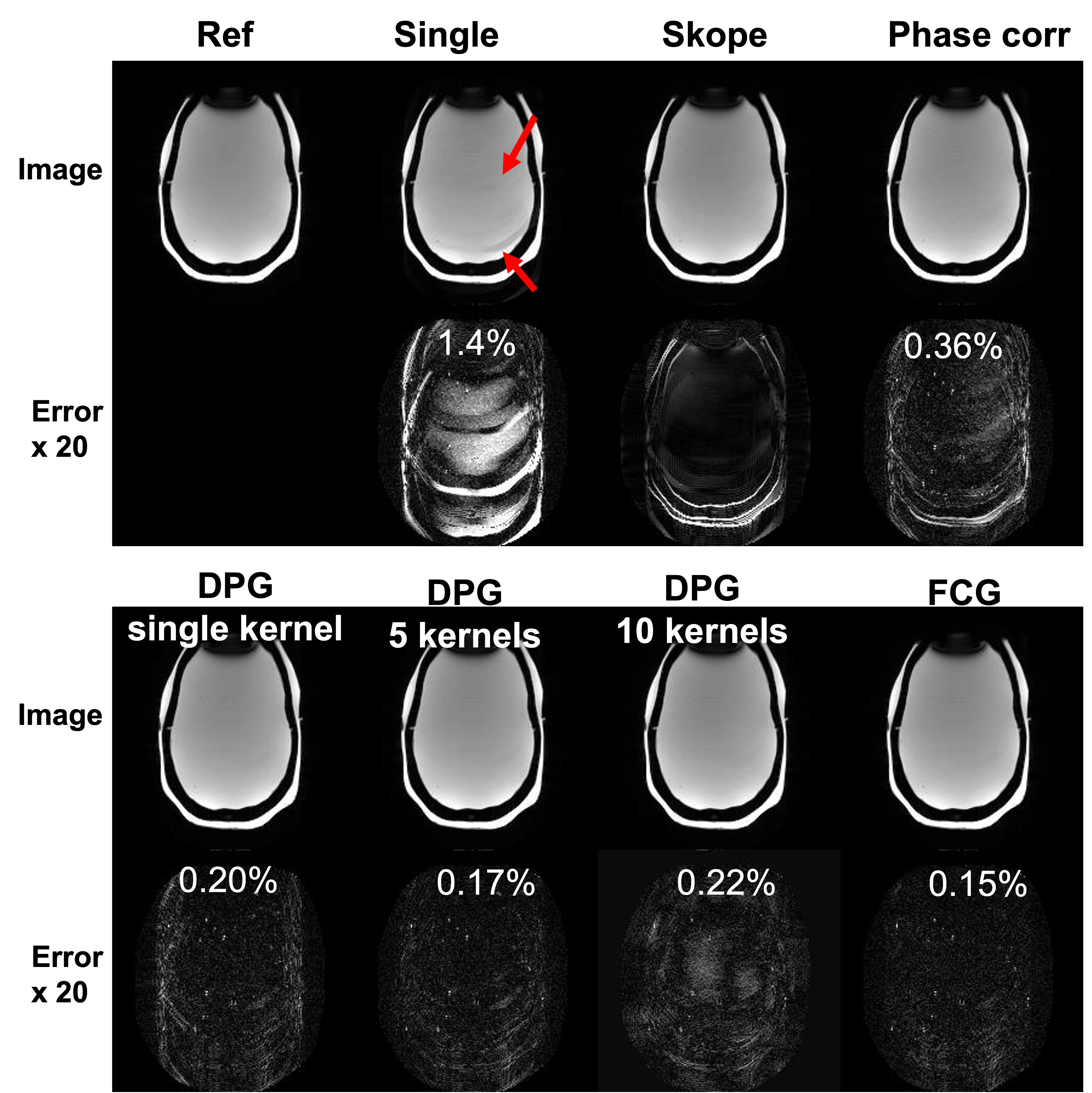
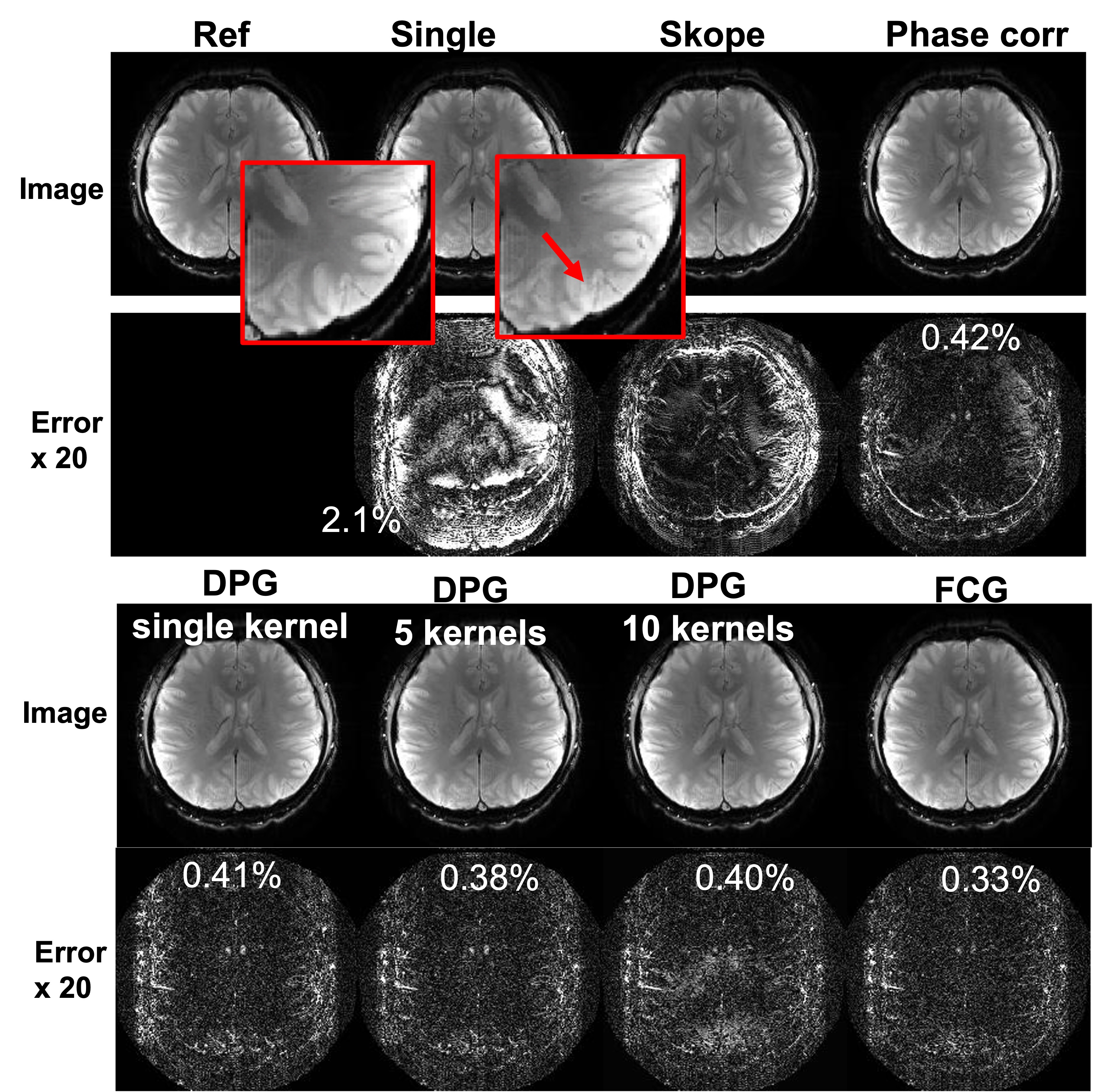
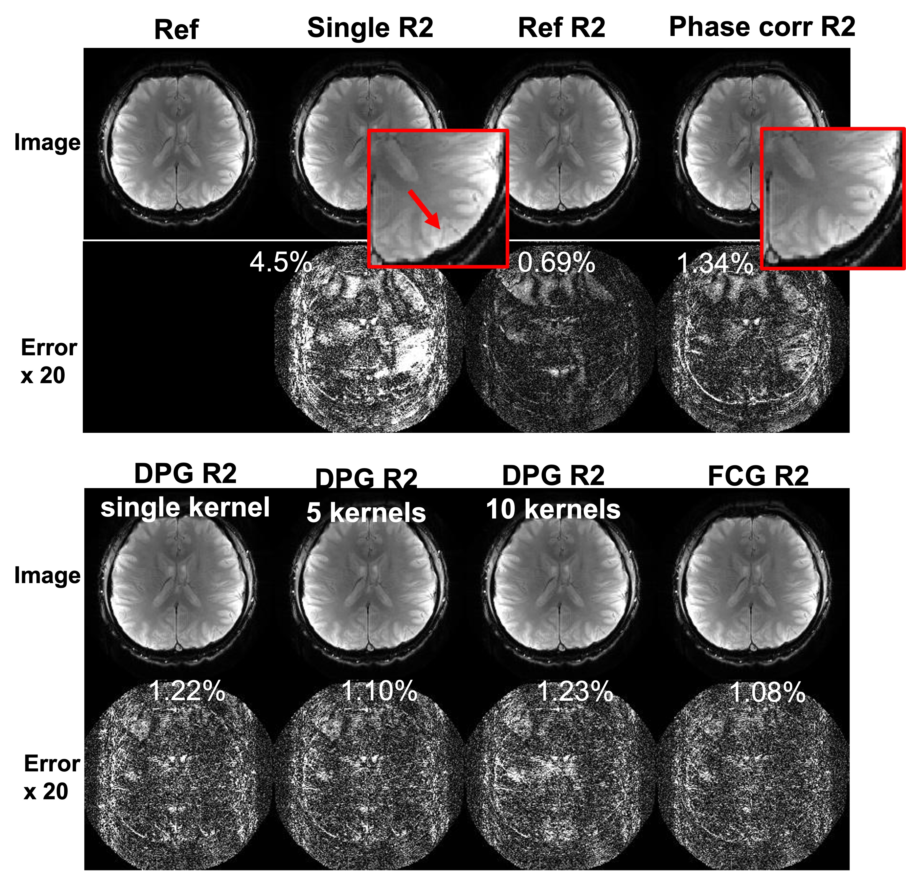
Figure 3 shows phantom results. DPA data is the reference. Skope also provided high quality images but has differences in the edges due to field drifting induced geometric distortion. Single polarity reconstruction produced very strong artifacts, while phase correction, DPG, and FCG can all largely reduce the artifacts and improve image quality. FCG resulting in the smallest error. DPG with 5 kernels produced best results in DPG approaches and is close to the FCG performance. The in vivo results in Figure 4 and the R=2 undersampling results in Figure 5 support the observation.Conclusion
FCG demonstrated the best image quality and smallest error in correcting field imperfection induced image artifacts. It has great potential to resolve the long-standing issue of EPI and can be extremely important for studies such as fMRI. Future work towards higher field strength, gradient performance and resolution (as Fig1C examples) will be conducted.Acknowledgements
This work is partially supported by R01MH116173, R01EB019437, U01EB025162, P41EB030006, R01EB033206, U24NS129893References
1. Stehling, Michael K., Robert Turner, and Peter Mansfield. "Echo-planar imaging: magnetic resonance imaging in a fraction of a second." Science 254.5028 (1991): 43-50.
2. Poustchi-Amin, Mehdi, et al. "Principles and applications of echo-planar imaging: a review for the general radiologist." Radiographics 21.3 (2001): 767-779.
3. Schmitt, Franz, Michael K. Stehling, and Robert Turner. Echo-planar imaging: theory, technique and application. Springer Science & Business Media, 2012.
4. Yamashita, Yasuyuki, Yi Tang, and Mutsumasa Takahashi. "Ultrafast MR imaging of the abdomen: echo planar imaging and diffusion‐weighted imaging." Journal of Magnetic Resonance Imaging 8.2 (1998): 367-374.
5. Kwong, K. K. "Functional magnetic resonance imaging with echo planar imaging." Magnetic resonance quarterly 11 (1995): 1-1.
6. Ahn, C B, and Z H Cho. “A New Phase Correction Method in NMR Imaging Based on Autocorrelation and Histogram Analysis.” IEEE Transactions on Medical Imaging 6, no. 1 (1987): 32–36.
7. Xiang, Qing‐San, and Frank Q. Ye. "Correction for geometric distortion and N/2 ghosting in EPI by phase labeling for additional coordinate encoding (PLACE)." Magnetic Resonance in Medicine: An Official Journal of the International Society for Magnetic Resonance in Medicine 57.4 (2007): 731-741.
8. Hoge, W. Scott, and Jonathan R. Polimeni. "Dual‐polarity GRAPPA for simultaneous reconstruction and ghost correction of echo planar imaging data." Magnetic resonance in medicine 76.1 (2016): 32-44.
9. Dietrich, Benjamin E., et al. "A field camera for MR sequence monitoring and system analysis." Magnetic resonance in medicine 75.4 (2016): 1831-1840.
10. Huber R, Stirnberg R, Feinberg DA, Ma SJ, Ehses P, Gulban OF, et al. “Low spatial-frequency ripple artifacts in layer-fMRI EPI: Identification, cause, and mitigation strategies with Dual-polarity readout”. ISMRM 2023, P1149.
11. Abraham D, Nishimura M, Liao C, Cao X, Setsompop K. “Non-cartesian Reconstruction Using an Implicit Representation of GROG”. https://arxiv.org/abs/2310.10823
12. Wang N, Brackenier Y, Liao C, Iyer SS, Cao X, Haldar J, et al. “Spherical Echo-Planar Time-resolved Imaging (sEPTI) for 3D highly-accelerated, distortion-free, time-resolved whole-brain T2* mapping”. ISMRM 2023, P 0119
13. Osowski, Stanislaw, Krzysztof Siwek, and Tomasz Markiewicz. "MLP and SVM networks-a comparative study." Proceedings of the 6th Nordic Signal Processing Symposium, 2004. NORSIG 2004. IEEE, 2004.
Figures