1255
Basis function compression for compact representation of high spatial orders of field variation using field monitoring1Medical Biophysics, Western University, London, ON, Canada, 2Centre for Functional and Metabolic Mapping, Western University, London, ON, Canada, 3Lawson Health Research Institute, London, ON, Canada, 4Center for Cognitive and Neurobiological Imaging, Stanford University, Stanford, CA, United States, 5Department of Electrical Engineering, Stanford University, Stanford, CA, United States
Synopsis
Keywords: Artifacts, Artifacts, Field Monitoring
Motivation: Field monitoring using field probes has shown to inaccurately estimate higher order field variations on a high-performance gradient system using the conventional fitting procedure.
Goal(s): To develop and validate a new fitting approach for field monitoring measurements for improved higher order field characterizations on complex MRI systems.
Approach: Perform a calibration scan by moving probes around imaging volume to accurately characterize field variations, then compress this data to preserve important field information, with the purpose of applying this information to new scans.
Results: Quantitative phantom results and qualitative in-vivo diffusion images show significantly improved image quality when using the proposed fitting method.
Impact: This work presents a new method for accurately calculating higher order field monitoring measurements on a head-only MRI scanner, resulting in substantially improved image quality. This may be useful for other research centers that also utilize complex, high-performance MRI systems.
Introduction
Field deviations arising from eddy currents,1 heating,2 and mechanical vibrations3 can be measured using field monitoring (FM) and be incorporated into image reconstructions to improve image quality. An FM system comprised of 16 field probes can characterize spatial variations up to 3rd-order using spherical harmonic basis functions. However, for high-performance head gradients, 16 probes may not sufficiently capture the spatial field variations, especially when probes are not optimally arranged or are outside the imaging volume.4 In this work, we propose and test a method that performs a system calibration scan using more probes to accurately characterize the field dynamics up to higher orders, which is then used to create a compression matrix5 that compresses FM data acquired with the original 16 probe arrangement to fewer basis functions made from linear combinations of the spherical harmonics.Methods
Scans were performed on a 7T (Siemens MRI Plus) equipped with an AC-84II head gradient (80-mT/m and 400-T/m/s max slew rate). A spherical phantom and healthy volunteer’s brain were scanned using a diffusion-weighted single-shot spiral acquisition: FOV = 192 x 192 mm2, in-plane resolution = 1.5 x 1.5 mm2, slice thickness = 3 mm, number of slices = 10, TE/TR = 33/2500 ms, R= 2, b = 0 s/mm2 acquisitions = 1, diffusion directions = 6, b-value = 1000 s/mm2. FM was performed concurrently using a Skope Clip-On Camera with 16 probes integrated into an RF head coil.6 For the calibration, FM measurements from different probe locations were performed on an identical sequence by advancing the scanner bed across 14 positions in 1 cm increments. Probe data from this scan was used to generate a synthetic probe array consisting of 41 probes. A 4th-order fit was then performed with this new data (Eq. 1; k = basis function coefficients; P+ = pseudoinverse of probing matrix; φ = phase of probes),7 after which a compression matrix (C) was devised from the singular value decomposition (SVD) of the coefficients (over all slices, time points and diffusion directions), as per Equations 2 and 3. Compression was performed on 2nd to 4th order coefficients (21 terms) down to 5 “custom” basis functions described by $$$\hat{P}$$$ (Eq. 4). Fitting of 1st-order and compressed basis functions was then performed simultaneously using 16 probes (Eq. 5).$$$k_{\text{calib}}(t)=P_{\text{calib}}^+ φ_{\text{P,calib}}(t) (1), $$$ $$$ k_{\text{calib}}(t)=UΣV^T (2), $$$ $$$C ≡ U^T (3), $$$ $$$\hat{P}_{\text{orig}}= CP_{\text{orig}} (4), $$$ $$$\hat{k}_{\text{orig}}(t)=\hat{P}_{\text{orig}}^+ φ_{\text{P,orig}}(t)(5)$$$
Images were reconstructed in MATLAB using an in-house reconstruction toolbox called matMRI,8 with both the described compressed and conventionally fit FM data. B0 and coil sensitivity maps were also included in an expanded encoding model.9 In-vivo diffusion-weighted images (DWI) and fractional anisotropy (FA) maps were compared qualitatively, while phantom images were compared quantitatively by evaluating the normalized-root-mean-squared-error (NRMSE) of DWI across all directions and slices relative to ground truth b0 images. Standard error was also calculated across diffusion directions.
Results
Comparison of NRMSE values showed that the lowest NRMSE and standard error was exhibited when images were informed with compressed coefficients, as opposed to conventional 1st and 2nd-order fits (Fig. 1). Similarly, in-vivo single-direction DWI (Fig. 2) and FA maps (Fig. 3) exhibited significantly reduced blurring and improved FA integrity, respectively, when using compressed 4th-order fits.Discussion
Qualitative and quantitative improvements in images and diffusion metrics when using compressed coefficients suggest that the proposed method enables higher spatial order fits. Here, 4th-order fits were shown even though 16 probes only allow fitting up to 3rd-order using spherical harmonics. Providing the calibration fitting procedure phase data from more probes and alternative positions permits the calculation of robust higher-order field dynamics, likely due to the surplus of probes that improves the conditioning of the least-squares problem, and also samples the linear gradient region. Applying the weights determined by the compression matrix to other probe data effectively translates the significance of select spherical harmonics to this data. Additionally, truncating the singular values reduces the number of basis functions that the original 16 probes fit, improving the probing matrix's conditioning. The number of singular values used was heuristically determined (Fig. 4). Future work will investigate automatic singular value thresholds. Also, including more diffusion directions in the calibration scan may be needed to improve robustness and generalizability of calibration data (Fig. 5). Furthermore, as this technique has only been applied to spiral trajectories, future work will evaluate the method’s ability to characterize other trajectories such as EPI.Conclusion
A new method for measuring field dynamics was presented, resulting in improved quality of DWI and diffusion metrics. This enables accurate FM for high-performance MRI systems.Acknowledgements
Authors wish to acknowledge funding from Canada Foundation for Innovation, NSERC Discovery Grant, Canada Research Chairs, Ontario Research Fund, BrainsCAN-the Canada First Research Excellence Fund award to Western University, and the NSERC PGS D program. Finally, authors would like to thank Mr. Trevor Szekeres for assisting with data acquisition.
References
1. Boesch C, Gruetter R, Martin E. Temporal and spatial analysis of fields generated by eddy currents in superconducting magnets: optimization of corrections and quantitative characterization of magnet/gradient systems. Magn Reson Med. 1991;20:268-284.
2. Ebel A, Maudsley AA. Detection and correction of frequency instabilities for volumetric 1H echo-planar spectroscopic imaging. Magn Reson Med. 2005;53:465-469.
3. Wu Y, Chronik BA, Bowen C, Mechefske CK, Rutt BK. Gradient-induced acoustic and magnetic field fluctuations in a 4T whole-body MR imager. Magn Reson Med. 2000;44:532-536.
4. , , . A correction algorithm for improved magnetic field monitoring with distal field probes. Magn Reson Med. 2023;90:2242-2260.
5. Zhang T, Pauly JM, Vasanawala SS, Lustig M. Coil compression for accelerated imaging with Cartesian sampling. Magn Reson Med. 2013;69:571-582.
6. Gilbert KM, Dubovan PI, Gati JS, Menon RS, Baron CA. Integration of an RF coil and commercial field camera for ultra high-field MRI. Magn Reson Med. 2021;87:2551-2565.
7. Barmet C, De Zanche N, Pruessmann KP. Spatiotemporal magnetic field monitoring for MR. Magn Reson Med. 2008;60:187-197.
8. Varela-Mattatall G, Dubovan PI, Santini T, Gilbert KM, Menon RS, Baron CA. Single-shot spiral diffusion-weighted imaging at 7T using expanded encoding with compressed sensing. Magn Reson Med. 2023;90:615-623.
9. Wilm BJ, Barmet C, Gross S, et al. Single-shot spiral imaging enabled by an expanded encoding model: demonstration in diffusion MRI. Magn Reson Med. 2017;77:83-91.
Figures
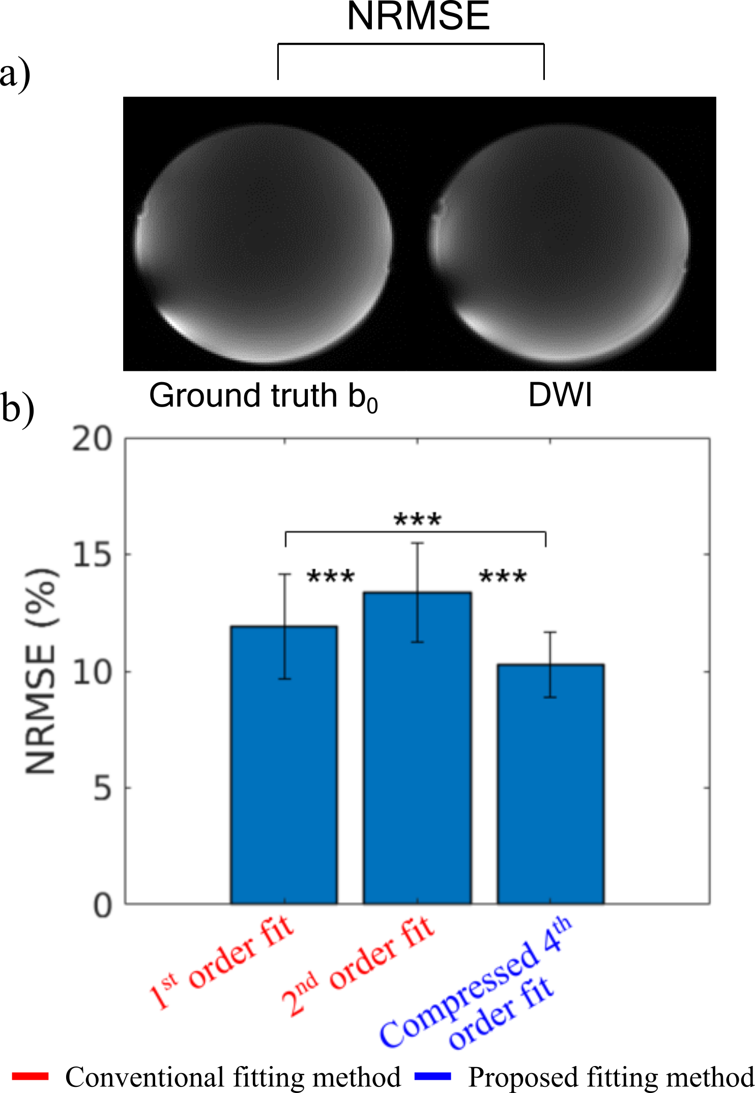
Figure 1 (a) Reconstructed ground truth b0 image and single-direction DWI of a phantom. Normalized-root-mean-squared-error (NRMSE) was calculated relative to b0 images over all 10 slices and 6 diffusion directions. NRMSE values averaged over all slices and directions for reconstructions informed with conventional 1st, 2nd, and new compressed 4th-order field dynamic fits. Error bars represent standard deviation of the mean across diffusion directions. p < 0.001
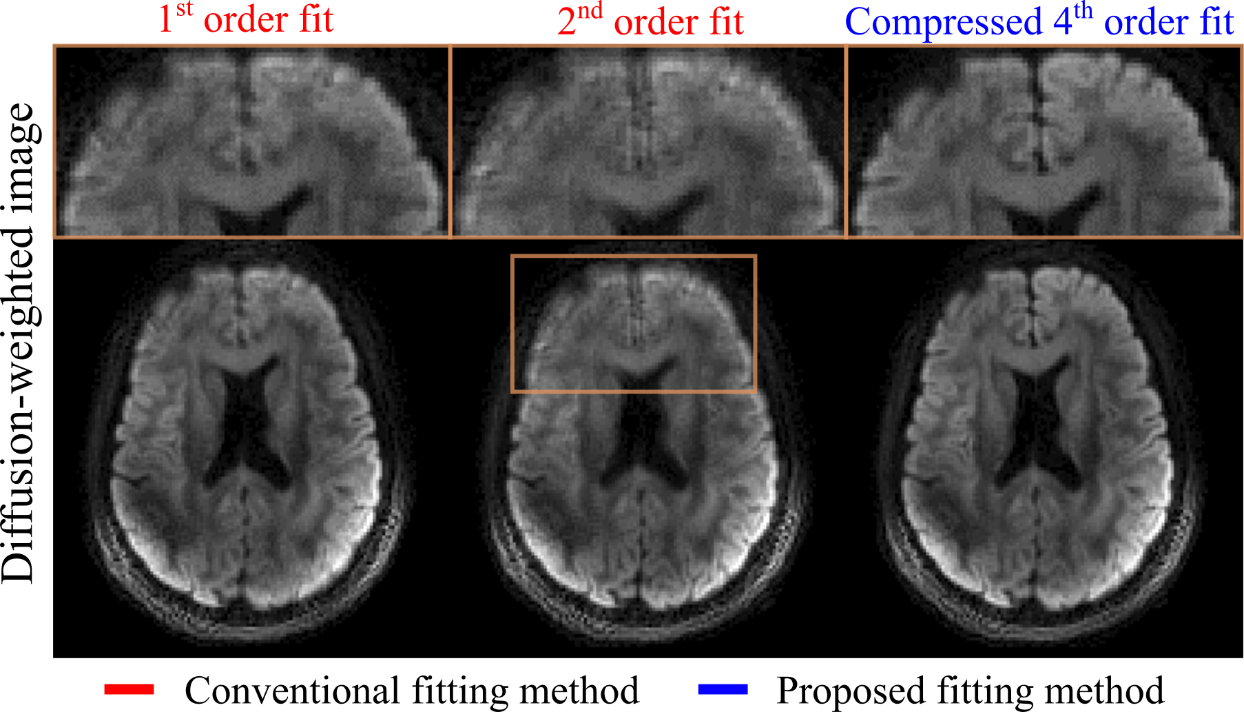
Figure 2 Single-direction diffusion-weighted images (DWI) reconstructed with 1st, 2nd, and new compressed 4th-order field dynamic fits. Zoom-in illustrates a reduction in blurring when implementing the proposed field dynamic fitting method, despite using the same number of total basis functions (9 basis functions) as the conventional 2nd-order fit, which is poorly conditioned.
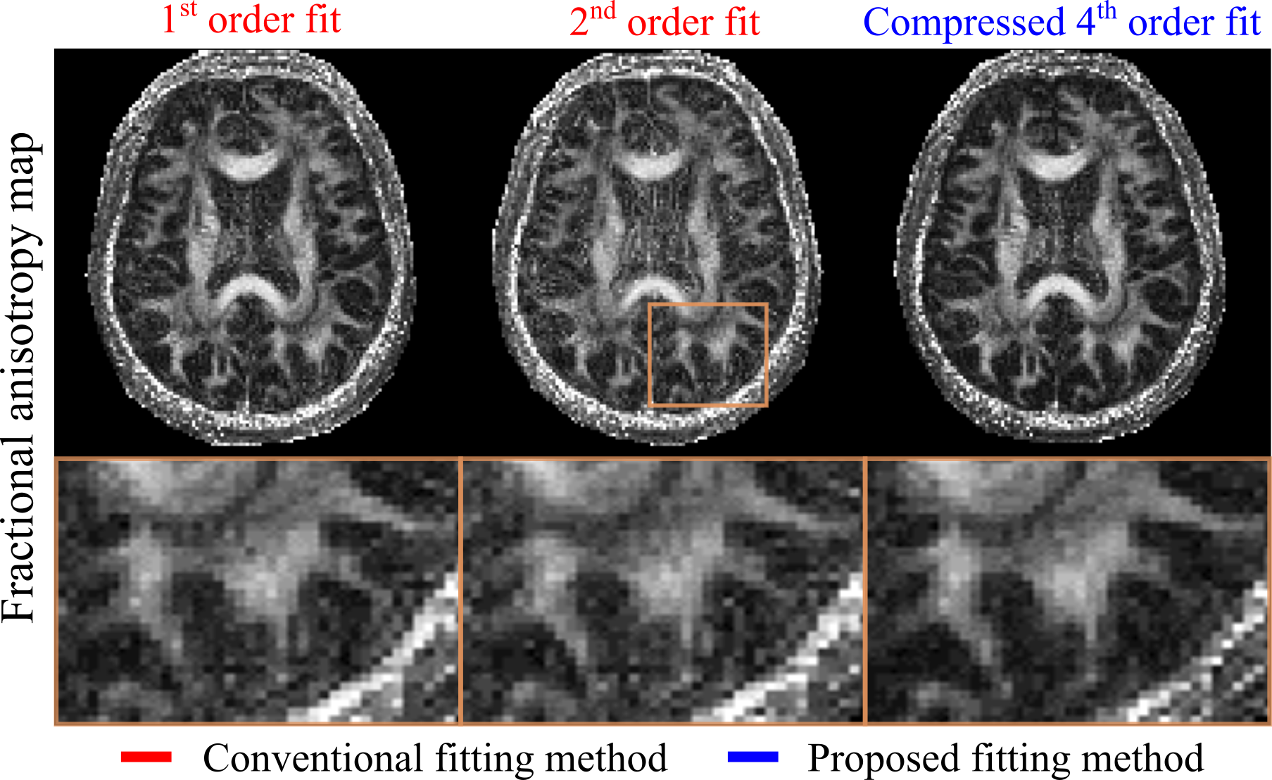
Figure 3 Computed fractional anisotropy (FA) map from DWI reconstructed with 1st, 2nd, and new compressed 4th-order field dynamic fits. Zoom-in highlighting the noise reduction and improved white matter delineation when implementing the proposed field dynamic fitting method, despite using the same number of total basis functions (9 basis functions) as the conventional 2nd-order fit, which is poorly conditioned.
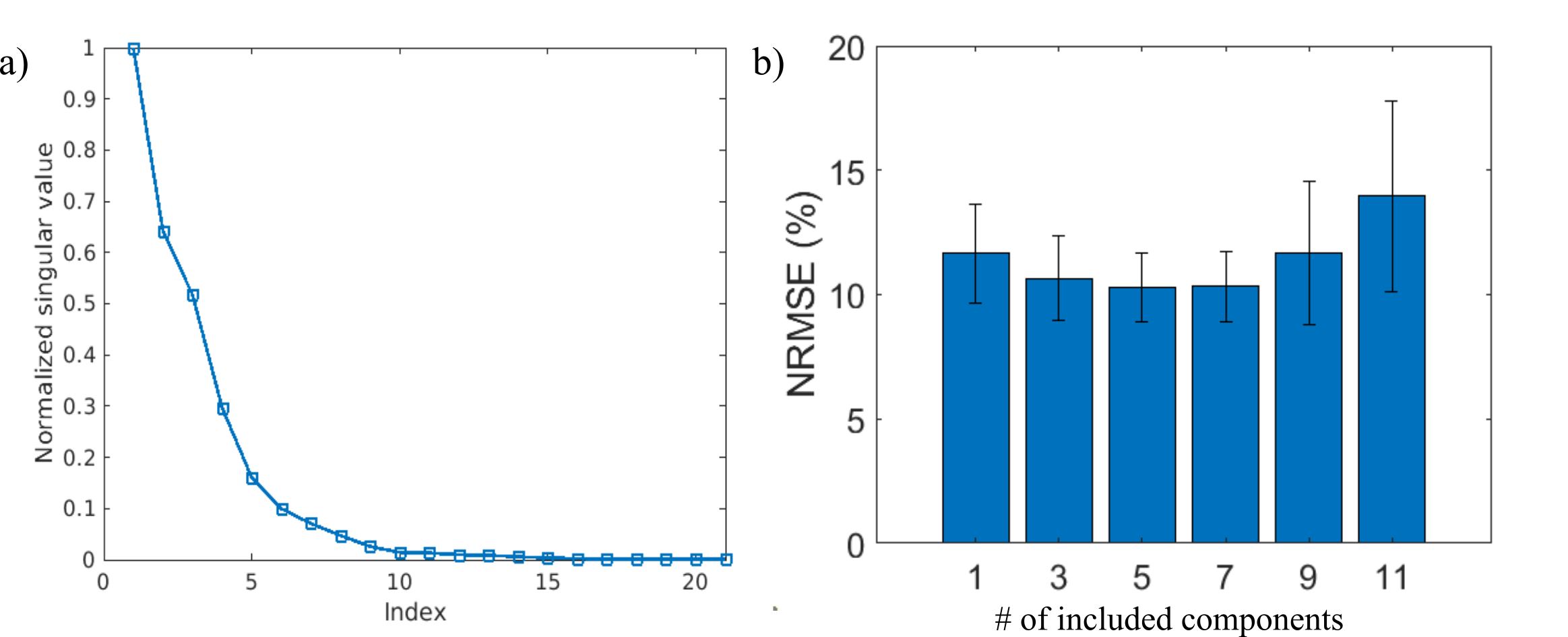
Figure 4 (a) Normalized singular values for all SVD components for the calibrated coefficient data. (b) Average NRMSE analysis when truncating the compression matrix C to the listed number of components. Incorporating few components leads to larger reconstruction errors due to a loss of substantial information, while preserving too many components, and therefore basis function terms, negatively impacts the probing matrix conditioning. This motivates using a moderate number of components, which ends up producing the lowest NRMSE and standard error, i.e. best image quality.
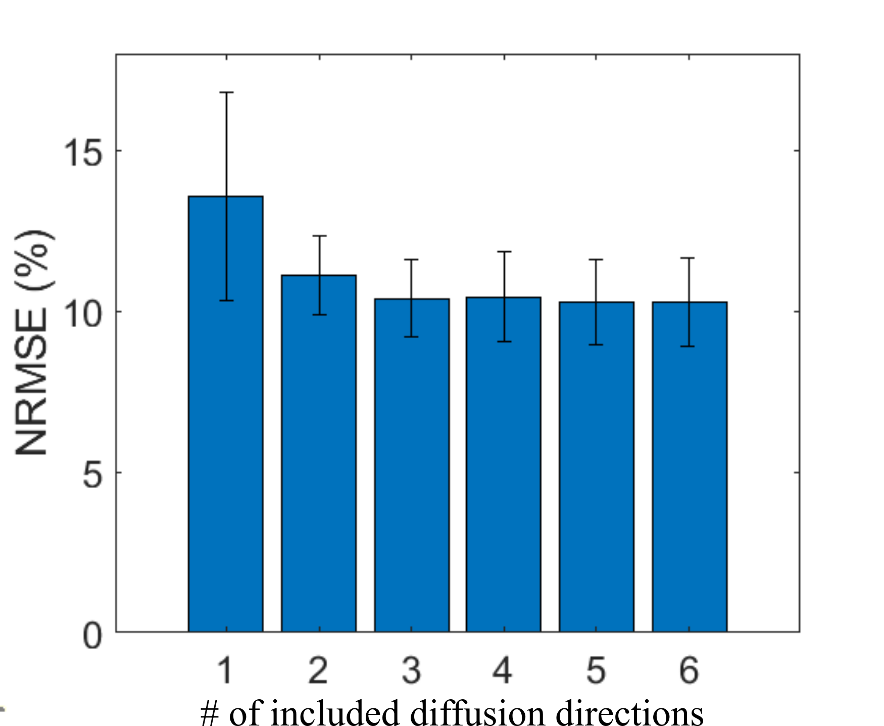
Figure 5 Average NRMSE values for DWI reconstructed with calibration data that was supplied with data from the listed number of diffusion directions. Providing a subset of diffusion directions prior to compression significantly reduces the NRMSE compared to supplying a single direction, as the compression weightings become more generalized. This is followed by an incremental reduction in NRMSE as more directions are included. However, it may be necessary to supply even more directions in order to accurately describe the basis functions across many arbitrary diffusion directions.