1254
Sequence Adaptive B1+ and B0 Field-imperfections Estimation (SAFE) for enhanced MRF quantification1Department of Radiology, Stanford University, Stanford, CA, United States, 2Department of Electrical Engineering, Stanford University, Stanford, CA, United States
Synopsis
Keywords: Artifacts, Machine Learning/Artificial Intelligence
Motivation: B1+ and B0 field-inhomogeneities can significantly reduce accuracy and robustness of MRF’s quantitative parameter estimates. Additional B1+ and B0 calibration scans can mitigate this but add scan time and cannot be applied retrospectively to previously collected data.
Goal(s): Here, we proposed a calibration-free sequence-adaptive deep-learning framework, to estimate and correct for B1+ and B0 effects of any MRF sequence.
Approach: We demonstrate its capability on arbitrary MRF sequences at 3T, where no training data were previously obtained.
Results: Such approach can be applied to any previously-acquired and future MRF-scans. The flexibility in directly applying this framework to other quantitative sequences is also highlighted.
Impact: Proposed method can estimate B1+ and B0 maps without calibration scan and be applied to arbitrary MRF sequence without new training data. It can be used retrospectively to improve quality of parameter maps of any previously-acquired or future MRF data.
Introduction
Magnetic Resonance Fingerprinting1 (MRF) is a widely-used efficient multiparameter mapping approach. However, significant reduction in accuracy and repeatability of the estimated tissue parameters can occur as a subject-and-scan-specific B1+ and B0 field inhomogeneity2,3. B1+ and B0calibration scans can be acquired to correct this issue, but cost of added scan time and inability to be applied retrospectively to previously collected data (as these field inhomogeneity vary between scan sessions). B0 estimation/correction via deep learning has been proposed4,5 for correction of non-cartesian acquisition. However, field map estimation and correction from highly under sampled time-series MRF data has not been attempted. Herein, we proposed a sequence adaptive deep learning framework (SAFE) to jointly estimate B1+, B0, distortion free T1, T2 and Proton Density (PD) maps from subspace coefficients in 3T MRF. Moreover, we demonstrate that our technique is seamlessly adaptable to any 3T MRF sequence and provide accurate field maps with no additional training data.Methods
Deep-learning-based B1 and B0 estimation: SAFE estimates field maps directly from the MRF subspace coefficient maps, with auxiliary task of generating (B1+ and B0 corrected) parameter maps added to improve the performance of the network. The structure is inspired by the U-Net9, SSIM was employed on the brain-masked area (parenchyma). Due to the smoothness of B1+ and B0, we down-sampled inputs to 2mm for training.Sequence-agnostic capability: SAFE relies on a set of previously-acquired T1, T2, PD, B1+, B0quantitative maps as training data, that can be acquired using any quantitative imaging approach. This data is then used to simulate the reconstructed subspace coefficient maps of the MRF sequence of interest for use as input to the network, where such simulation carefully accounts for B1+ and B0 effects on the coefficient maps through use of B1+-corrected dictionary model and spatial blurring of B0 in the spiral time-segmented modelling.
Data Acquisition all in vivo data were acquired using GE Premier 3T system with 32ch reception.
Calibration scan The ground-truth B1+, B0 maps were collect via Physical6 sequence with 1mm isotropic resolution.
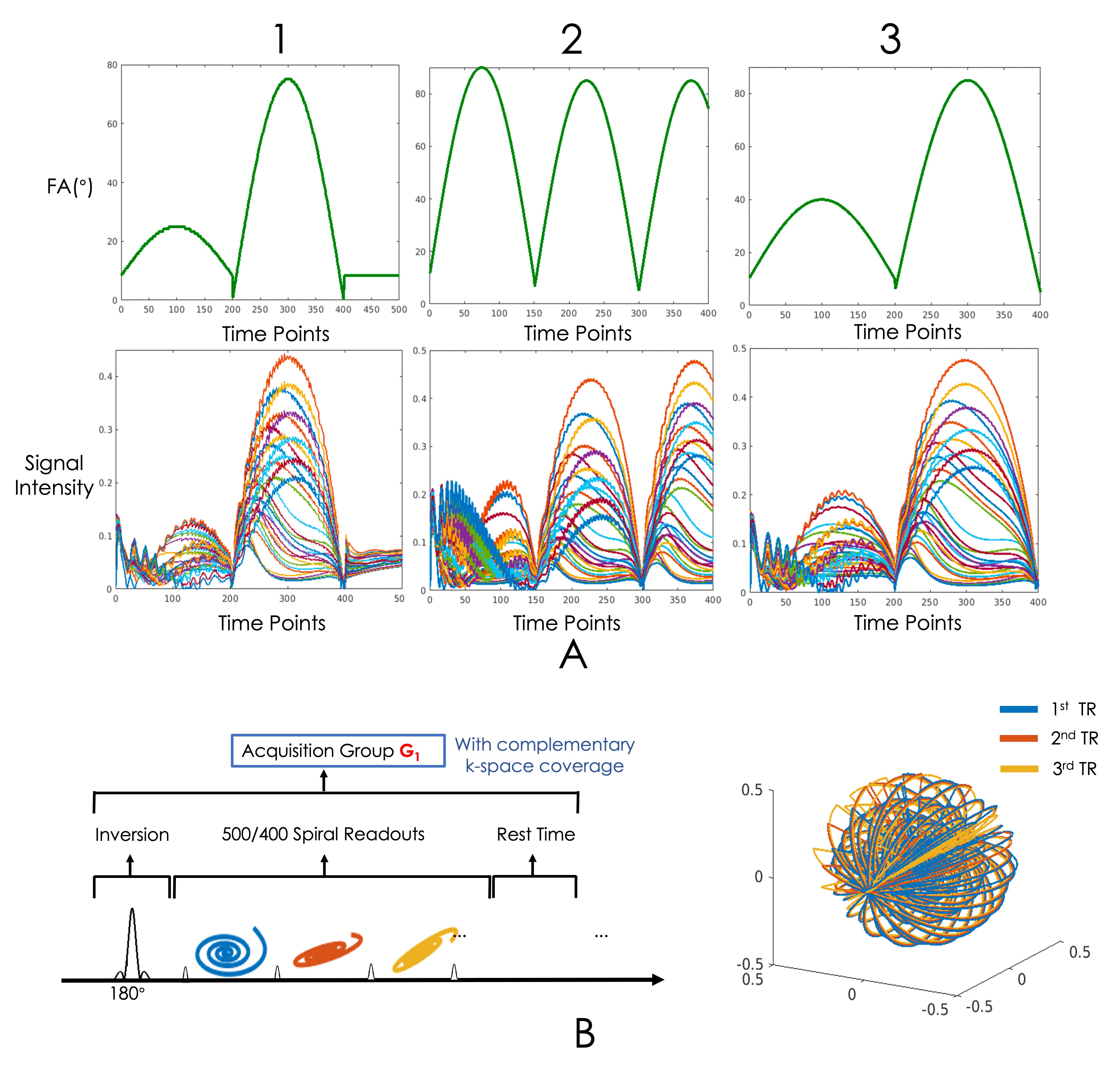
MRF acquisition Data were acquired on three unique MRF sequences, all utilizing the 3D-SPI-MRF7 acquisition. The flip-angle-trains for the three sequences (seq1,2,3) are shown in Figure 2A; with seq1 using an acquisition train with 500 TRs and 5.38ms spiral readouts, while seq2 and 3 uses 400TRs and 9ms spiral readouts. 7 healthy adults were scanned with seq to provide high-quality gold-standard 1mm isotropic resolution whole-brain data. Data were collected on two additional subjects using seq2 and 3, along with additional calibration scans for validation.
Ground truth generation for training data synthesis Subspace coefficient reconstruction and dictionary matching were performed to extract ground truth parameter maps from seq1 data. B0 and B1+ correction is performed via MFI8 and B1+-corrected dictionary matching respectively.
Application In addition to validation performed on healthy adult volunteers, we applied our network on on 8–13-year-old children’s data acquired at a collaborating site using seq1 from a longitudinal study for brain development .
Results
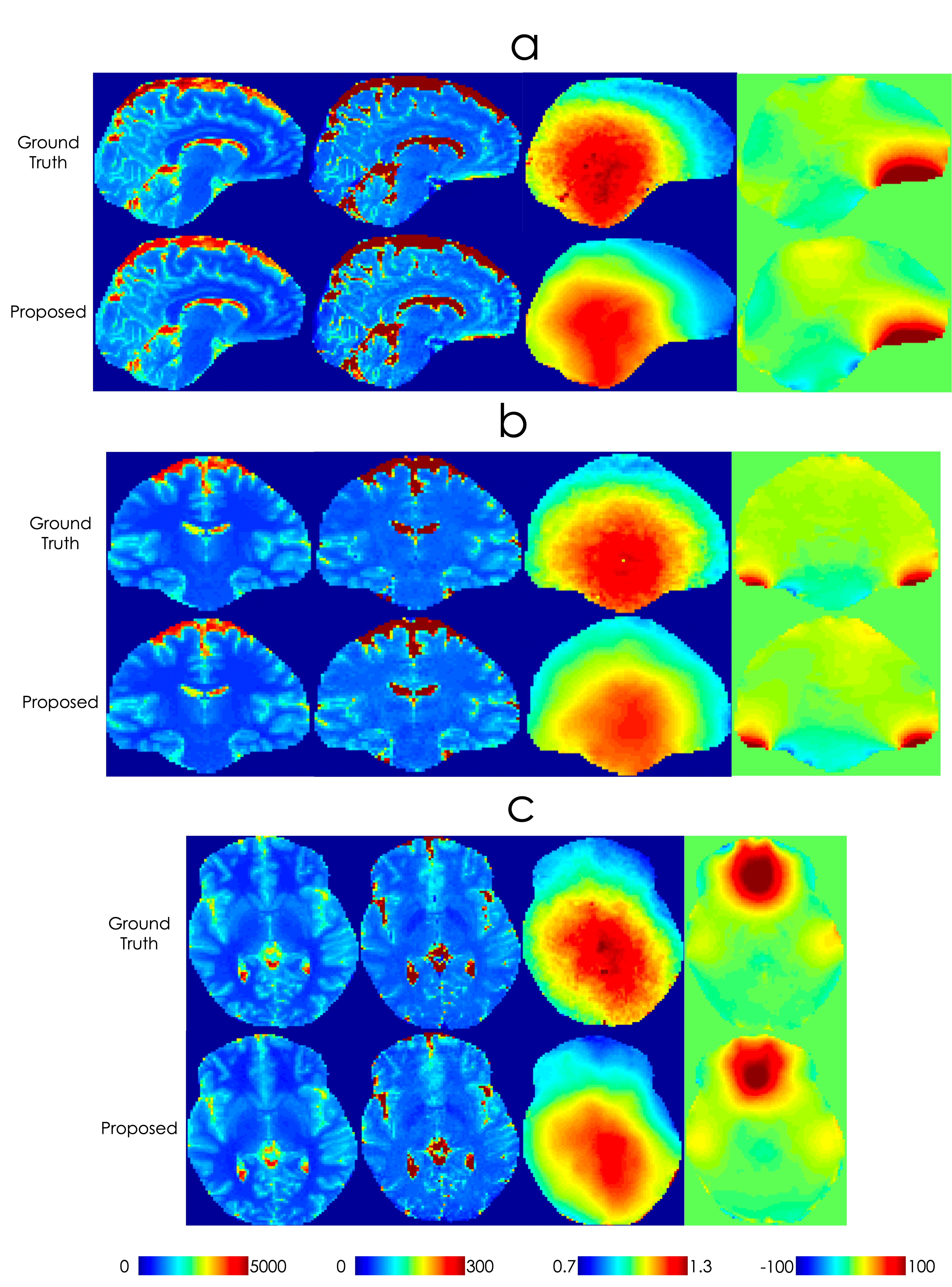
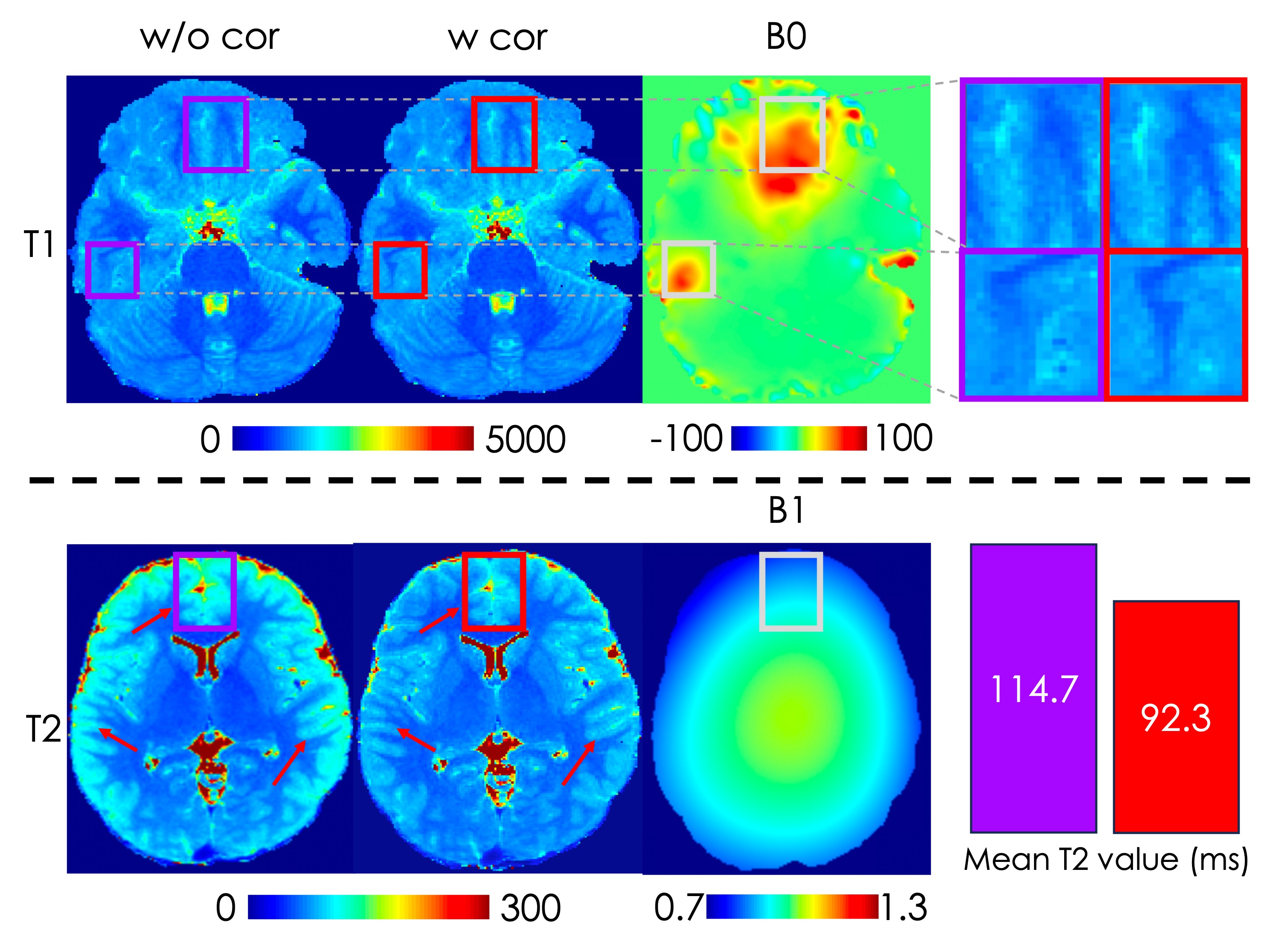
Fig3 shows representative field predictions and parameter estimates for seq1 obtained from the network in comparison to ground truth maps. The predicted field maps have high correspondence to ground truth maps, with some smoothing effect on B1+ map compared to ground truth obtain via the PhysiCal sequence.Fig4 shows the importance of B1+ and B0 corrections, in mitigating B0 blurring artifacts and T2 bias in region of low B1+. Here, the incorporation of B1 information caused a reduction in the estimated T2 value by 24.2% in the highlighted region.
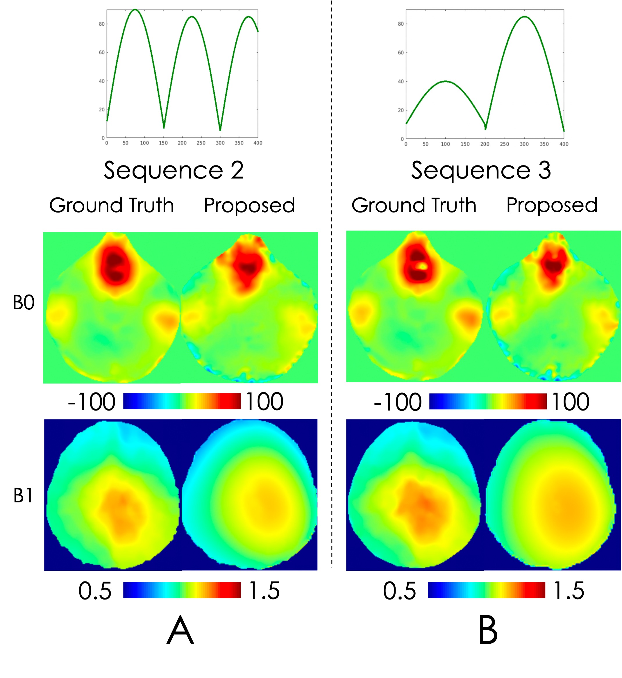
Fig5 shows the application of SAFE on sequence 2 and 3, where high quality field predictions are also obtained.
Discussion
SAFE exhibits the capability for high-quality estimation of B1+ and B0 maps across different MRF sequences. While using the convolutional layer allowed for capitalizing on spatial relations between voxels, larger receptive field improve network accuracy due to field map dependency on proximal regions.Conclusion
We demonstrate the ability of SAFE for robust and accurate B1+ and B0 field estimations on arbitrary MRF sequences. It should be feasible to estimate other field/system imperfections and apply to other quantitative imaging sequences such as EPTI10 and MR multitasking11. For applications to other magnetic field strengths and/or other organs, to get robust results, it will be important to train on B1+, B0, T1, T2 and PD that are in distribution to those applications, which can be obtained similarly here a good quantitative sequence for the specific application. Data augmentation approaches here could also further improve the robustness of our framework.Acknowledgements
This work was supported by NIH research grants: R01MH116173, R01EB019437, U01EB025162, P41EB030006, R01EB033206, U24NS129893.
References
1. Ma D, Gulani V, Seiberlich N, Liu K, Sunshine JL, Duerk JL, Griswold MA. Magnetic resonance fingerprinting. Nature. 2013 Mar 14;495(7440):187-92. doi: 10.1038/nature11971. PMID: 23486058; PMCID: PMC3602925.
2. Chen Q, Cao X, Liao C, et al. “Towards accurate and repeatable 1mm isotropic whole-brain MRF quantification using a 1-minute scan with optimized processing pipeline”. ISMRM 2023 P3477.
3. Ma D, Coppo S, Chen Y, et al. Slice profile and B1 corrections in 2D magnetic resonance fingerprinting[J]. Magnetic resonance in medicine, 2017, 78(5): 1781-1789.
4. Goyeneche A, Ramachandran S, Wang K, et al. “ResoNet: Physics Informed Deep Learning based Off-Resonance Correction Trained on Synthetic Data”. ISMRM 2022 P0555.
5. Salifu M, Haskell M, Noll D. “Estimating B0 changes in Oscillating Steady State Imaging (OSSI) using an Artificial Neural Network”. ISMRM 2022 P2352.
6. Iyer S, Liao C, Li Q, et al. “PhysiCal: A rapid calibration scan for B0, B1+, coil sensitivity and Eddy current mapping”. ISMRM 2020 P0661
7. Cao X, Ye H, Liao C, et al. Fast 3D brain MR fingerprinting based on multi‐axis spiral projection trajectory[J]. Magnetic resonance in medicine, 2019, 82(1): 289-301.
8. Ostenson J, Robison RK, Zwart NR, Welch EB. Multi-frequency interpolation in spiral magnetic resonance fingerprinting for correction of off-resonance blurring. Magn Reson Imaging. 2017 Sep;41:63-72. doi: 10.1016/j.mri.2017.07.004. Epub 2017 Jul 8. PMID: 28694017; PMCID: PMC5612382.
9. Ronneberger O, Fischer P, Brox T. U-net: Convolutional networks for biomedical image segmentation[C]//Medical Image Computing and Computer-Assisted Intervention–MICCAI 2015: 18th International Conference, Munich, Germany, October 5-9, 2015, Proceedings, Part III 18. Springer International Publishing, 2015: 234-241.
10.Wang F, Dong Z, Reese T G, et al. Echo planar time‐resolved imaging (EPTI)[J]. Magnetic resonance in medicine, 2019, 81(6): 3599-3615.
11.Christodoulou AG, Shaw JL, Nguyen C, Yang Q, Xie Y, Wang N, Li D. Magnetic resonance multitasking for motion-resolved quantitative cardiovascular imaging. Nat Biomed Eng. 2018 Apr;2(4):215-226. doi: 10.1038/s41551-018-0217-y. Epub 2018 Apr 9. PMID: 30237910; PMCID: PMC6141200.
Figures
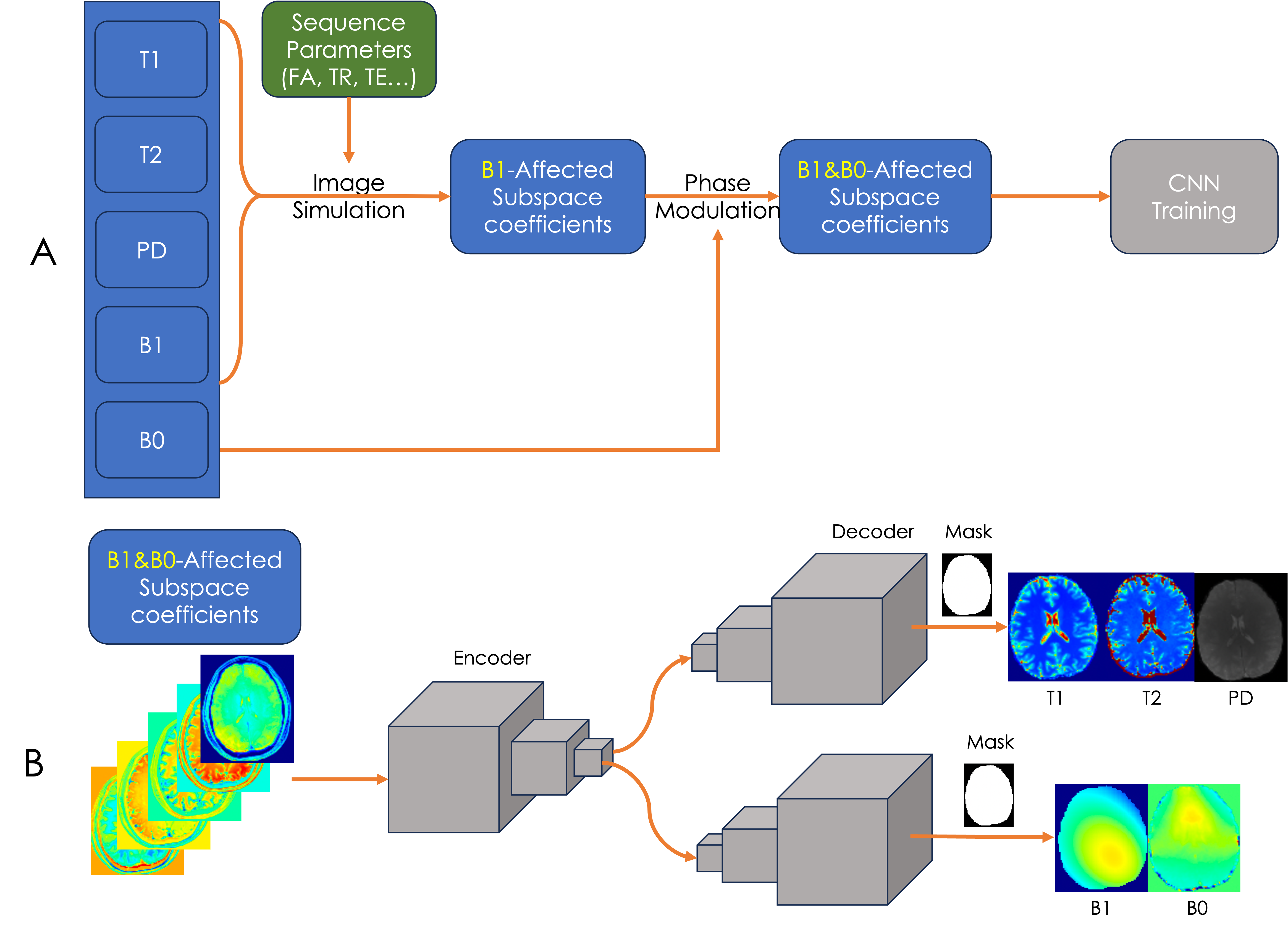
(B) Detailed network training structure. The network block consists of convolutional, instance normalization and activation layers, with skip connections between encoders and decoders.