1251
Dynamic Estimation of Respiration-Induced B0 Inhomogeneities in OSSI fMRI: A Novel Framework Using FIDNavs and SENSE Maps1Biomedical Engineering, University of Michigan, Ann Arbor, MI, United States
Synopsis
Keywords: Artifacts, System Imperfections: Measurement & Correction, Artifacts, Physiological noise correction, FID navigators, fMRI
Motivation: In OSSI fMRI, temporal fluctuations in the B0 field, predominantly resulting from respiration, can induce changes to the steady state, subsequently diminishing the temporal signal-to-noise ratio (tSNR)
Goal(s): Our objective is to develop an efficient technique for estimating respiration-induced B0 variations in OSSI fMRI
Approach: We have used Free Induction Decay (FID) frequency offset to estimate the first-order field inhomogeneities. A novel element in our approach lies in adopting a spatial encoding strategy for these FIDs, drawing on the geometric centroids of each coil's sensitivity profile
Results: Our initial findings indicates comparable results between our FIDNavs method and image-based field map method.
Impact: This approach allows for a rapid measurement of B0 variations, thus facilitating faster real-time corrections. This method bypasses the lengthy process of calibration or the need for reference images.
Introduction
Oscillating Steady State Imaging (OSSI) is a $$$ T_2^*$$$ weighted MRI sequence that employs quadratic RF phase increments in combination with balanced gradients1. This approach generates a high SNR signal exhibiting an oscillation period defined by $$$n_c\times TR$$$. By averaging over $$$n_c$$$ periods, it is possible to eliminate oscillations and mitigate $$$B_0$$$-dependent banding artifacts, enhancing the utility of OSSI for high-resolution functional MRI (fMRI). However, OSSI sensitivity to physiological noise-induced $$$B_0$$$ field variations, particularly from respiration, presents a significant challenge. This sensitivity arises from the non-linear dependence of the OSSI signal phase on off-resonance frequency, leading to a complex, temporal response that is not well-corrected by traditional gradient echo (GRE) based off-resonance correction methods2. FID navigators (FIDNavs) seem a fitting choice for addressing this issue due to their modest technological requirements when integrated with OSSI. Nevertheless, one hurdle is that the FID represents the volume integral of the MRI signal across the excited volume, obscuring the spatial distribution of $$$B_0$$$ inhomogeneities. Present techniques aiming to correct $$$B_0$$$ field fluctuations using FIDNavs necessitate an additional calibration scan or a reference image3, which can be time-consuming. Given these limitations, our research aims to investigate the efficacy of leveraging FID frequency offsets for estimating first-order shim inhomogeneity coefficients. This approach spatially encodes the FIDs via the geometric centroids of the sensitivity maps.Methods
Our method uses the FID frequency offset calculated from FIDNavs acquired at two different echo times and spatial data derived from the intensity-weighted centroids of coil sensitivity maps. The FID frequency offsets are a reliable indicator for identifying any local magnetic field deviations. At the same time, the geometric centroids of each coil's sensitivity maps offer the necessary spatial encoding (Figure 1C). As a result, this approach effectively addresses any spatial information inadequacy present in the FID signals.Our mathematical approach is shown in Figure 1D. We estimated the zeroth- and first-order coefficients by solving the inverse problem using weighted least squares. Coefficients estimated with our method are compared against those obtained from conventional spherical harmonics (SH) fitting to field maps acquired from double-echo spiral images.
[OSSI Sequence]
This approach was evaluated using a stack of spirals OSSI sequences with a double-echo readout (Figure 1A). An additional 1ms long FID was placed before and after the spiral readout. The double-echo spiral was used to generate field maps for comparison with our FIDNav results. Scan parameters are shown in the table in Figure 1B.
[Phantom Validation]
A phantom was scanned by changing the X and Y gradient settings on the scanner. Specifically, currents to the X and Y gradients were manually increased by a factor of two every 20s. This led to a step-wise change in the local magnetic field.
[In vivo Validation]
OSSI fMRI images were acquired where the subject was asked to perform a finger-tapping task (20s on/off for 200s) under regular and deep breathing conditions.
Results
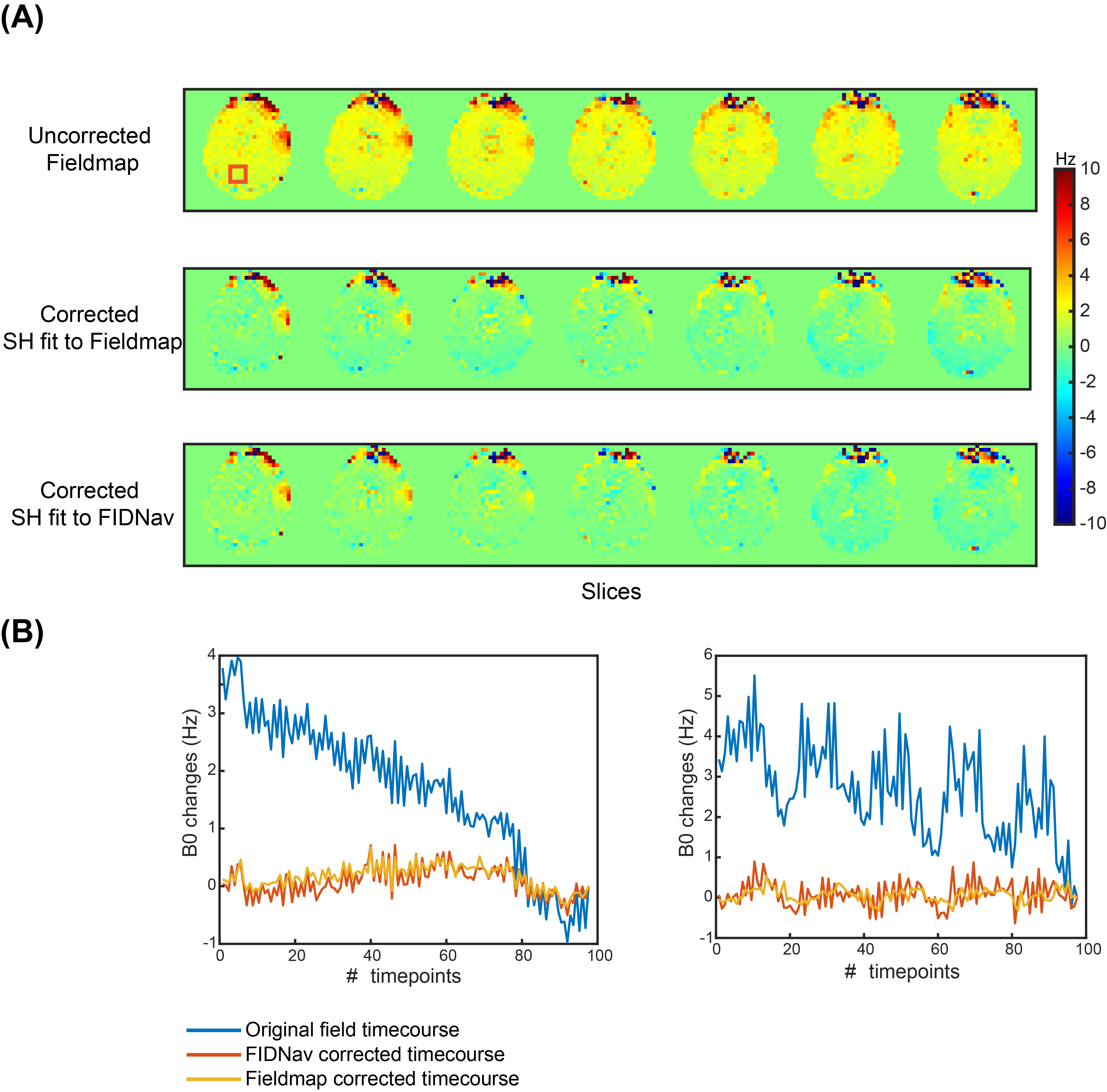
In Figure 2A, we compare the frequency offset obtained from the FIDNavs for all coils with the field maps from the double-echo spiral readout. In Figure 2B, we compare the inhomogeneity coefficients estimated from the FIDNavs with those calculated from the field map during the phantom experiments.Figure 3A compares the uncorrected field map and $$$B_0$$$ field correction achieved using the FIDNavs and field map coefficients across all slices. Voxel time courses are in Figure 3B.
In Figure 4, we compare the uncorrected field map with $$$B_0$$$ field correction based on the FIDNavs and field map coefficients. This is shown across different fMRI time series and breathing conditions.
Discussion & Conclusion
We have introduced a $$$B_0$$$ estimation method that leverages FID frequency offsets, spatially encoded using sensitivity maps, for calculating respiration-induced changes in OSSI fMRI.Our initial results demonstrate a near-optimal performance compared to the traditional image-based field map methods, with only minor residual errors observed. These may originate from gradient issues, as spiral trajectories are particularly sensitive to gradient system imperfections. Therefore, our subsequent attention will be directed toward characterizing and correcting these gradient system imperfections.
Importantly, this method circumvents the need for additional reference images or time-consuming calibration scans, paving the way for rapid, real-time estimation of dynamic $$$B_0$$$ variations. Based on these promising findings, subsequent research endeavors will be dedicated to refining and optimizing this methodology.
Acknowledgements
We wish to acknowledge the support of NIH Grants U01EB026977.References
[1] Shouchang Guo and Douglas C Noll. Oscillating steady-state imaging (ossi): A novel method for functional mri. Magnetic Resonance in Medicine,84(2):698–712, 2020.
[2] Amos A Cao and Douglas C Noll. A retrospective physiological noise correction method for oscillating steady-state imaging. Magnetic Resonance in Medicine, 85(2):936–944, 2020.
[3] Tess E Wallace, Onur Afacan, Tobias Kober, and Simon K Warfield. Rapid measurement and correction of spatiotemporal B0 field changes using fid navigators and a multi-channel reference image. Magnetic resonance inmedicine, 83(2):575–589, 2020.
Figures

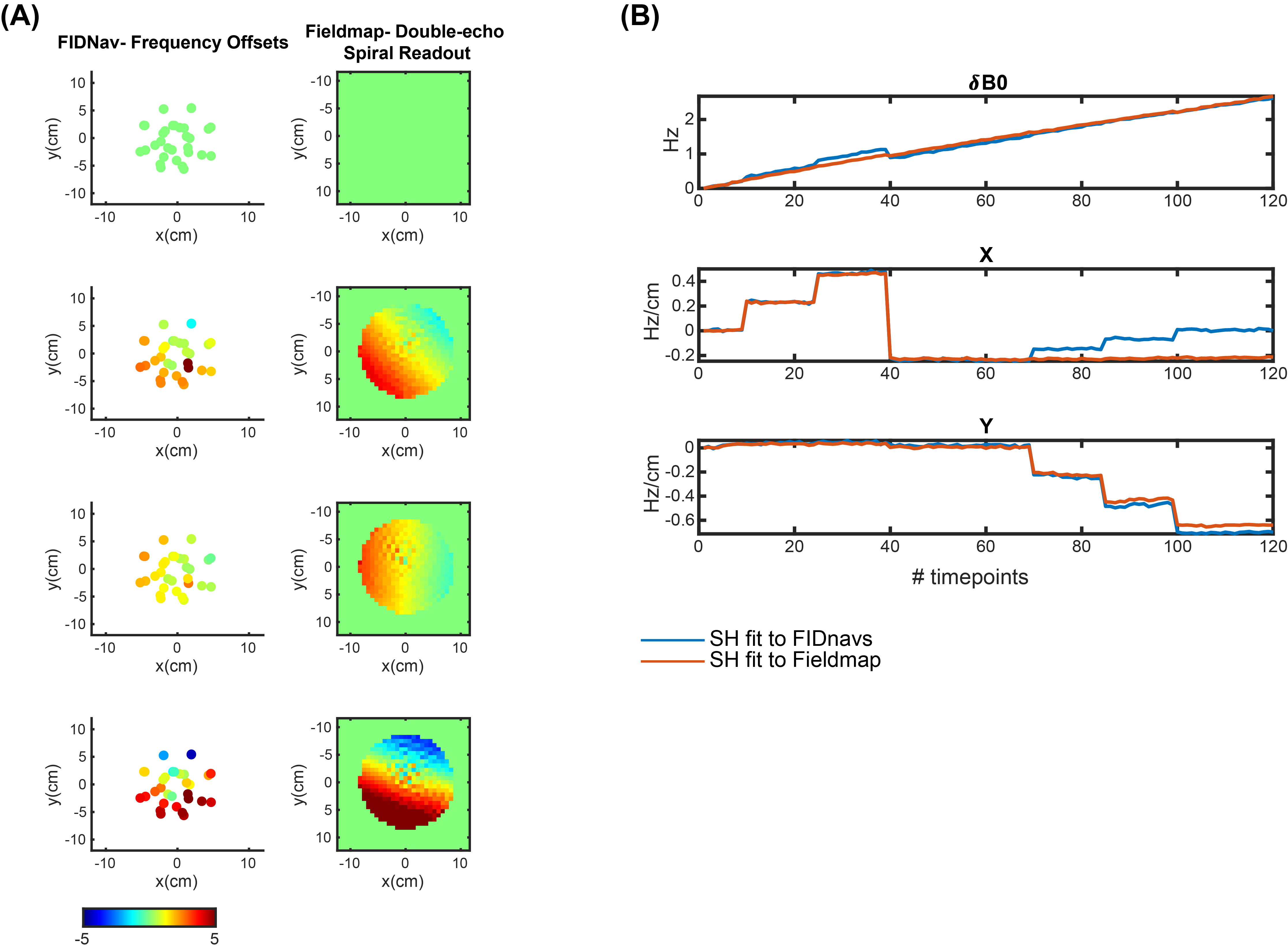
The phantom experiments show similarities between frequency offsets obtained from the FIDNavs and those estimated from the field map, as seen in (A). Exceptions were apparent with a few low-sensitivity coils, which consequently received less weighting during the least squares approximation of the coefficients.(B) shows the estimated coefficients after manual changes to the X and Y gradient settings. Here, the FIDNav coefficients demonstrated a close correlation with the field map coefficients, with a marginal exception observed in the X coefficients.