1250
Joint suppression of cardiac bSSFP cine banding and flow artifacts based on twofold phase-cycling and a dual-encoder neural network1Shanghai Jiao Tong University, Shanghai, China, 2United Imaging Healthcare Co., Ltd, Shanghai, China
Synopsis
Keywords: Artifacts, Artifacts, Cardiac function, Cine
Motivation: Cardiac bSSFP cine imaging suffers from banding and flow artifacts caused by the off-resonance. Although fourfold phase cycling suppresses the banding artifacts, it invokes flow artifacts and prolongs the scan.
Goal(s): To develop a twofold phase-cycling sequence with a neural-network-based reconstruction for a fast and joint suppression of banding and flow artifacts in cardiac cine imaging.
Approach: We compared the method with standard bSSFP and regular phase cycling in the left ventricle and atrium in 10 healthy subjects.
Results: Needing only 10 heartbeats, the proposed method robustly suppressed both artifacts in the presence of anatomical variations.
Impact: Banding and flow artifacts are common in bSSFP cine imaging, especially with cardiac devices or high-field MR. The proposed method provides a robust and practical tool for suppression of them and improves the reliability of cine MRI.
Introduction
Removing banding and flow artifacts from bSSFP cine imaging is desirable for accurate assessments of the left ventricular (LV)1,2 and left atrial (LA)3,4 function. Banding artifacts can be suppressed by fourfold phase-cycling (4PC)5–8. However, the method not only quadruples the scan time but also exacerbates flow artifacts by shifting dark bands into the flow regions9–12. Partial dephasing13 has been combined with 4PC to reduce both flow and banding artifacts14. However, this sequence needed a breath-hold of 21 heartbeats, and residual artifacts were still reported. A neural network has been reported as a post-processing method to reduce the two artifacts for standard bSSFP cine15. Here we propose a novel method that combines a twofold phase-cycled bSSFP cine sequence and a neural-network-based reconstruction to achieve a robust suppression of the two artifacts in an efficient 10-heartbeat scan.Methods
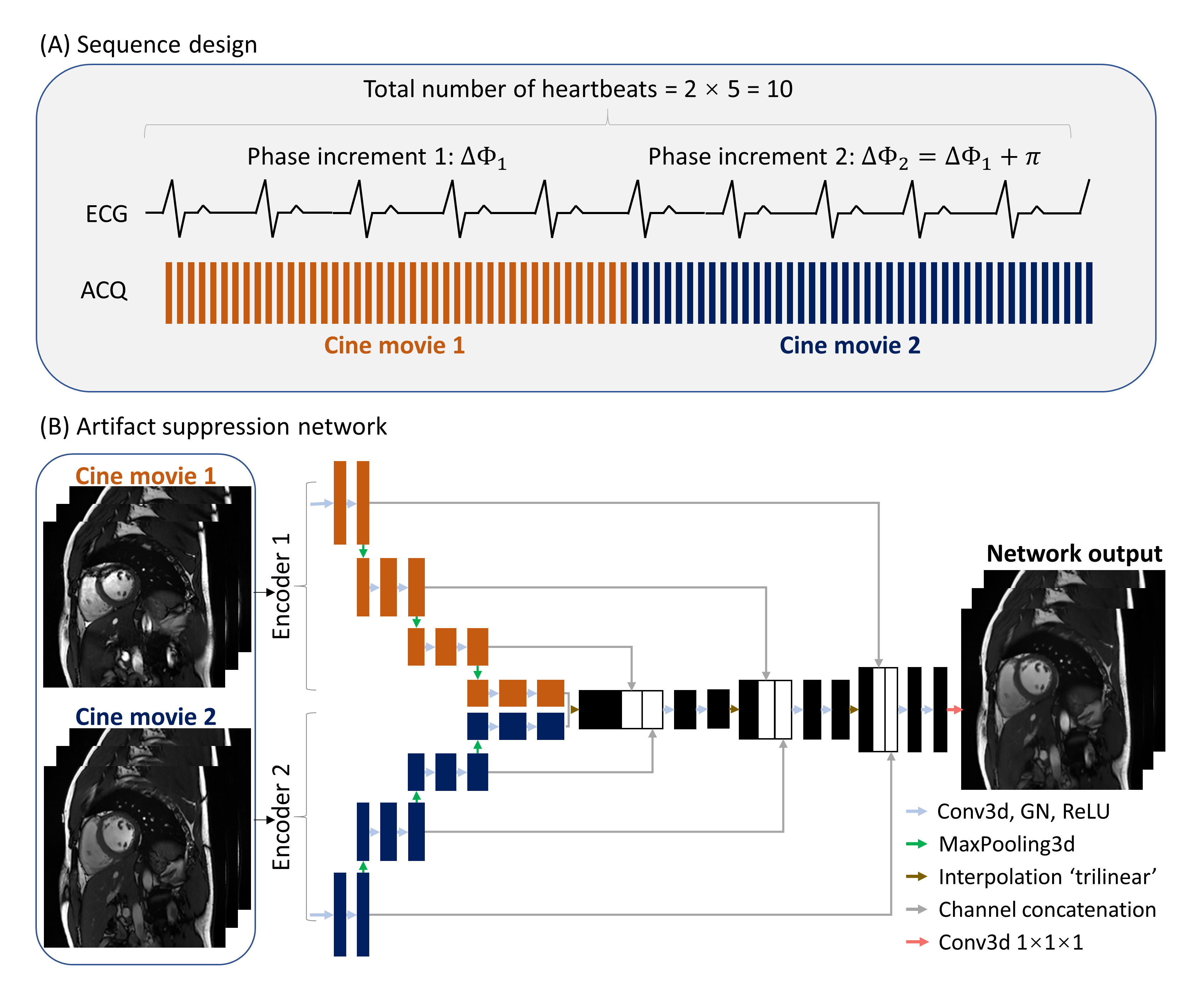
Figure 1 shows a schematic of the proposed method. The imaging sequence sequentially acquires two cine movies with half-cycle-apart RF phase increments. Since acquiring each movie takes 5 heartbeats, the entire sequence lasts 10 heartbeats. The reconstruction is based on a 3-dimensional (2D+time) dual-encoder U-Net, which differs from standard U-Net16 in that two separate encoders are involved, each processing a single movie in the input. The dual-encoder architecture improved the capability of the network to encode variable artifact patterns of the two input movies, leading to a better artifact suppression performance.To train the network, we scanned 18 healthy subjects in a 3T scanner (uMR 790, United Imaging Healthcare, Shanghai, China) with standard torso and spine coils. The institutional review board approved the study, and all subjects provided written informed consent. For each subject, we acquired 3 short-axis LV cine movies, each with 12 different RF phase increments uniformly spaced between 0° and 360°, generating 6 pairs of movies with half-cycle-apart frequencies. Together, 324 pairs of LV movies were generated to train the network. The training labels were provided by Short-range Phase Cycling (SPC)15, which linearly combines movies of phase increments 120°, 150°, 180°, 210°, and 240° and has a better balance between suppressing the banding artifacts and preventing incurrence of new flow artifacts.
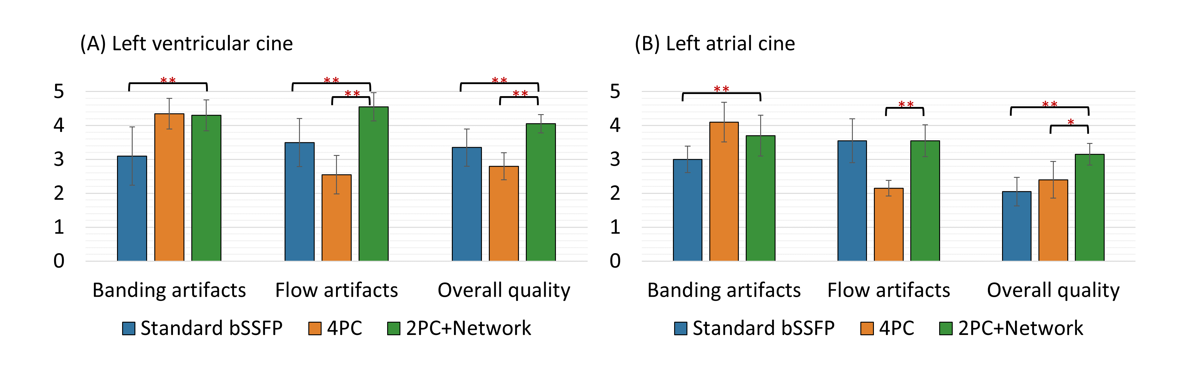
To test the proposed cine imaging method, we applied it to another group of 10 healthy subjects. For each subject, we acquired 3 short-axis LV slices and 2 transversal LA slices with 6 different combinations of phase increments. We compared our method with standard bSSFP and 4PC (phase increments=0°, 90°, 180°, and 270°) qualitatively by 2 experienced readers using a 5-point Likert scale via Wilcoxon signed-rank test.
Results
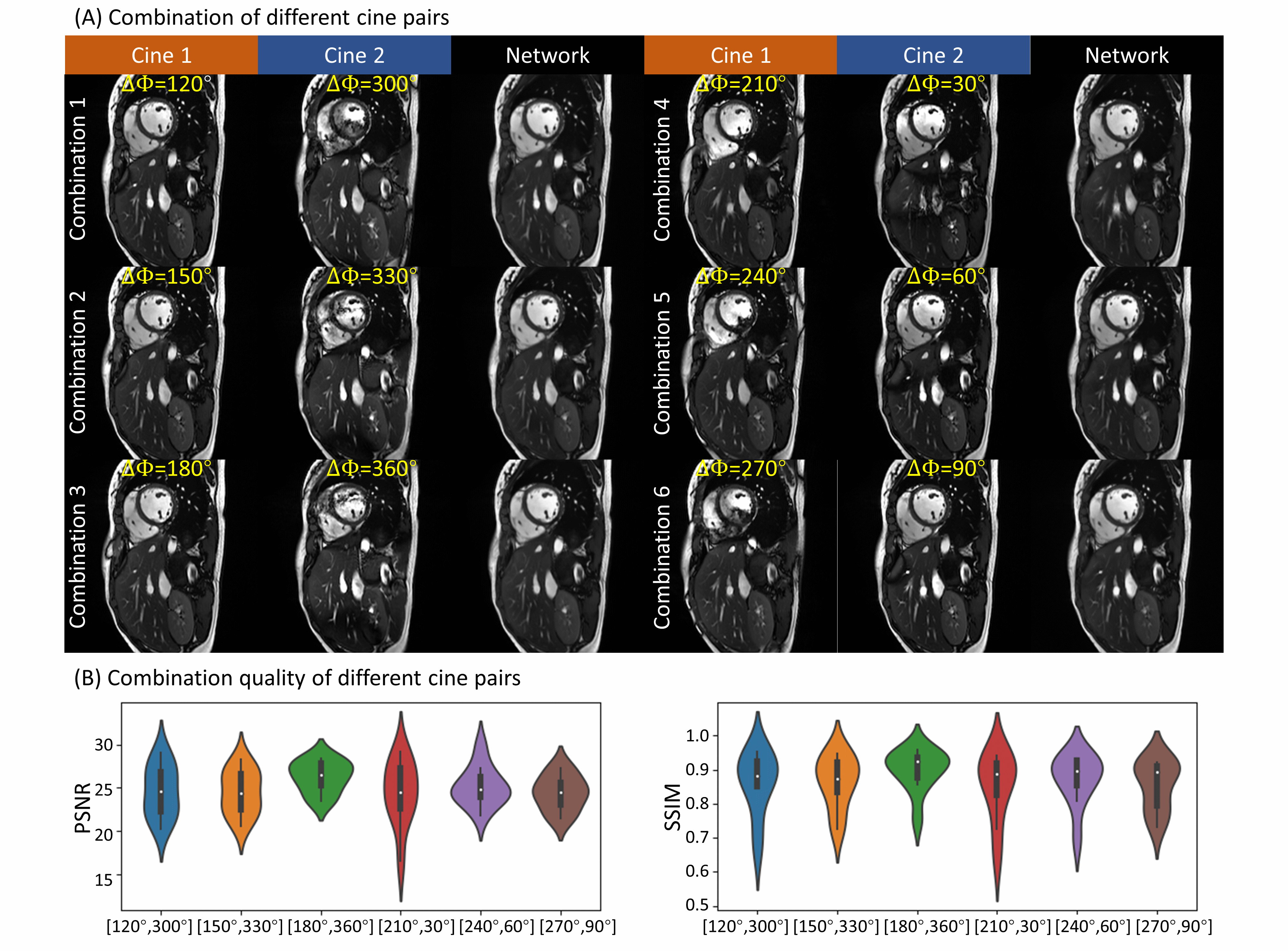
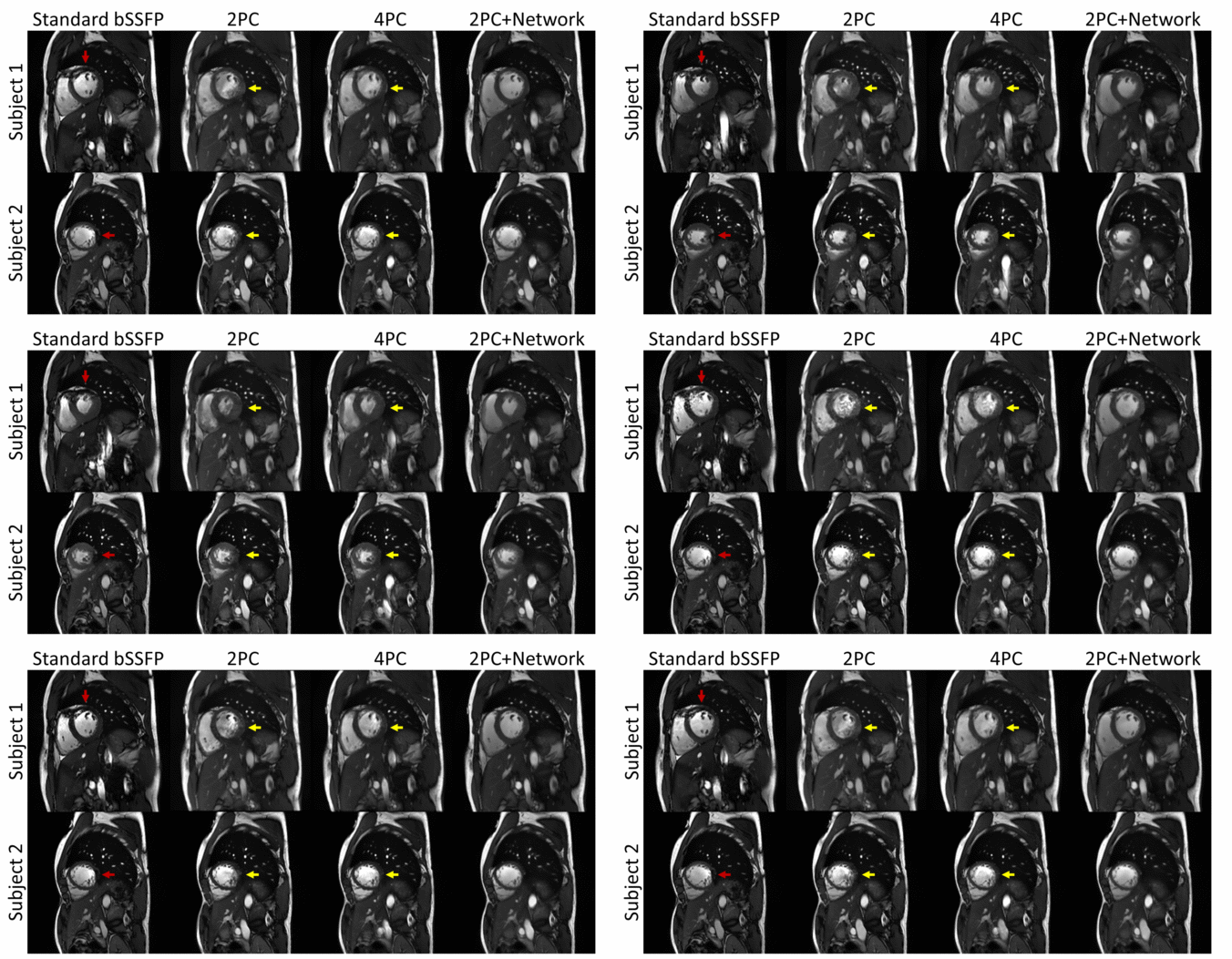
Figure 2 shows reconstruction results of the technique for 6 different phase combinations. The network suppressed the artifacts in the original images. Similar PSNRs and SSIMs were obtained at different phase combinations, suggesting that the method was robust against shifts of the off-resonance. In the following, we fixed the input phase increments to be 270° and 90°.Figure 3 shows the standard bSSFP, 2PC, 4PC, and the proposed method in the LV of two subjects. The standard cine had banding and flow artifacts. While 2PC and 4PC reduced the banding artifacts, they also enhanced the flow artifacts. On the contrary, our method achieved a joint reduction of both artifacts.
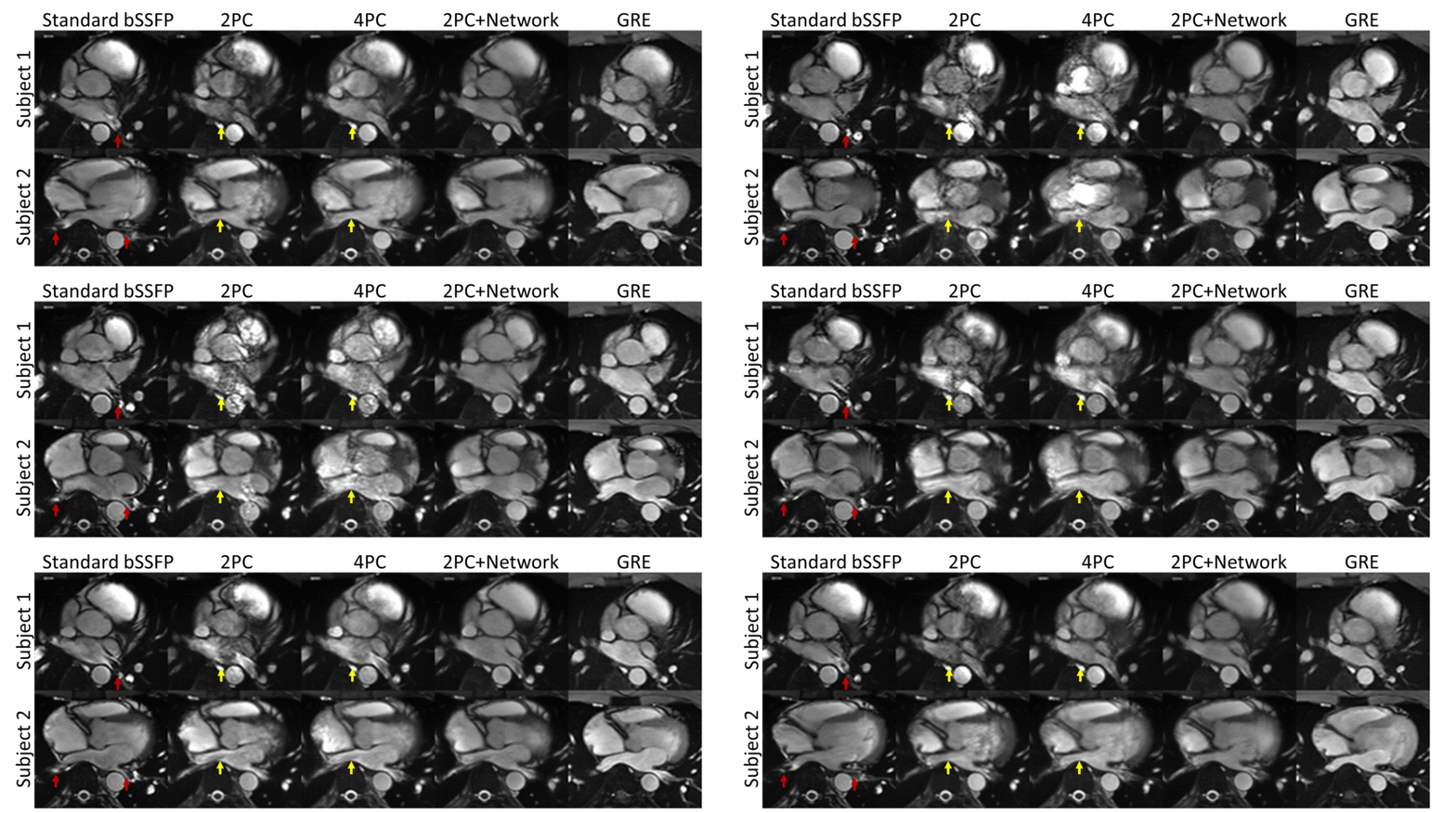
Figure 4 compares the previous four methods and GRE cine in the LA of two subjects. The banding artifacts obscured the pulmonary veins in the standard bSSFP cine. While this was improved by 2PC and 4PC, the flow artifacts severely worsened their image qualities. The proposed method achieved the best artifact suppression and good conspicuity of the pulmonary veins, similar to the GRE cine. Importantly, our method was not trained in the left atrium, indicating its robustness against anatomical changes.
Figure 5 shows qualitative comparisons of the standard bSSFP, 4PC, and our method in both the LV and LA. Our method significantly reduced banding artifacts relative to the standard bSSFP, and flow artifacts relative to 4PC, and significantly improved the overall image quality relative to both methods.
Discussion and Conclusions
The proposed 2PC method with network-based reconstruction achieved a joint suppression of banding and flow artifacts and manifested a good generalizability against anatomical variations. Furthermore, the scan time of the method was only half of 4PC. Although still longer than the standard cine, 10 heartbeats is a reasonable time for breath-holding and can be further reduced by acceleration techniques. Clinical translation of the method is warranted and may render cine imaging a more robust means for cardiac functional assessment.Acknowledgements
No acknowledgements found.References
1. Schär M, Kozerke S, Fischer SE, Boesiger P. Cardiac SSFP imaging at 3 Tesla. Magn Reson Med. 2004;51(4):799-806. doi:10.1002/mrm.20024
2. Scheffler K, Lehnhardt S. Principles and applications of balanced SSFP techniques. Eur Radiol. 2003;13(11):2409-2418. doi:10.1007/s00330-003-1957-x
3. Hu P, Stoeck CT, Smink J, et al. Noncontrast SSFP pulmonary vein magnetic resonance angiography: Impact of off-resonance and flow. J Magn Reson Imaging. 2010;32(5):1255-1261. doi:10.1002/jmri.22356
4. Robb JS, Hu C, Peters DC. Interleaved, undersampled radial multiple-acquisition steady-state free precession for improved left atrial cine imaging. Magn Reson Med. 2020;83(5):1721-1729. doi:10.1002/mrm.28036
5. Vasanawala SS, Pauly JM, Nishimura DG. Linear combination steady-state free precession MRI. Magn Reson Med. 2000;43(1):82-90. doi:10.1002/(SICI)1522-2594(200001)43:1<82::AID-MRM10>3.0.CO;2-9
6. Bangerter NK, Hargreaves BA, Vasanawala SS, Pauly JM, Gold GE, Nishimura DG. Analysis of multiple-acquisition SSFP. Magn Reson Med. 2004;51(5):1038-1047. doi:10.1002/mrm.20052
7. Jung KJ. Synthesis methods of multiple phase-cycled SSFP images to reduce the band artifact and noise more reliably. Magn Reson Imaging. 2010;28(1):103-118. doi:10.1016/j.mri.2009.05.045
8. Çukur T. Accelerated Phase-Cycled SSFP Imaging With Compressed Sensing. IEEE Trans Med Imaging. 2015;34(1):107-115. doi:10.1109/TMI.2014.2346814
9. Markl M, Alley M t., Elkins C j., Pelc N j. Flow effects in balanced steady state free precession imaging. Magn Reson Med. 2003;50(5):892-903. doi:10.1002/mrm.10631
10. Storey P, Li W, Chen Q, Edelman RR. Flow artifacts in steady-state free precession cine imaging. Magn Reson Med. 2004;51(1):115-122. doi:10.1002/mrm.10665
11. Markl M, Pelc NJ. On flow effects in balanced steady-state free precession imaging: Pictorial description, parameter dependence, and clinical implications. J Magn Reson Imaging. 2004;20(4):697-705. doi:10.1002/jmri.20163
12. Lagerstrand K m., Plewes D b., Vikhoff-Baaz B, Forssell-Aronsson E. Flow-induced disturbances in balanced steady-state free precession images: Means to reduce or exploit them. Magn Reson Med. 2009;61(4):893-898. doi:10.1002/mrm.21656
13. Datta A, Cheng JY, Hargreaves BA, Baron CA, Nishimura DG. Mitigation of near-band balanced steady-state free precession through-plane flow artifacts using partial dephasing. Magn Reson Med. 2018;79(6):2944-2953. doi:10.1002/mrm.26957
14. Xiang J, Lamy J, Lampert R, Peters DC. Balanced Steady-State Free Precession Cine MR Imaging in the Presence of Cardiac Devices: Value of Interleaved Radial Linear Combination Acquisition With Partial Dephasing. J Magn Reson Imaging. 2023;58(3):782-791. doi:10.1002/jmri.28528
15. Chen Z, Gao J, Tang X, Hu C. A dual-stage partially interpretable neural network for joint suppression of bSSFP banding and flow artifacts in non-phase-cycled cine imaging. Int Soc Magn Reson Med ISMRM Annu Meet. 2023.
16. Ronneberger O, Fischer P, Brox T. U-Net: Convolutional Networks for Biomedical Image Segmentation. In: Navab N, Hornegger J, Wells WM, Frangi AF, eds. Medical Image Computing and Computer-Assisted Intervention – MICCAI 2015. Lecture Notes in Computer Science. Cham: Springer International Publishing; 2015:234-241. doi:10.1007/978-3-319-24574-4_28
Figures