1247
A Novel Deep Learning Denoising Algorithm for Neural Signal Recovery in fMRI Scanning1Department of Biomedical Engineering, National Yang Ming Chiao Tung University, Taipei, Taiwan, 2Department of Neurology of the Second Affiliated Hospital, Interdisciplinary Institute of Neuroscience and Technology, Key Laboratory of Medical Neurobiology of Zhejiang Province, Zhejiang University School of Medicine, Hangzhou, China, 3MOE Frontier Science Center for Brain Science and Brain-Machine Integration, State Key Laboratory of Brain-machine Intelligence, School of Brain Science and Brain Medicine, Zhejiang University, Hangzhou, China, 4College of Biomedical Engineering and Instrument Science, Zhejiang University, Hangzhou, China, 5Affiliated Mental Health Center & Hangzhou Seventh People's Hospital, Zhejiang University School of Medicine, Zhejiang University, Hangzhou 310000, China
Synopsis
Keywords: Artifacts, Artifacts
Motivation: While fMRI infers neural activity from hemodynamic changes, the relationship between the two remains to be further clarified. Simultaneous electrophysiological recordings (Ephy) and fMRI can provide additional insights into neurovascular coupling and brain function.
Goal(s): Our objective is to address the electromagnetic interference (EMI) noise in the simultaneous Ephy and fMRI recording.
Approach: A deep learning-based fully convolutional neural network (FCNN) was proposed to effectively eliminate EMI noise. Simulated neural signals and tactile-evoked neural signals were implemented for training and testing.
Results: FCNN significantly reducing EMI noises, maintaining spike waveform consistency and successfully retaining the most neural signals.
Impact: This research proposed a universal and robust denoising approach to address electromagnetic interference during simultaneous recording of neural signals and fMRI data, which will be relevant for understanding of neurovascular coupling and brain function.
INTRODUCTION
Many mysteries still shroud the relationship between cerebral hemodynamic changes and neural activity, a phenomenon termed neurovascular coupling. Functional MRI (fMRI) infers neural activity from blood flow and oxygenation levels [1-3]. However, the lack of direct neural activity evidence has made fMRI controversial. Simultaneous electrophysiological recording (Ephy) and fMRI is useful to investigate mechanism of neurovascular coupling and provides multiscale brain function insights [4, 5]. Nonetheless, this approach faces challenges including MRI image artifacts due to implant susceptibility and Ephy noise from electromagnetic interference (EMI), including radio-frequency pulses and gradient switches, during MR scanning. While some methods have been proposed to address these issues [4, 6], a universal and robust solution is needed. To tackle this, we introduce a deep learning denoising algorithm using a fully convolutional neural network (FCNN) [7], effectively restoring spike signals from EMI noise during 7T fMRI scanning.METHODS
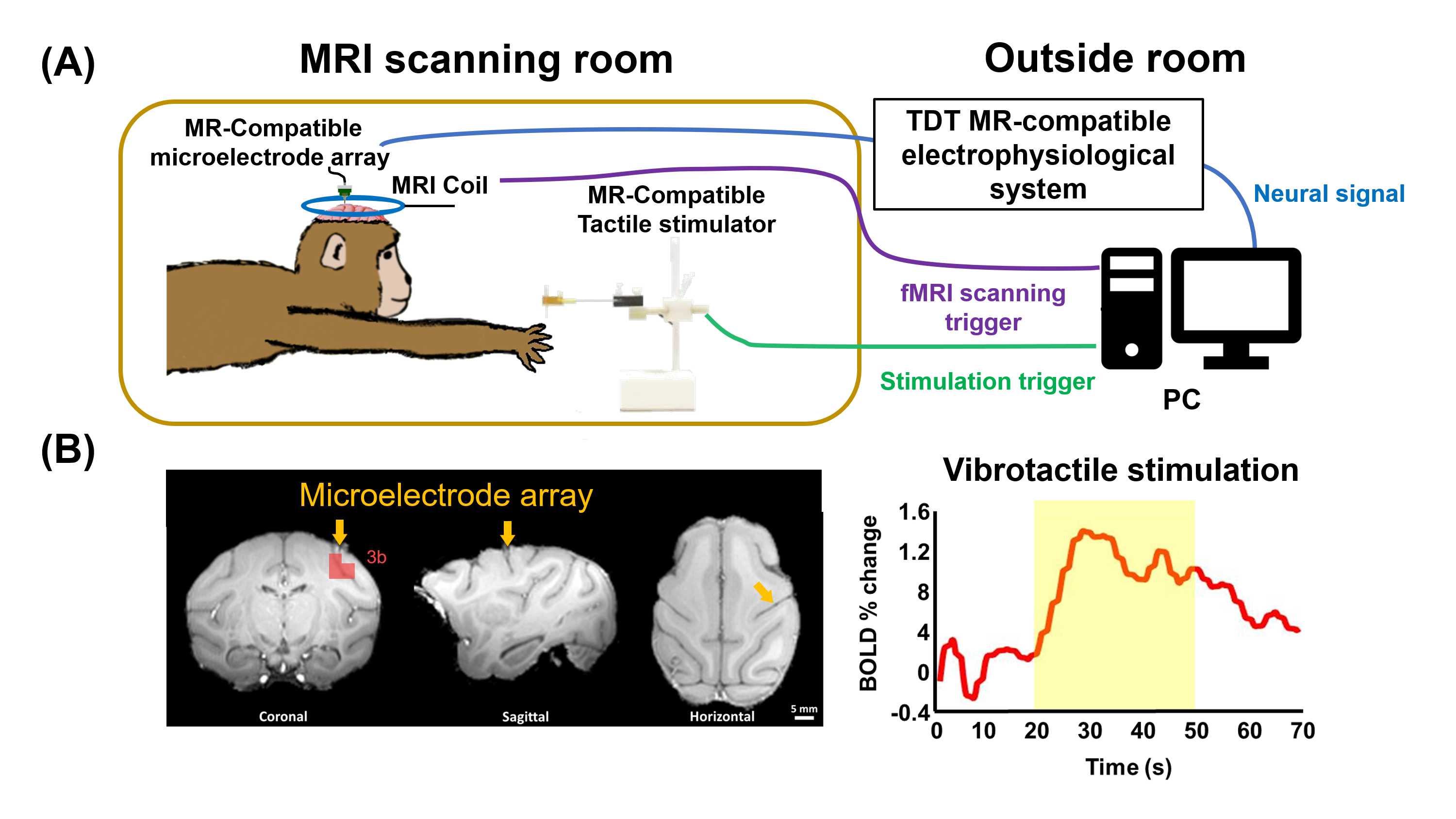
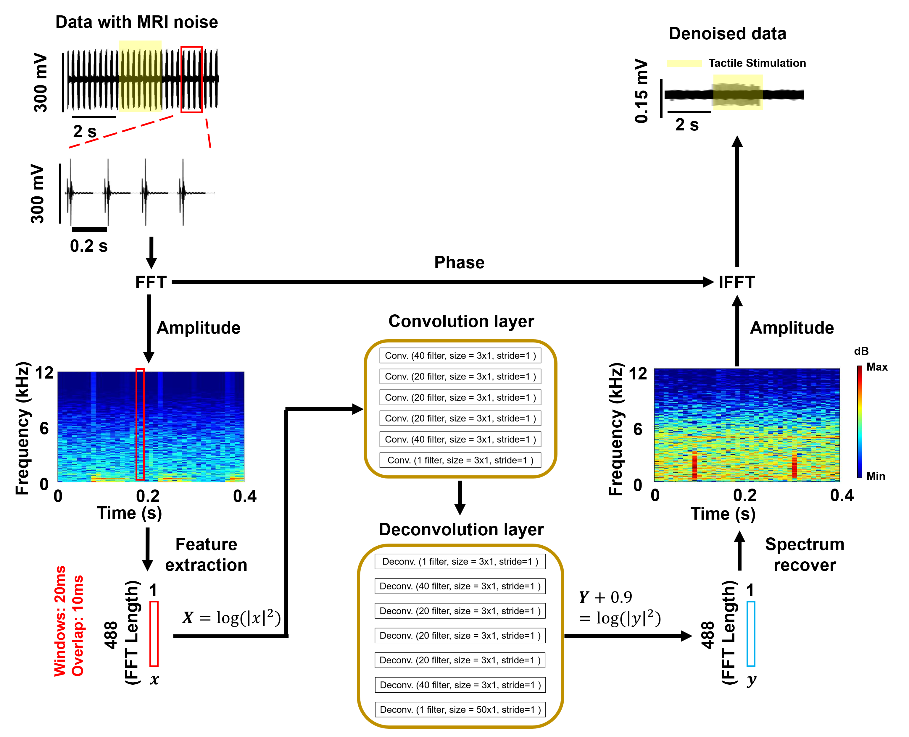
In this study, we conducted experiments on an adult female rhesus monkey weighing 3.0 kg (IACUC No. ZJU20190029). The animal was anesthetized with anesthetized with 0.2-0.5% isoflurane, ketamine (10 mg/kg bolus and 4 mg/kg/hr i.v.) and vecuronium bromide (50 ug/kg/hr i.v.) during fMRI experiments. To define the implant target, a digit map was obtained by vibrotactile stimulations on index, middle, and ring fingers, facilitated through an MR-compatible tactile stimulator (Figure 1A). The animal was anesthetized with 2% isoflurane, underwent craniotomy for the implantation of a lab-designed MR-compatible microelectrode array (16 channels) into digit-related area 3b (Figure 1B), and was placed in MR scanner. MRI data were acquired on a Siemens 7T Magnetom System, including structural images (T1-weighted TSE sequence, TR = 2530 ms, TE = 18 ms, BW = 100 Hz, voxel size: 0.5×0.5×1.0 mm3) and functional images (EPI sequence, TR = 2000 ms, TE = 24.2 ms, BW = 1710 Hz, voxel size: 1.5×1.5×1.5 mm3). Neural signals were recorded using a TDT MR-compatible electrophysiological system (Filter: 0.3 – 5 kHz, Sampling rate: 24 kHz). To mitigate EMI noise, we introduced an FCNN comprising six convolutional layers and seven deconvolutional layers (Figure 2).The neural signal was segmented into 20-ms time windows with a 10-ms sliding window and underwent fast Fourier transform (FFT) to obtain frequency amplitudes x and phases p. The amplitude x were transformed using Eq. 1 and used as input data X for FCNN.
$$$ X=\log(|x^{2}|) $$$ Eq. 1
After each layer’s output, data trimming (cropping) was applied to maintain the quantity of output data, and batch normalization expedited the neural network’s optimization speed. The output Y of the final deconvolutional layer was used to obtain the amplitude values y via Eq. 2:
$$$ Y+0.9=\log(|y^{2}|) $$$Eq. 2
Phases p and amplitudes y were reversed through inverse FFT (IFFT) to reconstruct the denoised signal Z(t), completing the denoising process.
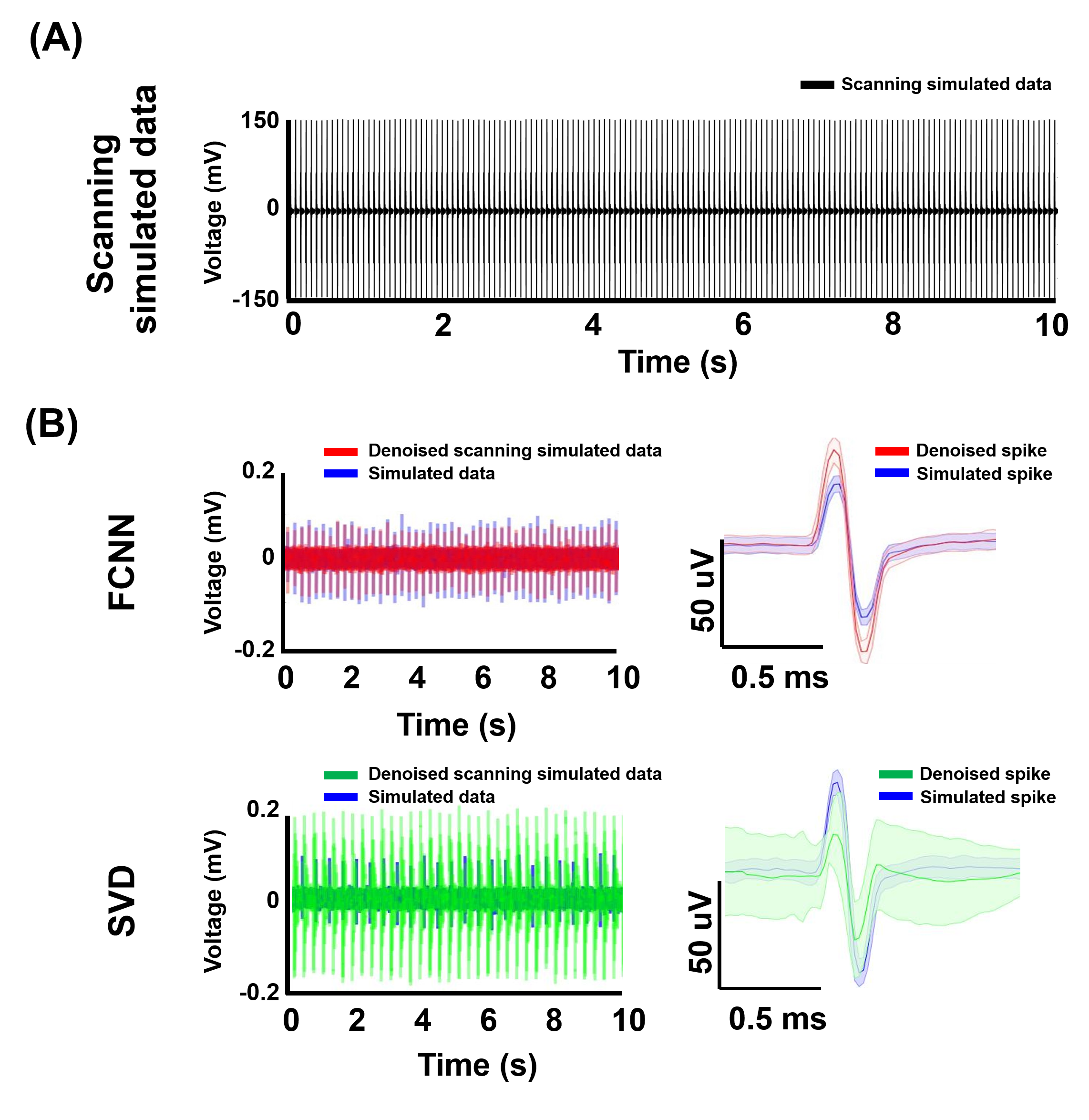
Three types of signals were recorded: 1) Simulated Data - simulated neural signals recoded outside the MRI scanner (10 mins, n=1); 2) Scanning Simulated Data - simulated neural signals and fMRI-induced EMI noises (10 mins, n = 3); 3) Tactile-evoked data - simultaneous recording of Ephy signals with fMRI induced by vibrotactile stimulation (90 s, n = 5). One Simulated Data and one Scanning Simulated Data were used for training, while two Scanning simulated data and Tactile-evoked Data were used for testing the denoising algorithm. Performance assessment included a comparison between Singular Value Decomposition (SVD) [6] and our proposed FCNN.
RESULT AND DISCUSSION
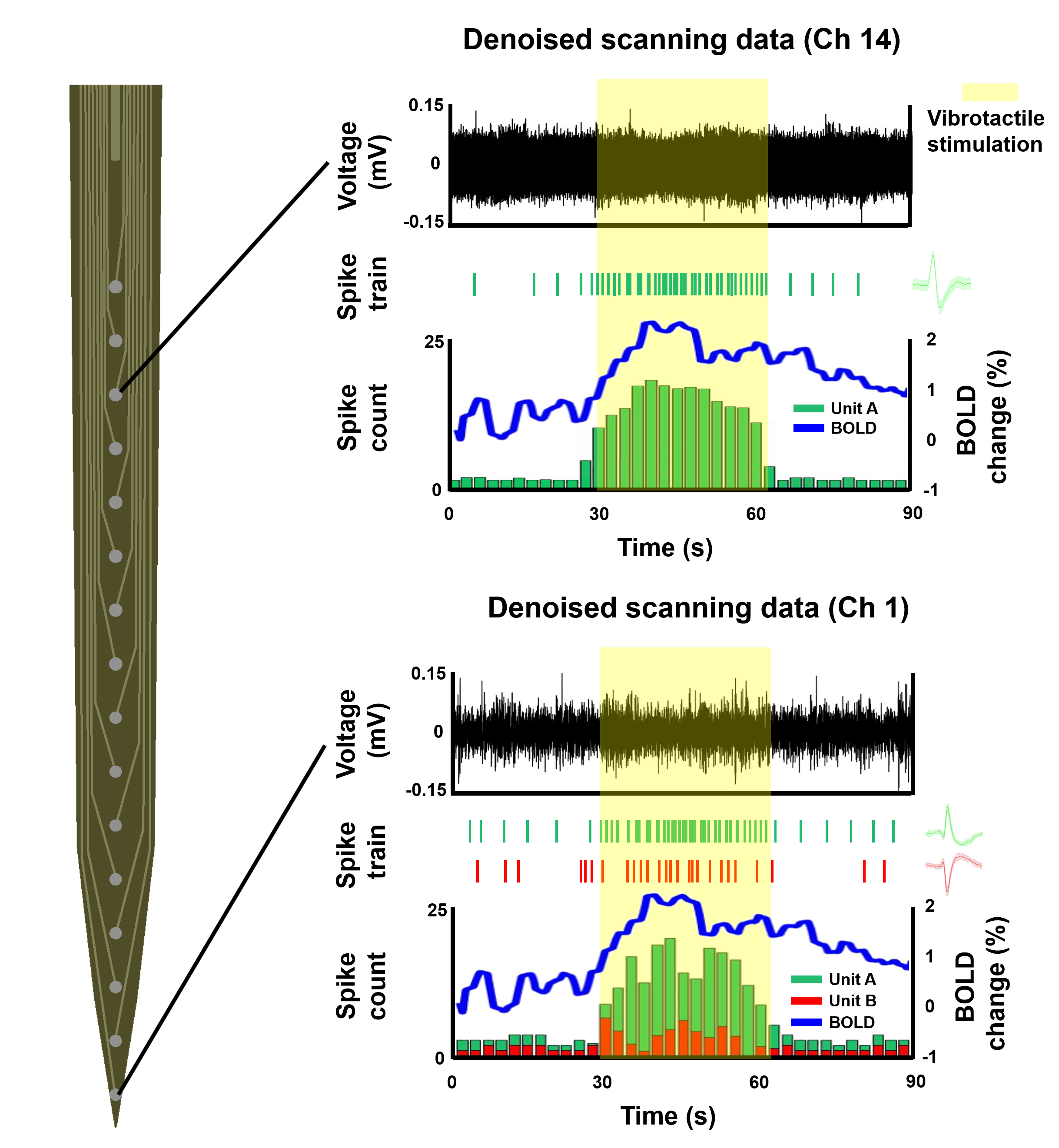
Figure 3 reveals fewer remaining EMI noises using FCNN compared to SVD. Spike sorting on denoised signals indicated significant alterations in spike waveforms by SVD, while FCNN maintained waveform consistency. Furthermore, spike loss rates were 3% for FCNN and 73% for SVD, indicating that FCNN retained most neural signals with significant restoration. Finally, we assessed FCNN's ability to restore Tactile-evoked data. Figure 4 shows two denoised neural signals (green and red) and BOLD signals (blue line) by tactile stimuli, confirming the precise recovery of neural signals by FCNN during fMRI. These results highlight the potential of the FCNN denoising algorithm as a valuable approach for simultaneous fMRI and neural activity research.CONCLUTION
This study introduces a novel approach using a FCNN to eliminate EMI noise in neural signals during fMRI. Our results show that the FCNN effectively reduces EMI noise while preserving neural signal consistency, suggesting its potential as a valuable tool for enhancing our understanding of neurovascular coupling and brain function.Acknowledgements
This research was supported by National Key R&D Program of China (2021YFF0702200), STI 2030-Major Projects (2021ZD0200401).References
1. S. Ogawa and T.M. Lee, Magnetic resonance imaging of blood vessels at high fields: in vivo and in vitro measurements and image simulation. Magnetic Resonance in Medicine, 1990. 16(1): 9-18.
2. S. Ogawa, R. Menon, D.W. Tank, et al., Functional brain mapping by blood oxygenation level-dependent contrast magnetic resonance imaging. A comparison of signal characteristics with a biophysical model. Biophysical Journal, 1993. 64(3): 803-812.
3. S. Ogawa, D.W. Tank, R. Menon, et al., Intrinsic signal changes accompanying sensory stimulation: functional brain mapping with magnetic resonance imaging. Proceedings of the National Academy of Sciences, 1992. 89(13): 5951-5955.
4. N.K. Logothetis, J. Pauls, M. Augath, et al., Neurophysiological investigation of the basis of the fMRI signal. Nature, 2001. 412(6843): 150-157.
5. P.V. Karmarkar, Implantable MRI compatible stimulation leads and antennas and related systems and methods. 2013; Google Patents.
6. C.E. Cruttenden, W. Zhu, Y. Zhang, et al., Toward completely sampled extracellular neural recording during fMRI. IEEE Transactions on Medical Imaging, 2022. 41(7): 1735-1746.
7. Y. Wang, Y. Jiang, and J. Lan, Fcnn: An efficient intrusion detection method based on raw network traffic. Security Communication Networks, 2021. 2021: 1-13.
Figures