1246
Two distinct thalamic subtypes associated with cognitive impairment in first-episode schizophrenia1Department of Radiology, West China Hospital of Sichuan University, Chengdu, China, 2Research Unit of Psychoradiology, Chinese Academy of Medical Sciences, Chengdu, China, 3Huaxi MR Research Center (HMRRC), West China Hospital of Sichuan University, Chengdu, China
Synopsis
Keywords: Psychiatric Disorders, Brain, thalamic nuclei
Motivation: Schizophrenia is characterized by high heterogeneity, with the core symptom of cognitive impairment. However, it is unclear that whether the patterns of thalamic nuclei volume alterations is associated with cognitive function in schizophrenia.
Goal(s): To investigate the pattern and heterogeneity of thalamic nuclei alterations and cognition in schizophrenia.
Approach: The cluster analysis (K-means++) was used to identify the subtype of schizophrenia. And machine learning model was developed to validate the reproducibility of subtypes.
Results: We uncovered two markedly distinct neuroanatomical subtypes of schizophrenia. One of the subtypes characterized as widespread decrease in thalamic nuclei and severe cognition deficit.
Impact: These findings could help to identify biological targets related to the treatment of schizophrenia, and improve the functional outcomes in patients with schizophrenia.
Introduction
Schizophrenia is characterized by high heterogeneity, with the core symptom of cognitive impairment[1]. Several neuroimaging studies showed that alterations of the thalamic nuclei were associated with cognitive impairment in schizophrenia[3, 4], indicating thalamic nuclei play an important role in the pathology mechanism of schizophrenia[2]. So far, there is no study to investigate whether the heterogeneity of thalamic nuclei is associated with the pattern of cognitive impairment in schizophrenia. Thus, we intended to investigate the subtypes of schizophrenia using semi-supervised machine learning methods to discover various patterns of thalamic nuclei alterations.Methods
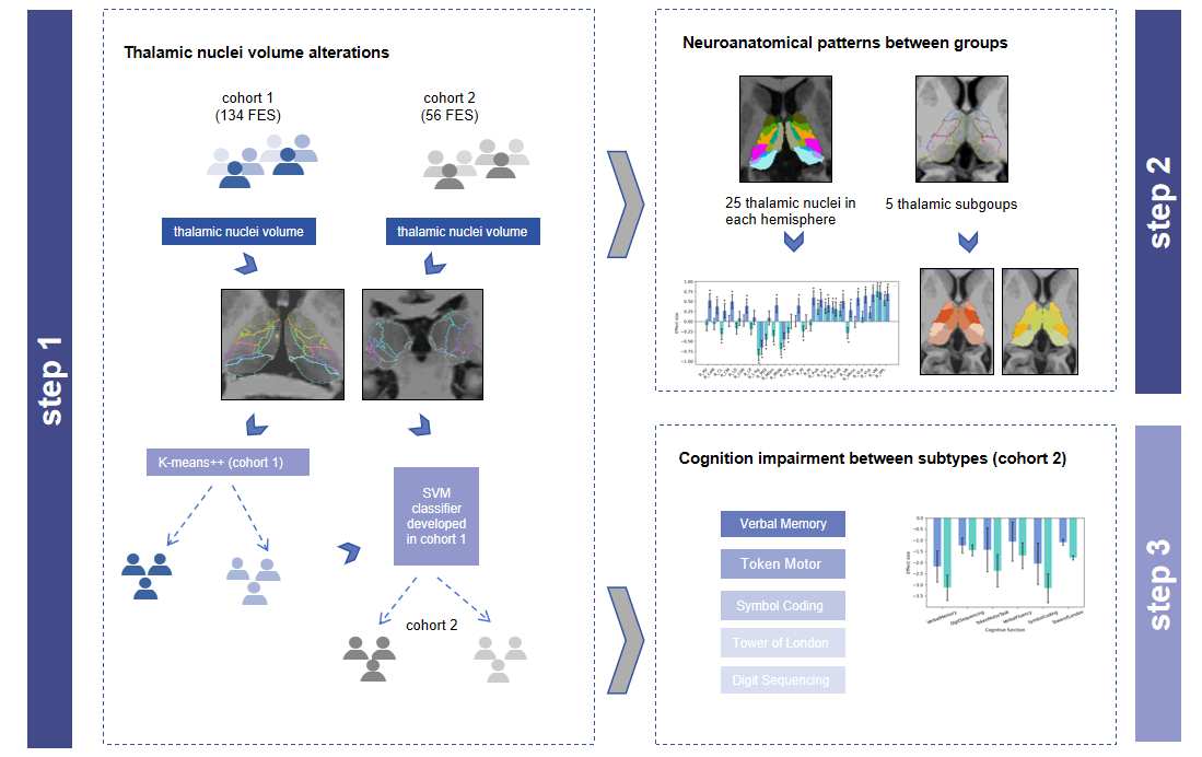
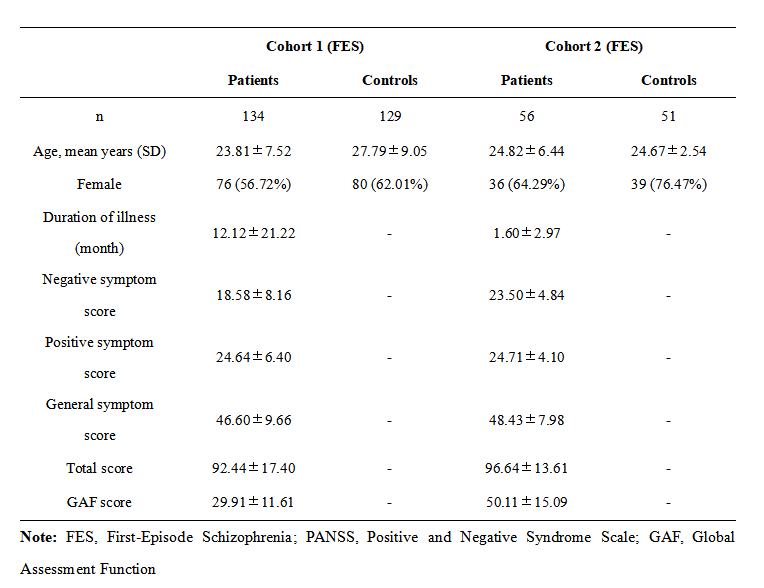
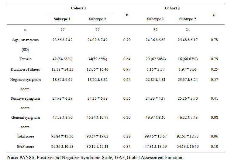
We recruited 190 first-episode schizophrenia patients (FES) and 180 healthy controls (HCs) from 2 cohorts. Cohort 1 included 134 FES and 129 HCs and cohort 2 consisted of 56 FES and 51 HCs. For each participant, high-resolution T1-weighted data was obtained, and the thalamic nucleus were segmented into 25 nuclei in each brain hemisphere by Freesurfer. Then, nuclei volume from 134 individuals with FES (cohort 1) were evaluated using cluster analysis to test for heterogeneity. Then, a subtype of FES was identified based on their patterns of thalamic nuclei volume alterations. After the subtype of FES determined in cohort 1, a support vector machine (SVM) model developed was used to discriminate the subtype in cohort 2. We compared the structual alteration, cognition and symptom patterns between groups.Results
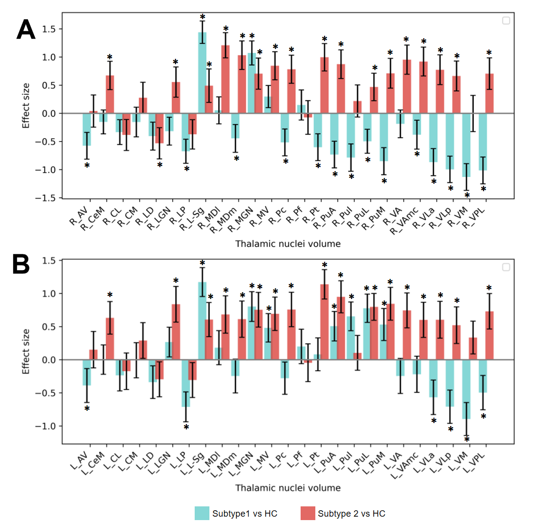
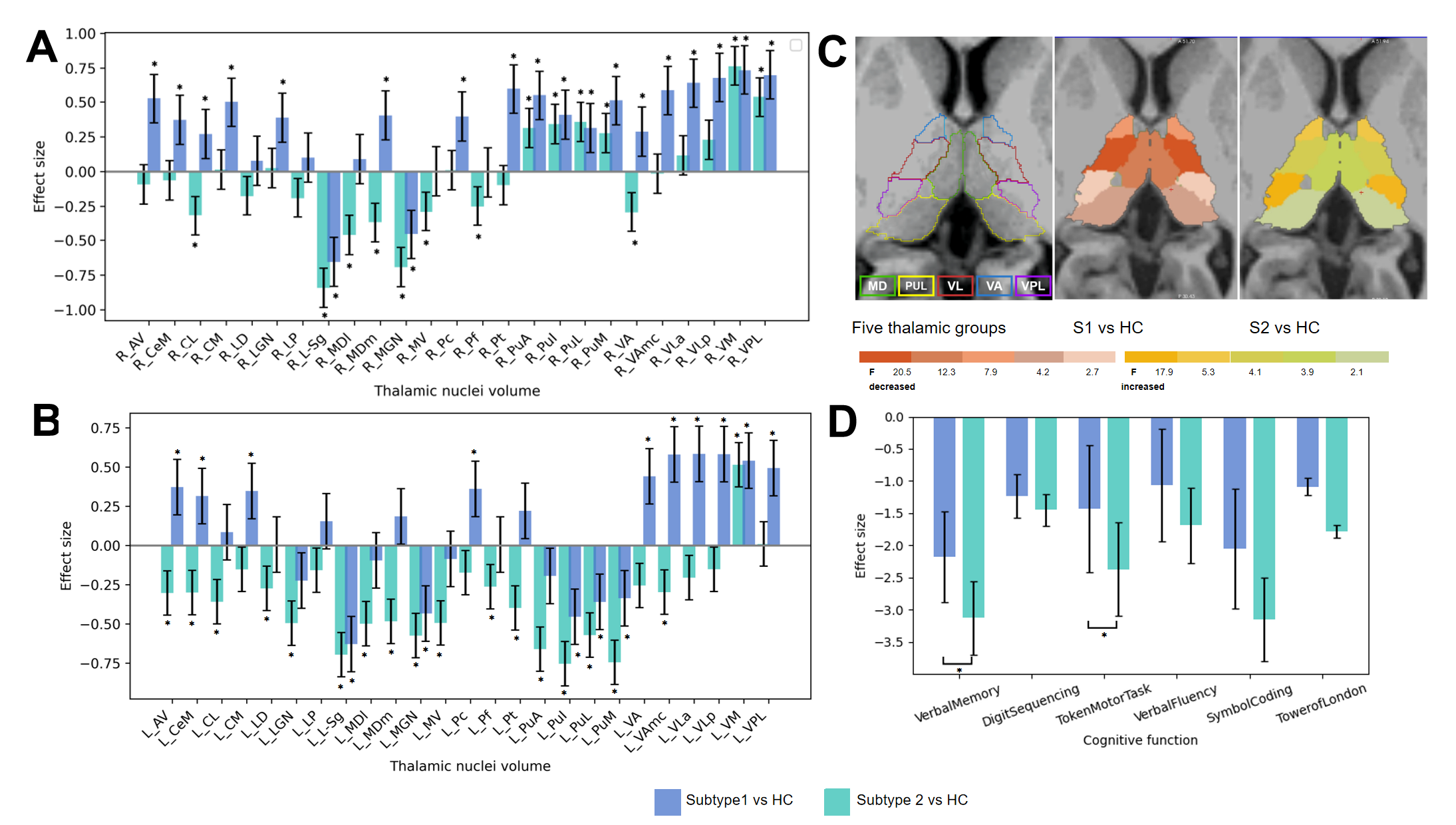
In cohort 1, FES patients were classified into two subtypes. Subtype 1 (57%) was characterized as widespread thalamic nuclei deficits, while Subtype 2 (43%) displayed large area of thalamic nuclei increased in volume. We also combined the thalamic nuclei into five groups according to the methods of Anna et al[5], and the thalamic subgroup showed a global volume decreased in subtype 1. Cohort 2 were classified into two subtypes according to the classifier developed in cohort 1. The classification rates in these two cohorts were similar (approximately 50% each). The subtypes in cohort 2 presented similar patterns of thalamic nuclei volume alterations. And Subtype 1 (57%) was characterized as widespread thalamic nuclei deficits, while Subtype 2 (43%) displayed large scale of thalamic nuclei volume increased (Fig.3. A, B). Moreover, in subgroup analysis, Subtype 1 showed a global volume decreased, especially in ventrolateral (VL) nuclei subgroup (Fig.3. C). We also explored the patterns of cognition impairment between subtypes. And Subtype 1 presented more severe cognition deficits, significantly in Vertical Memory and Token Motor Task (Fig.3. D). There was no significant difference in the severity of symptoms between subtypes in these two cohorts.Discussion and conclusion
In this study, we uncovered two markedly distinct neuroanatomical subtypes of schizophrenia. One of the subtypes characterized as widespread decrease in thalamic nuclei and severe cognition deficits. These findings indicated that the patterns of thalamic nulei alterations may be the potential biomarkers for predicting cognitive impairment and functional outcomes in patients with schizophrenia. Furthermore, it may provide new insights for targeted treatment in cognition impairment of schizophrenia.Acknowledgements
This study is supported by grants from Chengdu Science and Technology Office, major technology application demonstration project (2022-YF09-00062-SN & 2022-GH03-00017-HZ), and China Postdoctoral Science Foundation (2022M722270).References
[1] McCutcheon R A, Keefe R, McGuire P K. Cognitive impairment in schizophrenia: aetiology, pathophysiology, and treatment[J]. Mol Psychiatry, 2023,28(5):1902-1918.
[2] Fryer S L, Ferri J M, Roach B J, et al. Thalamic dysconnectivity in the psychosis risk syndrome and early illness schizophrenia[J]. Psychol Med, 2022,52(13):2767-2775.
[3] Perez-Rando M, Elvira U, García-Martí G, et al. Alterations in the volume of thalamic nuclei in patients with schizophrenia and persistent auditory hallucinations[J]. Neuroimage Clin, 2022,35:103070.
[4] Mørch-Johnsen L, Jørgensen K N, Barth C, et al. Thalamic nuclei volumes in schizophrenia and bipolar spectrum disorders - Associations with diagnosis and clinical characteristics[J]. Schizophr Res, 2023,256:26-35.
[5] Huang A S, Rogers B P, Sheffield J M, et al. Thalamic Nuclei Volumes in Psychotic Disorders and in Youths With Psychosis Spectrum Symptoms[J]. Am J Psychiatry, 2020,177(12):1159-1167.
Figures