1243
Effects of bright light therapy on cingulate cortex dynamic functional connectivity and neurotransmitter activity in subthreshold depression1First Affiliated Hospital of Jinan University, Guangzhou, China, 2MR Research, GE Healthcare, Beijing, China, Guangzhou, China
Synopsis
Keywords: Psychiatric Disorders, fMRI (resting state)
Motivation: Bright light therapy (BLT) is one of the effective interventions for subthreshold depression, but its neural mechanism is still unclear.
Goal(s): The goal of this double-blind, randomized, placebo-controlled clinical trial was to assess the correlation between BLT and the dynamic functional connectivity (dFC) changes in the cingulate cortex along with distribution of specific neurotransmitters in subthreshold depression.
Approach: A double-blind randomized controlled trial
Results: BLT alleviates depressive symptoms and changes the cingulate cortex dFC variability in subthreshold depression. And pre-treatment dFC variability of the cingulate cortex could be used as a biomarker for improved BLT treatment in subthreshold depression.
Impact: BLT alleviates depressive symptoms and changes the cingulate cortex dFC variability in subthreshold depression, which raises the possibility that pre-treatment dFC variability of the cingulate cortex could be used as a biomarker for improved BLT treatment in subthreshold depression.
Background
Subthreshold depression is a state of mental sub-health with clinically relevant depressive symptoms that do not meet the criteria for major depressive disorder (MDD) or dysthymic disorder [1, 2]. It is a strong risk factor for the onset of major depressive disorder [3] and is associated with an increased burden of disease and suicide risk [4]. Therefore, early and effective interventions of subthreshold depression are vital to prevent depressive disorders and the associated burdens. Bright light therapy (BLT) combines multiple advantages such as direct availability, sufficient efficacy, low costs and high safety, thus being a promising treatment for seasonal and non-seasonal depression [5]. However, mechanisms underlying such efficacy of BLT are not fully clear. The cingulate cortex, which includes the anterior cingulate cortex (ACC), middle cingulate cortex (MCC) and posterior cingulate cortex (PCC), is emerging as a crucial region correlated with treatment outcome for repetitive transcranial magnetic stimulation [6, 7] and antidepressant treatment response [8, 9] in depression. In seasonal affective disorder, light therapy reduced serotonin transporter binding[10] and changed regional cerebral blood flow[11] in the cingulate cortex, which correlated with improvement of mood symptoms. However, few studies have reported neuroimaging biomarker that may assist in predicting the treatment outcome of light therapy in nonseasonal subthreshold depression. An emerging approach, dynamic FC (dFC) analysis, may index changes in macroscopic neural activity patterns underlying critical aspects of cognition and behavior [12]. Recent evidence has shown the dFC may be more informative than static indices in classification[13, 14] and prediction[15, 16] in mood disorders. Unfortunately, no study has investigated dFC changes after BLT in subthreshold depression. Of note, monoaminergic neurotransmission has been proposed to play an important role in the mechanisms of the action of light therapy [17]. Monoaminergic system that includes serotonin (5-HT), norepinephrine (NE) and dopamine (DA) has been reported to exert influence on brain circuits concerned by the regulation of mood [18]. Disturbances in the brain serotonin systems might play a key role in the pathogenesis of seasonal affective disorder and that light therapy may compensate for the underlying deficit [19]. However, the neurobiological mechanisms behind the antidepressant effect of light therapy are still not well-elucidated. The goal of this double-blind, randomized, placebo-controlled clinical trial was to assess the correlation between BLT and the dFC changes in the cingulate cortex along with distribution of specific neurotransmitters in subthreshold depression.Methods
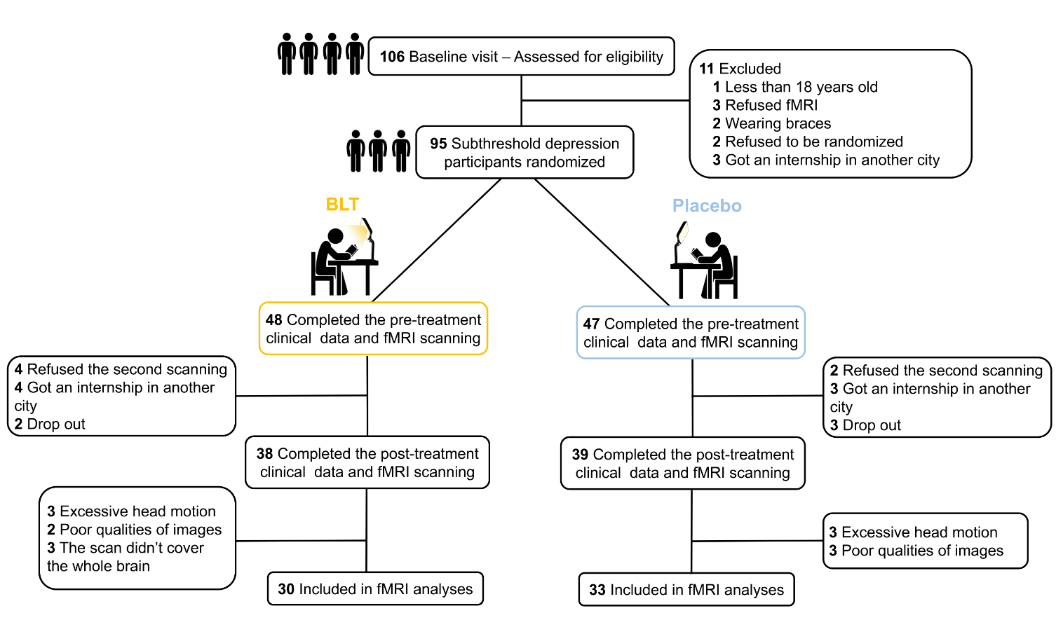
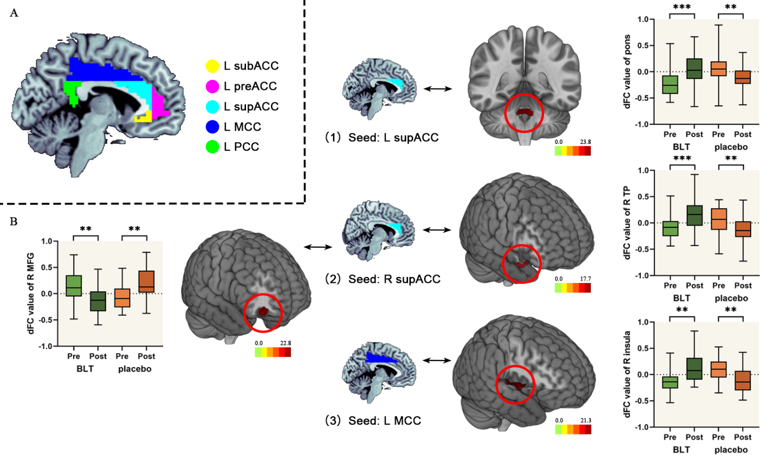
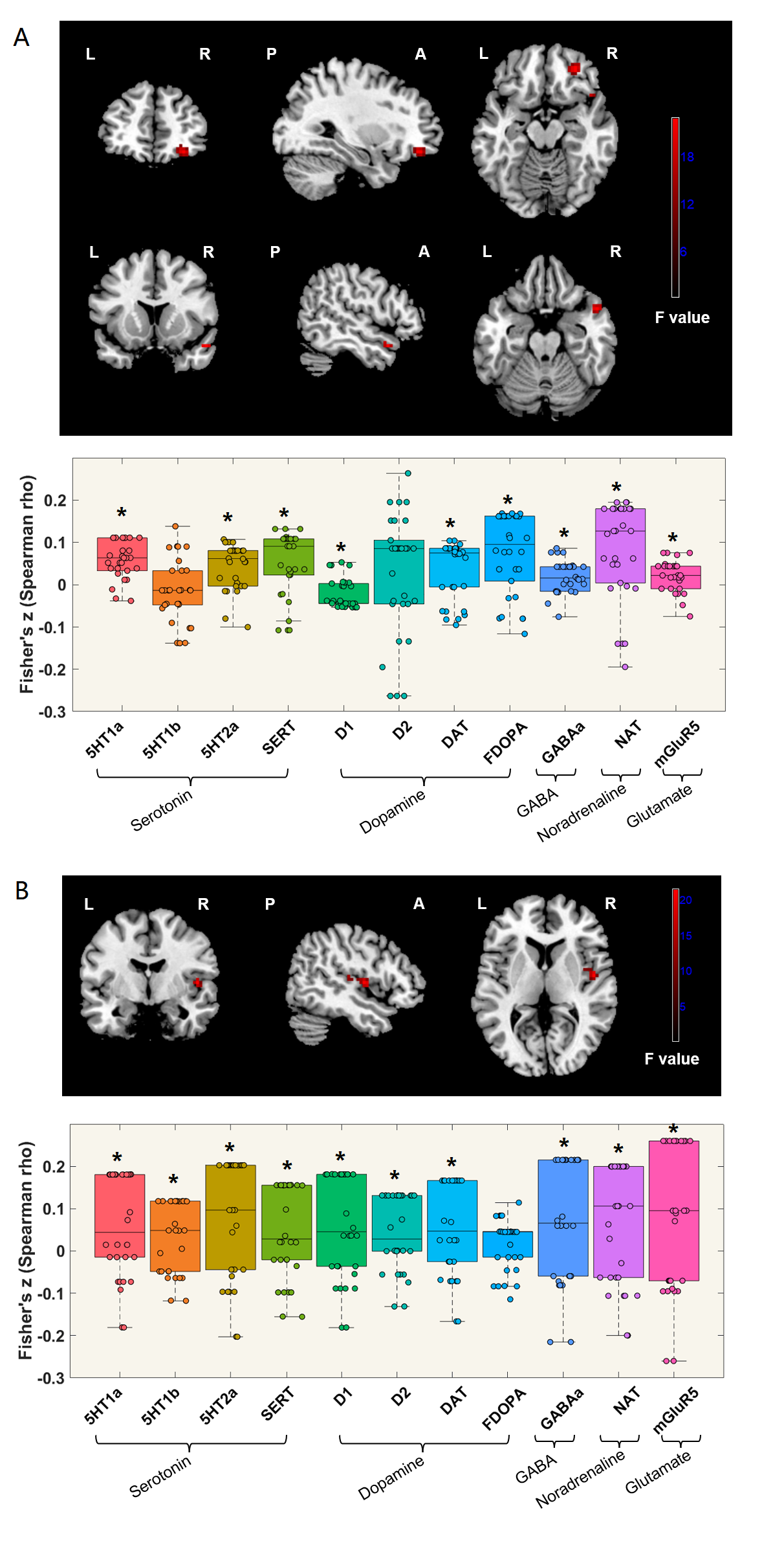
Participants with subthreshold depression were randomly assigned to either the BLT group (N = 38) or the placebo group (N = 39). The primary outcome was Hamilton Depression Rating Scale (HDRS), and secondary outcomes were Centre for Epidemiologic Studies Depression Scale (CESD) and Hamilton Anxiety Scale (HAMA), which were assessed before and after 8 weeks. The participants also underwent resting-state functional magnetic resonance imaging before and after 8 weeks. The subgenual, pregenual and the supracallosal anterior cingulate cortex (subACC, preACC, supACC), middle cingulate cortex (MCC) and posterior cingulate cortex (PCC) seed-based whole-brain dFC analysis was conducted. Besides, a multivariate regression model was adopted to predict HDRS and CESD scores changes after BLT. Furthermore, JuSpace toolbox was used to calculate the associations between dFC (pre- and after interventions) and neurotransmitter activity in the BLT group.Results
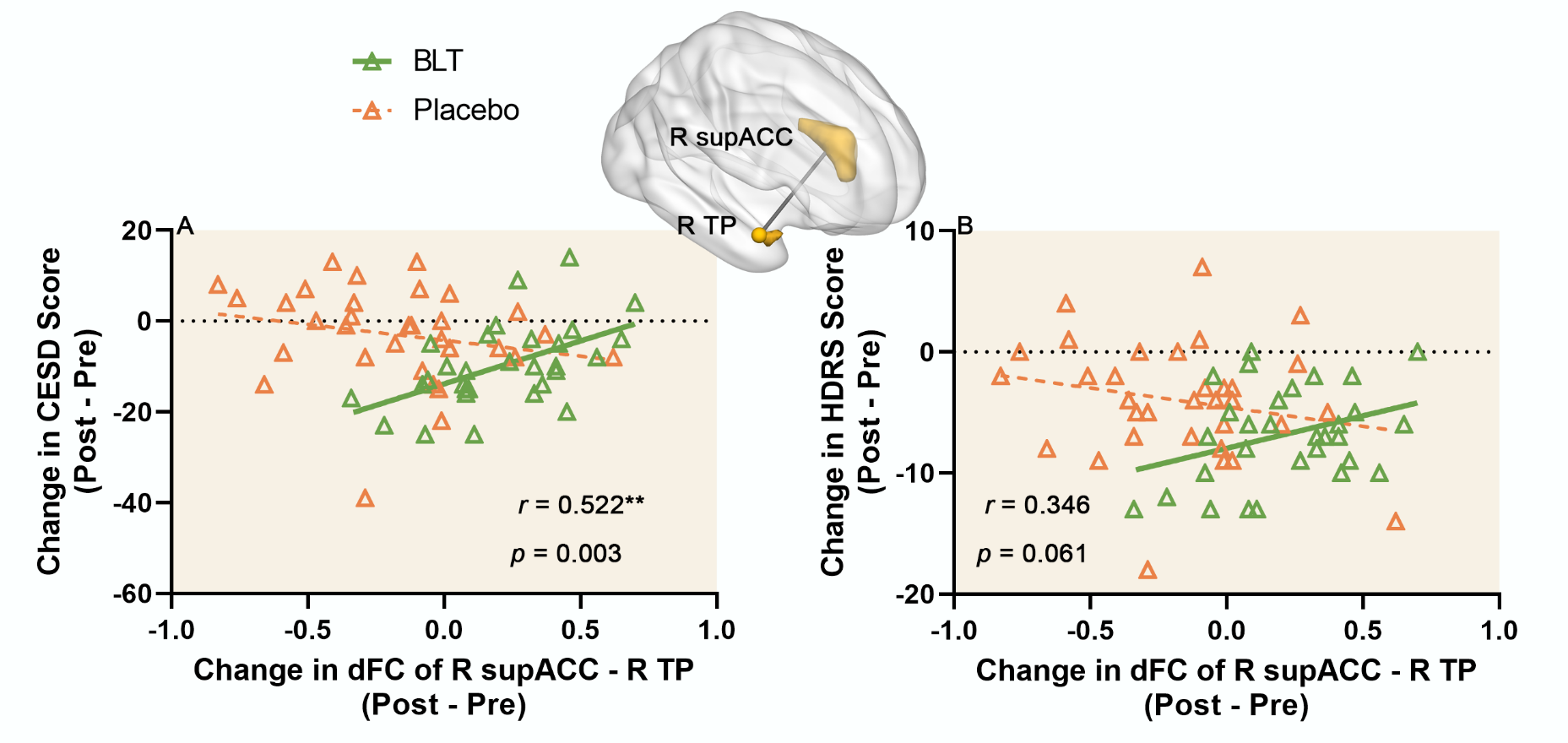
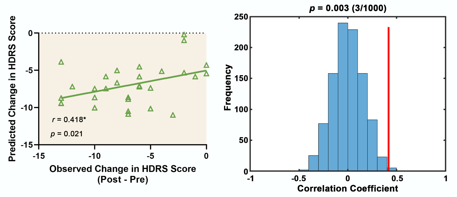
BLT group showed decreased CESD score (t = 3.195, p = 0.002) and HDRS score (t = 3.232, p = 0.002) from pre-treatment to post-treatment compared to the placebo group. Also, compared to baseline, BLT group showed increased dFC of the right supACC-right temporal pole (TP), left MCC-right insula, and left supACC-pons, and decreased dFC of the right supACC- right middle frontal gyrus (MFG) after intervention. Changes in dFC of the right supACC-right TP showed positive correlation with changes in CESD score (significant: r = 0.522, p = 0.003) and HDRS score (marginally significant: r = 0.346, p = 0.061) before and after BLT. Moreover, combining the baseline dFC variability of the cingulate cortex could predict HDRS changes in BLT. Finally, compared to baseline, the supACC and MCC dFC changes after BLT showed significant correlations with the serotonergic, dopaminergic, noradrenergic systems as well as the GABAergic and glutamatergic maps.Conclusions
These results suggested that BLT alleviates depressive symptoms and changes the cingulate cortex dFC variability in subthreshold depression, which raises the possibility that pre-treatment dFC variability of the cingulate cortex could be used as a biomarker for improved BLT treatment in subthreshold depression. Furthermore, dFC changes with specific neurotransmitter systems after BLT may underline the antidepressant mechanisms of BLT.Acknowledgements
The study was supported by grants from the National Natural Science Foundation of China (81671670, 81971597, and 82172530); National Key Research and Development Project (2020YFC2005700); Key-Area Research and Development Program of Guangdong Province (2020B1111100001). The funding organizations play no further role in study design, data collection, analysis and interpretation and paper writing.References
1. Xian, J., et al., Acupuncture for Subthreshold Depression: Study Protocol for a Randomized Controlled Trial. Front Psychiatry, 2021. 12: p. 772360.
2. Kroenke, K., When and How to Treat Subthreshold Depression. Jama, 2017. 317(7): p. 702-704.
3. Klein, D.N., et al., Subthreshold depressive disorder in adolescents: predictors of escalation to full-syndrome depressive disorders. J Am Acad Child Adolesc Psychiatry, 2009. 48(7): p. 703-710.
4. Balázs, J., et al., Adolescent subthreshold-depression and anxiety: psychopathology, functional impairment and increased suicide risk. J Child Psychol Psychiatry, 2013. 54(6): p. 670-7.
5. Pail, G., et al., Bright-light therapy in the treatment of mood disorders. Neuropsychobiology, 2011. 64(3): p. 152-62.
6. Ge, R., et al., Functional connectivity of the anterior cingulate cortex predicts treatment outcome for rTMS in treatment-resistant depression at 3-month follow-up. Brain Stimul, 2020. 13(1): p. 206-214.
7. Jing, Y., et al., Pregenual or subgenual anterior cingulate cortex as potential effective region for brain stimulation of depression. Brain Behav, 2020. 10(4): p. e01591.
8. Yi, S., et al., Neural activity changes in first-episode, drug-naïve patients with major depressive disorder after transcutaneous auricular vagus nerve stimulation treatment: A resting-state fMRI study. Front Neurosci, 2022. 16: p. 1018387.
9. Zhang, Y., et al., Functional impairment-based segmentation of anterior cingulate cortex in depression and its relationship with treatment effects. Hum Brain Mapp, 2021. 42(12): p. 4035-4047.
10. Harrison, S.J., et al., Light therapy and serotonin transporter binding in the anterior cingulate and prefrontal cortex. Acta Psychiatr Scand, 2015. 132(5): p. 379-88.
11. Vasile, R.G., et al., Changes in regional cerebral blood flow following light treatment for seasonal affective disorder: responders versus nonresponders. Biol Psychiatry, 1997. 42(11): p. 1000-5.
12. Hutchison, R.M., et al., Dynamic functional connectivity: promise, issues, and interpretations. Neuroimage, 2013. 80: p. 360-78.
13. Rashid, B., et al., Classification of schizophrenia and bipolar patients using static and dynamic resting-state fMRI brain connectivity. Neuroimage, 2016. 134: p. 645-657.
14. Cheng, B., et al., Abnormal dynamics of resting-state functional activity and couplings in postpartum depression with and without anxiety. Cereb Cortex, 2022. 32(24): p. 5597-5608.
15. Shunkai, L., et al., Alterations of insular dynamic functional connectivity and psychological characteristics in unmedicated bipolar depression patients with a recent suicide attempt. Psychol Med, 2022: p. 1-12.
16. Xue, S.W., et al., Abnormal Dynamic Functional Connectivity of the Left Rostral Hippocampus in Predicting Antidepressant Efficacy in Major Depressive Disorder. Psychiatry Investig, 2022. 19(7): p. 562-569.
17. Neumeister, A., et al., Monoaminergic function in the pathogenesis of seasonal affective disorder. Int J Neuropsychopharmacol, 2001. 4(4): p. 409-20.
18. Hamon, M. and P. Blier, Monoamine neurocircuitry in depression and strategies for new treatments. Prog Neuropsychopharmacol Biol Psychiatry, 2013. 45: p. 54-63.
19. Neumeister, A., et al., Monoamine depletion in non-pharmacological treatments for depression. Adv Exp Med Biol, 1999. 467: p. 29-33.
Figures