1240
Cognition-related connectome gradient dysfunctions of thalamus and basal ganglia in drug-naïve first-episode major depressive disorder1Huaxi MR Research Center (HMRRC), Department of Radiology, West China Hospital of Sichuan University, Chengdu, China
Synopsis
Keywords: Psychiatric Disorders, Psychiatric Disorders, major depressive disorder, fMRI, functional gradient, subcortical structure, cognition
Motivation: The continuous spatial patterns of inter-region connectivity within the subcortical network still remain less well-understood in MDD.
Goal(s): Using functional gradient mapping, a novel approach to identify hierarchical organization of functional networks, we aim to evaluate multiscale subcortical gradients in MDD and their association with cognition.
Approach: Subcortical gradient alterations at the global-, system-, and subregion-levels and their relation to neuropsychological functioning were assessed in MDD patients relative to healthy controls.
Results: Principal gradient values were lower in thalamic and limbic systems but higher in basal ganglia in MDD. Interactions between thalamic and basal ganglia gradient alterations were implicated in MDD-related memory impairments.
Impact: Multiscale subcortical gradient alterations can enhance our understanding of MDD-related hierarchical disturbances in subcortical function and may provide useful clinical biomarkers for cognitive impairments in MDD.
INTRODUCTION
Functional abnormalities in subcortical networks are believed to be implicated in pathophysiology of clinical symptoms and cognitive impairments in patients with major depressive disorder (MDD) 1. While significant progress has been made in characterizing discrete large-scale functional network alterations in MDD patients, the continuous spatial patterns of inter-region connectivity, especially alterations in subcortical function, remain less well-understood. By introducing functional gradient mapping, a novel approach to depict spatial organization of brain function by capturing patterns of functional connectivity similarity 2, the present study evaluated subcortical gradients in MDD patients and their association with cognitive features.METHODS
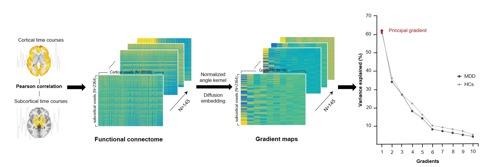
Utilizing functional gradient mapping approach (Fig. 1), we investigated the organization patterns and between-group differences in the principal subcortical gradient in 145 never-treated first-episode MDD patients and 145 healthy controls (HCs) across all subcortical voxels (global), three main systems (limbic, thalamic, and basal ganglia), subcortical structural subregions, and functional subregions related to different cortical functional networks. The degree of connectivity similarity and the relative spatial position of each subcortical regions along the principal gradient were represented by principal gradient values 3. We also examined the associations of significant gradient alterations with clinical and cognitive features of MDD patients and HCs, as well as the spatial and functional connectivity measurements. All reported P values were FDR corrected.RESULTS
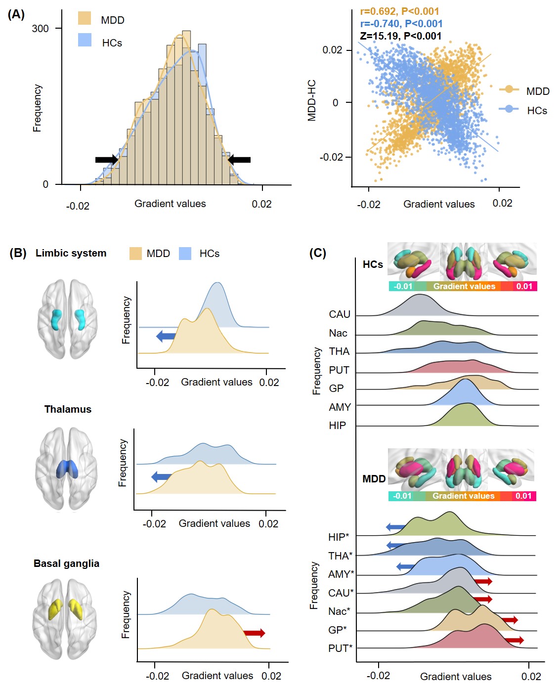
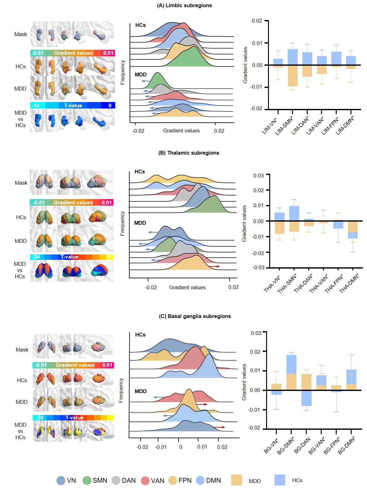
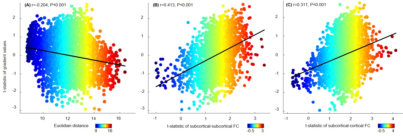
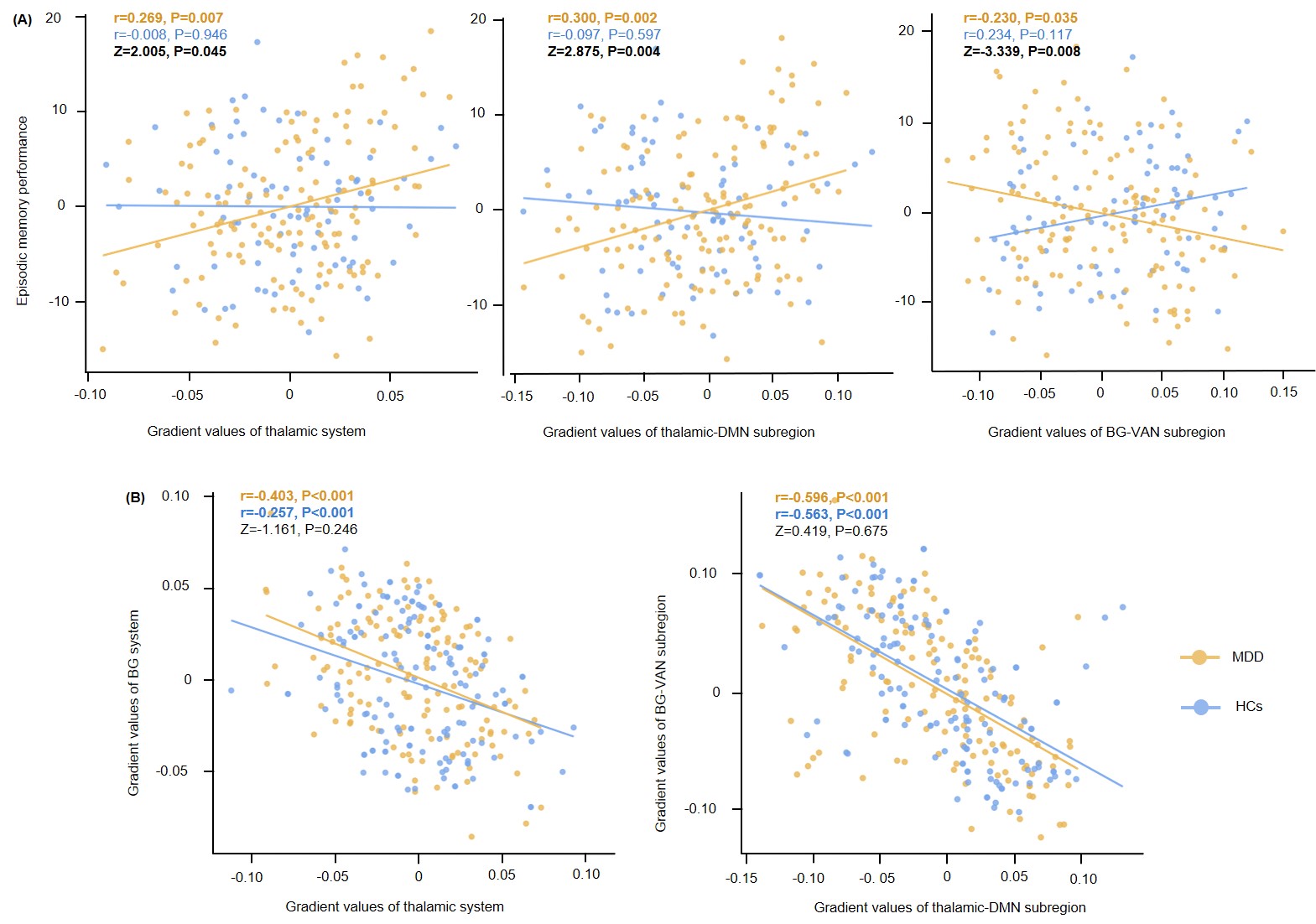
Overall, MDD patients showed a relatively compressed and disturbed gradient organization than HCs, with limbic and BG regions located at both ends ((K-S stat=0.06, P<0.001, Fig. 2A). Specifically, MDD patients had lower principal gradient values in thalamus (t=-5.972, P<0.001) and limbic system (t=-15.916, P<0.001) but higher values in BG (t=15.121, P<0.001) than HCs (Fig. 2B). These gradient alterations manifested as spatial rearrangements of gradient values within each respective structural (Fig. 2C) and functional subregions (Fig. 3) , which were further associated with intrinsic Euclidian distance (r=-0.204, P<0.001) and functional connectivity patterns (r=0.413, P<0.001) (Fig. 4). Furthermore, lower gradient values in thalamic subregion projecting to default mode network were associated with higher principal gradient values in BG subregion projecting to ventral attention network (r=-0.596, P<0.001), and these gradient alterations were correlated with poorer episodic memory performance in MDD patients (both P<0.05, Fig. 5).DISCUSSION
In addition to MDD-related cortical connectome gradient dysfunction revealed by previous study 4, we identified multiscale alterations in both organization and changing patterns of the principal subcortical gradient, which captured spatial disorganizations and functional disturbance of the subcortical-cortical connectome in MDD patients. Notably, opposing gradient alterations in thalamic and BG regions synergistically impact the episodic memory performance in MDD patients, reflecting the neuropathological mechanisms implicated in memory processing.CONCLUSION
Our findings revealed an internally differentiated and clinically relevant pattern of subcortical gradient dysfunction in MDD, which enhanced our understanding of MDD-related hierarchical disturbances in subcortical function and may provide potential neuro-biomarkers for cognitive impairments in MDD.Acknowledgements
Dr. John A. Sweeney served as a consultant to VeraSci.
References
1. Mulders PC, van Eijndhoven PF, Schene AH, Beckmann CF, Tendolkar I. Resting-state functional connectivity in major depressive disorder: A review. Neurosci Biobehav Rev. 2015;56:330-344.
2. Vos de Wael R, Benkarim O, Paquola C, et al. BrainSpace: a toolbox for the analysis of macroscale gradients in neuroimaging and connectomics datasets. Commun Biol. 2020;3(1):103.
3. Huntenburg JM, Bazin PL, Margulies DS. Large-Scale Gradients in Human Cortical Organization. Trends Cogn Sci. 2018;22(1):21-31.
4. Xia M, Liu J, Mechelli A, et al. Connectome gradient dysfunction in major depression and its association with gene expression profiles and treatment outcomes. Mol Psychiatry. 2022;27(3):1384-1393.
Figures