1235
Physics-informed and uncertainty-aware deep learning approach for liver PDFF quantification1Biomedical Imaging Center, Pontificia Universidad Catolica de Chile, Santiago, Chile, 2i-Health Millennium Institute for Intelligent Healthcare Engineering, Santiago, Chile, 3Faculty of Engineering, Universidad Alberto Hurtado, Santiago, Chile, 4Institute for Biological and Medical Engineering, Pontificia Universidad Catolica de Chile, Santiago, Chile, 5Department of Electrical Engineering, Pontificia Universidad Catolica de Chile, Santiago, Chile, 6Radiology Department, School of Medicine, Pontificia Universidad Catolica de Chile, Santiago, Chile, 7Institute for Mathematical & Computational Engineering, Pontificia Universidad Catolica de Chile, Santiago, Chile, 8Pediatric Gastroenterology and Nutrition Department, Division of Pediatrics, School of Medicine, Pontificia Universidad Catolica de Chile, Santiago, Chile, 9Nutrition & Dietetics. Department of Health Sciences; Faculty of Medicine, Pontificia Universidad Catolica de Chile, Santiago, Chile, 10Department of Nutrition, Diabetes and Metabolism. Faculty of Medicine, Pontificia Universidad Catolica de Chile, Santiago, Chile, 11Department of Medical Imaging and Radiation Sciences, Monash University, Melbourne, VIC, Australia
Synopsis
Keywords: Analysis/Processing, Fat, Proton Density Fat Fraction
Motivation: Most Deep Learning (DL) methods to estimate liver PDFF require reference results for training and can only calculate deterministic outputs with unknown uncertainty.
Goal(s): To estimate liver PDFF using a fully-unsupervised DL method for MR water-fat separation capable of quantifying uncertainty.
Approach: We propose a physics informed DL-based framework which can be trained purely on chemical shift-encoded MR images. Our method estimates stochastic R2* and Δf maps, enabling uncertainty quantification, which are then used to obtain stochastic water-only and fat-only components.
Results: Liver PDFF estimations showed good agreement with a reference technique, and uncertainty maps associated with imperfections in the considered physical model.
Impact: The proposed physics-informed DL model requires only MR data for training, which facilitates the data gathering process. Moreover, our uncertainty-aware approach can quantify the uncertainty associated to the final estimations, which may be of significant value in clinical practice.
Introduction
Proton Density Fat Fraction (PDFF), which could be estimated using Chemical Shift-Encoded (CSE) MR images, is a demonstrated biomarker that of Metabolic dysfunction-Associated Fatty Liver Disease (MAFLD)1.Previously proposed Deep Learning (DL)-based methods to estimate PDFF achieved shorter post-processing times and promising accuracy using fewer data than standard methods2–4. However, most of them required labeled training data with reference results, which are usually unreliable. Furthermore, to our knowledge, only a single work considered uncertainty quantification, but it required reference results for training5.
In this work, we propose a novel fully-unsupervised Physics-Informed and Uncertainty-Aware neural Network (PIUA-Net) to estimate liver PDFF from CSE-MR and the uncertainty maps of the resulting water-only and fat-only MR signals.
Theory
The pixelwise physical model that describes the CSE-MR signal at each echo (In) as a function of the water and fat proton densities (ρW, ρF), the R2* signal decay, and the off-resonance field (Δf) is the following:$$I_n = e^{-R_2^* \cdot TE_n}\cdot e^{i2\pi\Delta f\cdot TE_n}\cdot [\rho_W+\rho_F\sum_p \alpha_p\cdot e^{i2\pi f_{F,p}\cdot TE_n}]$$
where an a priori known 6-peak fat signal spectrum is considered (αp and fF,p are the frequency and relative amplitude of each peak, respectively)6.
Since the sum of the water and fat proton densities between brackets is linear, if R2* and Δf are known, the MR signal can be separated into water-only and fat-only components using a least squares method. Then the PDFF corresponds to the ratio ρF / (ρW + ρF).
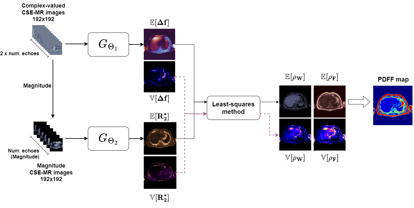
Methods
Our proposed framework considers two Convolutional Neural Networks (CNN) that estimate R2* and Δf maps, respectively. Each CNN has a U-Net architecture and considers multi-echo CSE-MR images as inputs and their corresponding parametric maps, with their uncertainties, as outputs in a two-channel array. In the case of Δf U-Net (GΘ1), the inputs are complex-valued MR images, while for R2* U-Net (GΘ2) the input are magnitude-only images (Figure 1).Due to its physics-informed nature, our PIUA-Net can be trained in a fully unsupervised manner, without requiring any reference MR water-fat separation results. To achieve a more stable training, we first trained GΘ1 during 20 epochs considering a simplified signal model with no signal decay (R2* = 1). After convergence, we trained GΘ2 for 5 epochs, and finally we jointly trained both CNNs for up to 60 epochs. The considered loss function was the mean squared error between the original and the reconstructed MR images, scaled by the corresponding uncertainties:
$$\mathcal{L}=\frac{||s-\hat{s}||}{\sigma}+log(\sigma)$$
We used a 5-fold cross-validation scheme considering an overall dataset of 190 subjects (4246 axial slices). The resulting PDFF, R2*, and Δf maps were compared to those obtained with iterative Graph Cuts7, using the mean absolute error (MAE). Three different versions of PIUA-Net were trained considering series of CSE-MR images with four, five, and six echoes.
Results
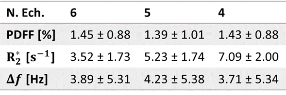
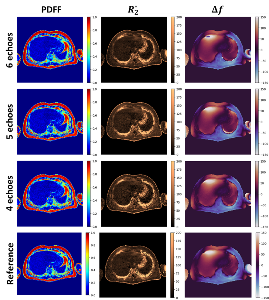
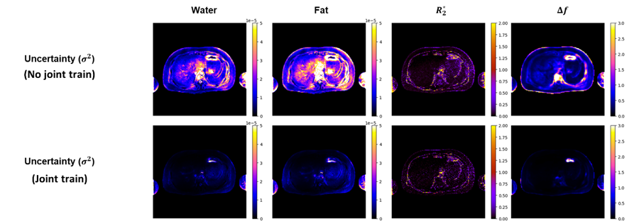
The MAE for PDFF, R2* and Δf maps, obtained using 6-, 5-, and 4-echo PIUA-Net’s versions, are shown in Table 1. As expected, the R2* error increased when the number of echoes was reduced. However, the PDFF and Δf errors remained stable despite the echo reduction.The PDFF, R2*, and Δf maps obtained for a specific testing subject are shown in Figure 2. Additionally, the uncertainty maps included in Figure 3 were obtained before and after the joint training process of GΘ1 and GΘ2.
Discussion
The use of CNNs to solve R2* and Δf, which was previously proposed8, was further extended to train them in a fully unsupervised manner. This novel feature represents a significant advantage with respect to supervised approaches since reference results are no longer necessary during training.Moreover, PIUA-Net also included the estimation of uncertainties. This feature enables the quantification of our method’s imperfections, as shown in Figure 4 where the observed Δf uncertainty before joint training was larger than after joint training. The higher uncertainty in the first case was due to the simplified MR signal model (with R2* = 1) that was assumed during the training stage.
Conclusion
Our proposed PIUA-Net framework is an accurate and efficient DL-based method to quantify liver PDFF using up to 4-echo CSE-MR images without sacrificing performance compared to reference 6-echo techniques. DL-based techniques are usually seen as “black-boxes”, and uncertainty quantification may represent a major advance towards an improved transparency that could lead to the adoption of DL-based PDFF quantification in clinical practice.Acknowledgements
This work was funded by Fondecyt 1200839. J.M. was funded by the National Agency for Research and Development (ANID) / Scholarship Program / DOCTORADO BECAS CHILE/2020 – 21210665. CT was funded by Fondecyt 1231535 and Millennium Institute for Intelligent Healthcare Engineering, iHEALTH (ICN2021_004),References
1. Idilman, I. S. et al. Hepatic steatosis: Quantification by proton density fat fraction with MR imaging versus liver biopsy. Radiology 267, 767–775 (2013).
2. Jafari, R. et al. Deep neural network for water/fat separation: Supervised training, unsupervised training, and no training. Magn Reson Med (2021) doi:10.1002/mrm.28546.
3. Liu, K. et al. Robust water–fat separation based on deep learning model exploring multi-echo nature of mGRE. Magn Reson Med 1–14 (2020) doi:10.1002/mrm.28586.
4. Meneses, J. P. et al. Liver PDFF estimation using a multi-decoder water-fat separation neural network with a reduced number of echoes. Eur Radiol (2023) doi:10.1007/s00330-023-09576-2.
5. Shih, S., Kafali, S. G., Calkins, K. L. & Wu, H. H. Uncertainty‐aware physics‐driven deep learning network for free‐breathing liver fat and R 2 * quantification using self‐gated stack‐of‐radial <scp>MRI</scp>. Magn Reson Med (2022) doi:10.1002/mrm.29525.
6. Hamilton, G. et al. In vivo characterization of the liver fat 1H MR spectrum. NMR Biomed 24, 784–790 (2011).
7. Hernando, D., Kellman, P., Haldar, J. P. & Liang, Z. P. Robust water/fat separation in the presence of large field inhomogeneities using a graph cut algorithm. Magn Reson Med 63, 79–90 (2010).
8. Meneses, J. P. et al. Reproducible DL-based approach for liver PDFF quantification. in ISMRM Annual Meeting (2023).
Figures