1229
Tailored dielectric shimming in MRI using machine learning - a feasibility study1Radiology, Weill Cornell Medicine, New York, NY, United States, 2GE Healthcare, Aurora, OH, United States
Synopsis
Keywords: Analysis/Processing, Shims
Motivation: Inhomogeneities of the MRI transmit field cause image shading and hinder diagnosis. In dielectric shimming, pads of high permittivity are used to recover signal in low intensity areas, but full-wave calculation of the resulting fields is too slow for real-time use at the scanner.
Goal(s): We study feasibility of using AI to rapidly predict the transmit field with dielectric pads.
Approach: An AI pipeline is trained using a small simulated data set for proof of concept.
Results: We obtain a structural similarity of 97% with a mean squared error of 0.02%, demonstrating feasibility and the potential for a real-time implementation in the future.
Impact: This work improves image shading and diagnostics. Dielectric shimming in particular and rapid calculation of electromagnetic fields in general especially apply to ultra-high field strengths such as 7T, 9.4T and 10.5T, where significant inhomogeneity is hindering proper evaluation.
Introduction
B1 inhomogeneity in MRI arises from the increasingly complex electromagnetic behavior of tissues at higher frequencies, causing artifacts and inconsistent signal intensity in the images, hampering diagnostic accuracy1,2. Dielectric shimming can be used to counteract these issues by strategically placing dielectric materials on the anatomy, thereby recovering signal in low-intensity regions and compensating for the inherent inhomogeneities3,4,5.Here, we propose an artificial intelligence (AI) software that predicts the B1 field when dielectric pads are used. The ultimate goal of this tool is to provide real-time guidance in an MR scan, delivering the optimal location, material, and shape/size for the dielectric pad(s).
Methods
A. Concept:We employed a deep learning pipeline that uses a B1 map representing the original transmit field as the input. We added the dielectric pad into this input image to encode its location and size. The relative dielectric permittivity was encoded by varying the brightness of the pad in the image. The output of the algorithm was a B1 map recorded when the dielectric pad was in place.
B. Data generation:
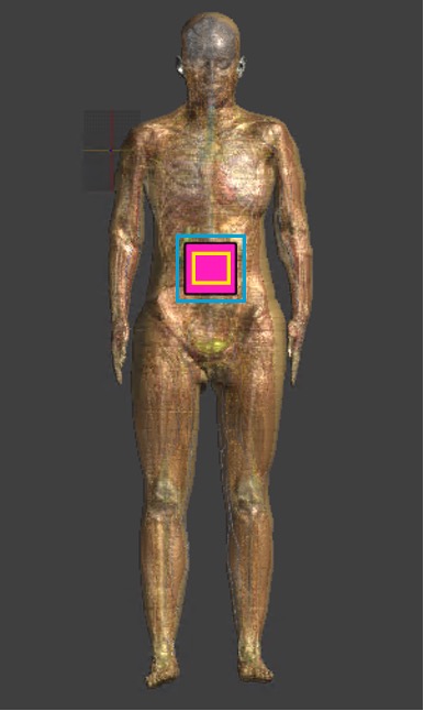
Input data was acquired from abdominal Sim4Life simulations (Zurich MedTech, Zurich, Switzerland) employing the Virtual Population (IT’IS Foundation, Zurich, Switzerland). A 3T birdcage body coil was interfaced with the virtual body model Ella. A dielectric pad (εr = 500 and σ = 0 in this first feasibility study) was added (Fig.1). A parametric sweep was performed with varied pad size (thickness=10mm, length=125±25mm, width=125±25mm) and location (±50mm, and ±50mm from isocenter along y and z), leading to 81 unique configurations. B1 maps with and without the pad were exported into MATLAB for post-processing.
C. Postprocessing
The simulated data was exported slice by slice into images of dimension 224x224.
Input: The dielectric pad was added into the images of the original B1 transmit field without the pad.
Output: The B1 maps when simulated with the pad were exported.
This dataset consisted of ~5,000 images and was split into 75%/15%/10% subsets for training, validation, and testing.
D. Network
We implemented a unet2D architecture using a cascade of convolutional filters with nonlinear reLU activation and input image sizes of 224x224. Training was performed using stochastic gradient descent (SGD) and Keras/Tensorflow (Google, Mountain View, California). Hyperparameters were optimized by minimizing root mean squared error (RMSE) loss on the validation datasets. Training was performed over 30 epochs (SGD) using a GeForce RTX 2080 Ti Graphics Processing Unit (GPU) (NVIDIA, Santa Clara, USA). Layers were randomly initialized using the He initialization.
E. Testing
Quantitative image quality comparisons were performed between the simulated images (simulated B1 field with dielectric pad) and the predicted B1 field using RMSE, MSE and structural similarity (SSIM), which unlike RMSE can evaluate perceptual image quality.
Results
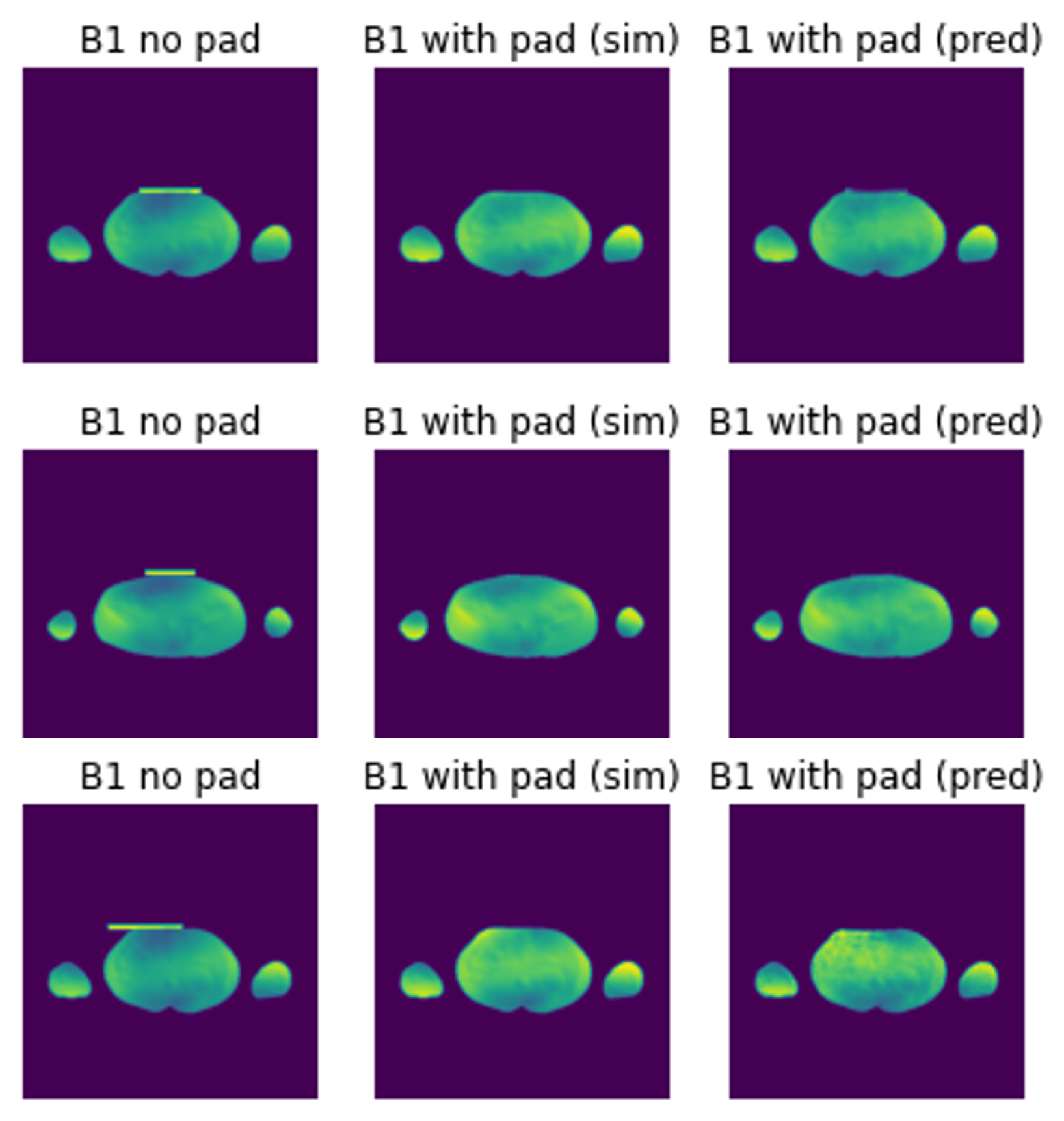
Qualitative examples of the predicted and simulated images are shown in Table 1, showing good visual agreement. Comparing the prediction to the simulation for the entire dataset, we obtained a mean squared error (MSE) of 0.020%±0.005% with a root mean squared error (RMSE) of 1.4%±0.2%. A structural similarity index (SSIM) of 97%±8% shows excellent agreement.Discussion and Conclusion
This pilot work shows proof of concept for a future real-time tool that can determine optimal dielectric pad configurations. In a future implementation, a 3D neural network will prove useful such that fields outside of the area with the dielectric pad can be tailored. This work only includes one body model, only the abdominal anatomy, and one dielectric permittivity – the expansion to a larger dataset is straightforward. A future real-time operation will also require an added optimization algorithm that evaluates different configurations of dielectric pads and reports the best suited arrangement for use by the MR technician. Currently, the input used is a B1 map – this is a map that can be scanned in practice. It might prove useful to use an anatomical image such as a localizer instead to avoid extra scan time. The localizer contains the image shading corresponding to the B1 transmit field inhomogeneity and is expected to perform well in an AI pipeline based on a similar tool created by our group for local SAR prediction6-10.Acknowledgements
This research work was supported by the National Institutes of Health (R01EB031820).References
- Ibrahim, T. S., Lee, R., Abduljalil, A. M., Baertlein, B. A., & Robitaille, P. M. (2001). Dielectric resonances and B1 field inhomogeneity in UHF magnetic resonance imaging: computational models and experiments. Magnetic Resonance Imaging, 19(2), 219-226.
- Collins, C. M., & Smith, M. B. (2001). Signal-to-noise ratio and absorbed power as functions of main magnetic field strength, and definition of "90°" RF pulse for the head in the birdcage coil. Magnetic Resonance in Medicine, 45(4), 684-691.
- Webb, A. G. (2011). Dielectric shimming to correct for RF inhomogeneities. NMR in Biomedicine, 24(8), 957-964.
- Katscher, U., Bornert, P., Leussler, C., & van den Brink, J. S. (2003). Transmit SENSE. Magnetic Resonance in Medicine, 49(1), 144-150.
- Van Gemert, J. H., Brink, W., Webb, A., & Remis, R. (2016). An efficient methodology for the analysis of dielectric shimming materials in magnetic resonance imaging. IEEE transactions on medical imaging, 36(2), 666-673.
- Gokyar, S., Zhao, C., Ma, S. J., & Wang, D. J. J. (2023). Deep learning-based local SAR prediction using B1 maps and structural MRI of the head for parallel transmission at 7 T. Magnetic resonance in medicine, 90(6), 2524–2538. https://doi.org/10.1002/mrm.29797
- Winkler, S. A., Saniour, I., Chaudhari, A., Robb, F., & Vaughan, J. T. (2020, December). MRSaiFE: tissue heating prediction for MRI: a feasibility study. In 2020 IEEE MTT-S International Microwave Biomedical Conference (IMBioC) (pp. 1-3). IEEE.
- Gokyar, S., Saniour, I., Robb, F., Nghiem, A., Kainz, W., Chaudhari, A. S., & Winkler, S. MRSaiFE: towards the real-time prediction of SAR in 3T and 7T MR RF coils-a feasibility study with 10 body models.
- Gokyar, S., Robb, F. J., Kainz, W., Chaudhari, A., & Winkler, S. A. (2021). MRSaiFE: an AI-based approach towards the real-time prediction of specific absorption rate. IEEE Access, 9, 140824-140834.
- Moghadam MC, Panjwani N, Motovilova E, Zhang M, Robb F, Hoang A, Afrin T, Winkler S.A. (2023). Optimized model architecture and generalization for deep learning-based SAR prediction (MRSaiFE). ISMRM 2023
Figures