1226
Feasibility of boosting SNR using cooled RF receive coils for low field human brain imaging at 72mT1Martinos Center for Biomedical Imaging, Massachusetts General Hospital, Charlestown, MA, United States, 2Radiology, Harvard Medical School, Boston, MA, United States
Synopsis
Keywords: Non-Array RF Coils, Antennas & Waveguides, Low-Field MRI, Cyrogenic RF receive coil, Portable MRI
Motivation: Cooled coils could provide an boost SNR for portable low field MRI as an alternative to increasing the field strength using larger, heavier magnets.
Goal(s): Test whether cooling RF receive coils with liquid nitrogen could provide an appreciable boost to signal-to-noise ratio (SNR) on low-field portable MRI scanners, where noise is dominated by copper losses in the coil.
Approach: A 3.04 MHz multi-turn surface coil was cooled to ~77K using liquid nitrogen and performance was assessed using bench Q measurements and imaging data.
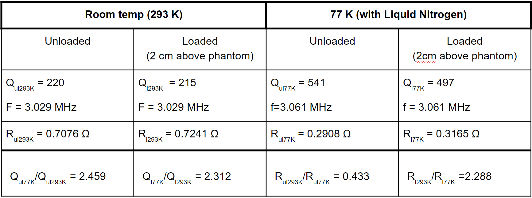
Results: The unloaded Q increases from 220 at 293K to 541 at 77K. SNR for the cooled coil increased 4.15x.
Impact: Liquid nitrogen cooled radiofrequency receive coils could improve image quality for low field portable MRI scanners, broadening the impact of these systems.
Introduction
Several portable permanent-magnet based MRI scanners have been recently introduced [1-4], but SNR can limit imaging performance at low field particularly for applications such as diffusion-weighted imaging (DWI). While external noise can be mitigated using adaptive noise cancellation [3,5-6], improved baseline SNR typically requires increased magnetic field strength, which comes at the cost of portability. Because SNR scales according to b1-/sqrt(R), an alternative way to improve SNR is to reduce the receive coil noise resistance R by cooling the copper coil conductor to cryogenic temperatures. As low-field receive coils are typically coil copper noise dominated (Q-ratio < 2), cooling the coil reduces the primary source of Johnson-Nyquist noise and improves the unloaded Q [8]. This stands in contrast to high field imaging, where we expect only limited benefit from cooling Rx coils since the Q-ratio is typically significantly greater than 2 and thus most of the noise comes from the body.Methods
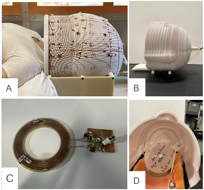
Coil: Figure 1 shows a surface 8-turn spiral surface coil wound from AWG 18 solid copper wire to 3.03 MHz and matched it to 50 Ohms by using a lattice balun network.Q value measurement: We measured the unloaded and loaded Q values of the coil on a network analyzer twice using a decoupled double probe S21. The Q’s were measured first at room temperature (293K) and then following immersion in a liquid nitrogen bath in a styrofoam container.
Frequency shift: We measure the resonant frequency of the coil in the liquid nitrogen bath. Then we removed the coil and measured the frequency every 20 seconds for 32 minutes.
Image acquisition: Images were acquired on an 72mT human brain scanner [9-10]. using a volumetric solenoid coil [11] for Tx and the 8-turn surface coil for Rx (Figure 1). The Tx and Rx coils were geometrically decoupled with the Tx coil positioned orthogonal to B0 as well as the main B1+ field from the Tx coil. The Rx coil was connected to a Tx/Rx switch and two stages of pre-amp (MITEQ 1583). The ball phantom (diameter 8cm) was filled with water 0.9% of NACL and 4 ml GAD/400 ml. Images were acquired using a 2D single-shot RARE imaging sequence using frequency-swept excitation and refocusing pulses [12]. The images were first acquired with the Rx coil at room temperature. Then the Rx coil was immersed in the bath with liquid nitrogen outside of the scanner for 5 minutes and placed back into the scanner, and images were acquired 2-3 minutes later.
Results & Discussion
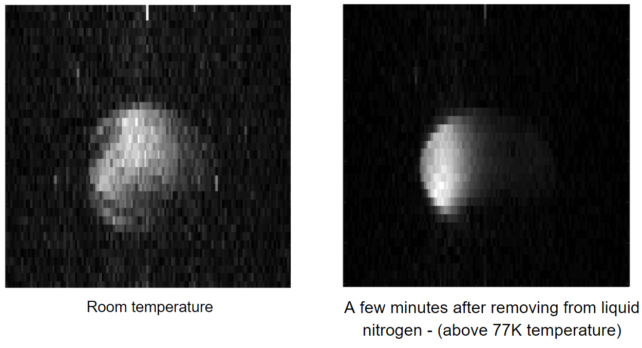
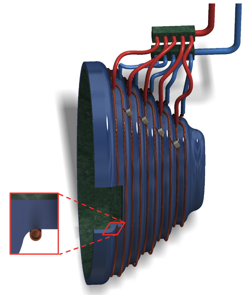
The first experiment showed 2.46x improvement in the Q value for the unloaded cooled coil (Q=541) vs. measurement at room temperature (Q=220). During warm-up, the resonant frequency decreased exponentially from 3.06 MHz at 77K to 3.03 MHz at room temperature (Figure 2). Figure 3 shows the imaging results with a much more conspicuous noise floor at 293K compared to 77K. The signal to noise ratio changed from 21.74 to 90.13 which gives us 4.15x improvement when the coil resistivity is decreased. Unfortunately, increasing the Q value of the coil also caused narrowing bandwidth, influencing image size.Several challenges remain before cooled coils can be used for brain imaging at low field. First, it is impractical to place a container with liquid nitrogen inside the scanner because it constrains the position of the coil. Second, the coil and body must be thermally insulated from one another. One potential solution to these problems is shown in Figure 4. A volumetric solenoid coil can be made of hollow conductor loops connected in series across tuning capacitors. Liquid nitrogen is pumped through the hollow conductor providing efficient heat dissipation. To avoid gas accumulation inside the conductor due to liquid nitrogen boiling, the loops are connected in parallel to inlet and outlet manifolds using PTFE tubes. In this approach, the volume of liquid in close proximity to the body is significantly reduced. The hollow conductor is mounted on a few millimeters of helmet substrate made from a thermally insulating between the coil and body in order to maximize the signal while still providing good thermal isolation.Conclusion
Experiments suggest that lowering receive coil temperature to 77K can significantly improve the coils sensitivity and image SNR for human brain scanners. Future work will focus on adaptations to make cooled coils safe and practical for human brain imaging experiments.Acknowledgements
This work was supported by grant R21-EB034865-01 and R01HD104649.References
1. Kimberly WT, Sorby-Adams AJ, Webb AG, et al. Nat Rev Bioeng. 2023. doi: 10.1038/s44222-023-00086-w.
2. Cooley CZ, McDaniel PC, Stockmann JP, Srinivas SA, Cauley SF, Śliwiak M, Sappo CR, Vaughn CF, … Wald LL, 2021. A portable scanner for magnetic resonance imaging of the brain. Nat Biomed Eng. 5(3):229-239. PMID: 33230306.
3. Liu Y, Leong ATL, Zhao Y, Xiao L, Mak HKF, Tsang ACO, Lau GKK, Leung GKK, Wu EX, 2021. A low-cost and shielding-free ultra-low-field brain MRI scanner. Nat Commun 2021 121. 12(1):1-14. PMID: 34907181.
4. O’Reilly T, Teeuwisse W, Winter L, Webb AG, 2019. The design of a homogenous large-bore Halbach array for low field MRI. Proc 27th Annu Meet ISMRM, Montr 2019.:272.
5. Srinivas SA, Cauley SF, Stockmann JP, Sappo CR, Vaughn CE, Wald LL, Grissom WA, Cooley CZ, 2021. External Dynamic InTerference Estimation and Removal (EDITER) for low field MRI. Magn Reson Med. PMID: 34480778.
6. Dyvorne et al, Freeing MRI from its Faraday cage with Interference Rejection, ISMRM 2021, 0749
7. McDaniel P, Cooley CZ, Stockmann JP, Wald LL, 2019. Numerically optimized design for a low-cost, lightweight 86mT whole-brain magnet. Proc 27th Annu Meet ISMRM, Montr 2019.:1466.
8. Frank Resmer, Hugh C. Seton, and James M.S. Hutchison. Cryogenic receive coil and low noise preamplifier for MRI at 0.01 T. Journal Magn. Res., 203;2010:57-65.
9. McDaniel P, Cooley CZ, Stockmann JP, Wald LL, 2019. Numerically optimized design for a low-cost, lightweight 86mT whole-brain magnet. Proc 27th Annu Meet ISMRM, Montr 2019.:1466.
10. Cooley CZ, Stockmann JP, Wald LL, 2021. A portable brain MRI scanner based on a 72 mT, 35 kg “Halbach-bulb” magnet and external gradient coil. Proc 29th Annu Meet ISMRM, virtual, 2021.:0559.
11. Sheng Shen, Zheng Xu, Neha Koonjoo, Matthew S. Rosen. Optimization of a close-fitting volume RF coil for brain imaging at 6.5 mT using linear programming. IEEE Trans. Biomed. Eng. 202012. Mohammadali Foroozandeh, Mathias Nilsson, Gareth A. Morris. Improved ultra-broadband chirp excitation. Journal of Magnetic Resonance, 302;2019:28-33.
Figures