1221
Optimization of Flexible Metasurfaces at 7T and in vivo B1+ Correction Effects1Center for Advanced Metabolic Imaging in Precision Medicine, Department of Radiology, University of Pennsylvania, Philadelphia, PA, United States, 2Magnetic Detection and Imaging group, TechMed Centre, University of Twente, Enschede, Netherlands, 3Department of Radiology, University of Pennsylvania, Philadelphia, PA, United States, 4Department of Neurology, University of Pennsylvania, Philadelphia, PA, United States
Synopsis
Keywords: New Devices, New Devices
Motivation: Ultra-high field imaging (≥7T) lacks transmit (B1+) field inhomogeneity due to the shortened RF wavelengths, often resulting in poor image quality. Novel metasurface designs have previously been effective at improving image quality at lower field strengths but have not yet been implemented at 7T.
Goal(s): To optimize and demonstrate a novel metasurface design for in vivo usage at 7T.
Approach: Empirical optimization and phantom testing produced the final design, while image enhancement was assessed via in vivo calf skeletal muscle imaging.
Results: The metasurface produced a 126.5% increase in image relative SNR and a 27.6% increase in transmit efficiency.
Impact: The work impacts 7T imaging by presenting a novel piece of hardware that can effectively improve image quality. Future work on this project will include further optimization via variable distributed capacitance across the metasurface.
Introduction
Transmit field (B1+) inhomogeneities continue to be a challenge at ultra-high fields (≥7T) due to "standing wave" effects caused by shortened radiofrequency (RF) wavelengths. Among the many methods aimed at overcoming this problem, passive high-permittivity materials have seen a high degree of success in the form of aqueous dielectric pads. However, they are limited in their corrective effect due to the limited permittivity of materials found in nature. One alternative solution implemented at lower field strengths1,2, that attempts to overcome these limitations is a 2-dimensional composite material, known as metasurfaces. However, there have been limited studies using metasurfaces or metamaterials for enhancing B1+ field at 7T3. In the work presented here, an optimized metasurface is demonstrated to correct B1+ inhomogeneities both in phantom and in vivo at 7T.Methods
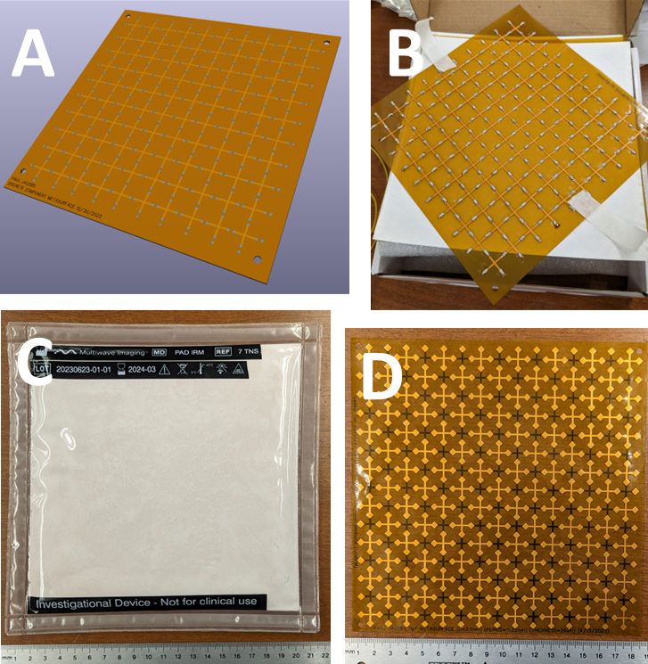
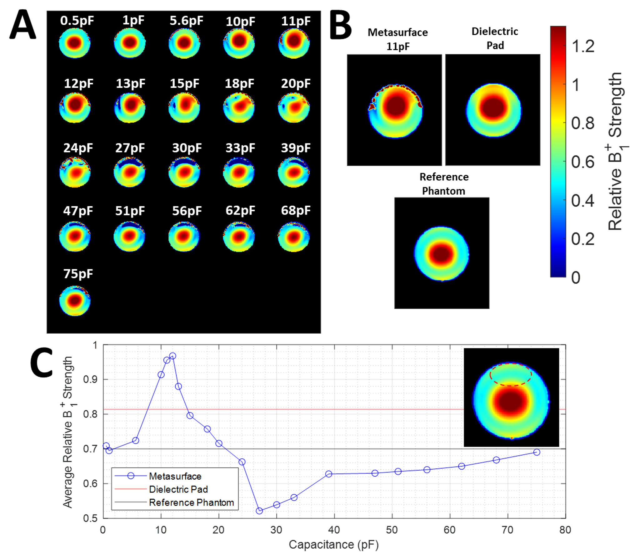
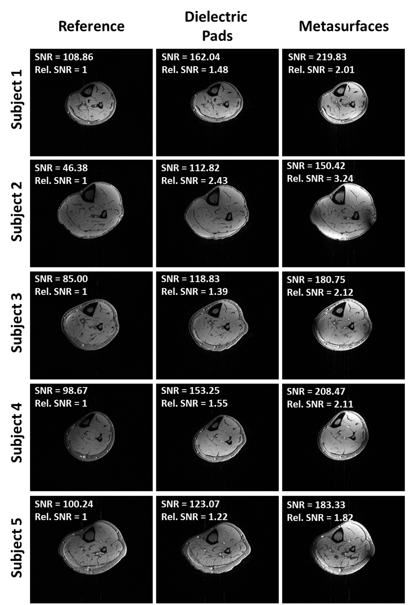
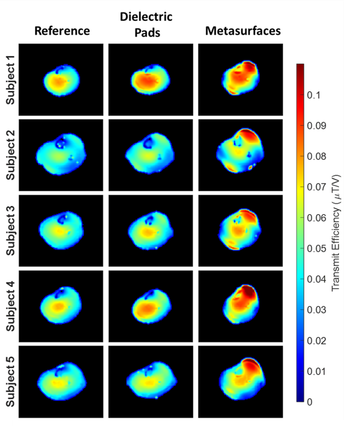
A prototype 18×18cm2 metasurface was designed as a single-layer flexible printed circuit board (PCB) in a 9×9 unit cell arrangement (unit cell size: 15mm, trace width: 1mm, board thickness: 83.5μm), as shown in Figure 1a and 1b. Initial optimization was performed by varying 220 discrete capacitors across 21 values (0.5-75pF) and performing B1+ mapping as described in Volz et al.4 on a saline phantom at 7T (MAGNETOM Terra, Siemens Healthcare) using a single-channel transmit/32-channel receive phased array proton head coil (Nova Medical). The optimized metasurface used for in vivo imaging had capacitors with a values of 11pF and an adjusted unit cell size of 11.25mm. Temperature safety testing was performed in a polyacrylic acid (PAA) phantom using fiber optic probes to determine if any heating occurred from the metasurfaces. In vivo images were acquired at 7T with metasurfaces placed on the top and bottom of the distal calf skeletal muscles across five healthy volunteers with written informed consent under an approved institutional regulatory board protocol. In vivo scans were performed using a single-channel transmit.28-channel receive phased array proton knee coil (Quality Electrodynamics). GRE images were acquired across a 12-slice slab with the following parameters: FA=30°, TR/TE=200/2.99ms, matrix size = 240×210, in-plane resolution = 1×1mm2, and slice thickness = 2mm. Transmit efficiency maps were also generated from relative B1+ maps (normalized to transmitter voltage) in which the average value was calculated across the calf to determine degree of enhancement. B0 maps were also acquired to determine if the metasurfaces introduced any off-resonant effects. Additionally, a set of conventional dielectric pads, composed of calcium titanate (CaTiO3) suspended in D2O (7TNS, Multiwave Imaging) with a relative permittivity of 110, were used as a comparison in both phantom and in vivo5, as seen in Figure 1c.Results
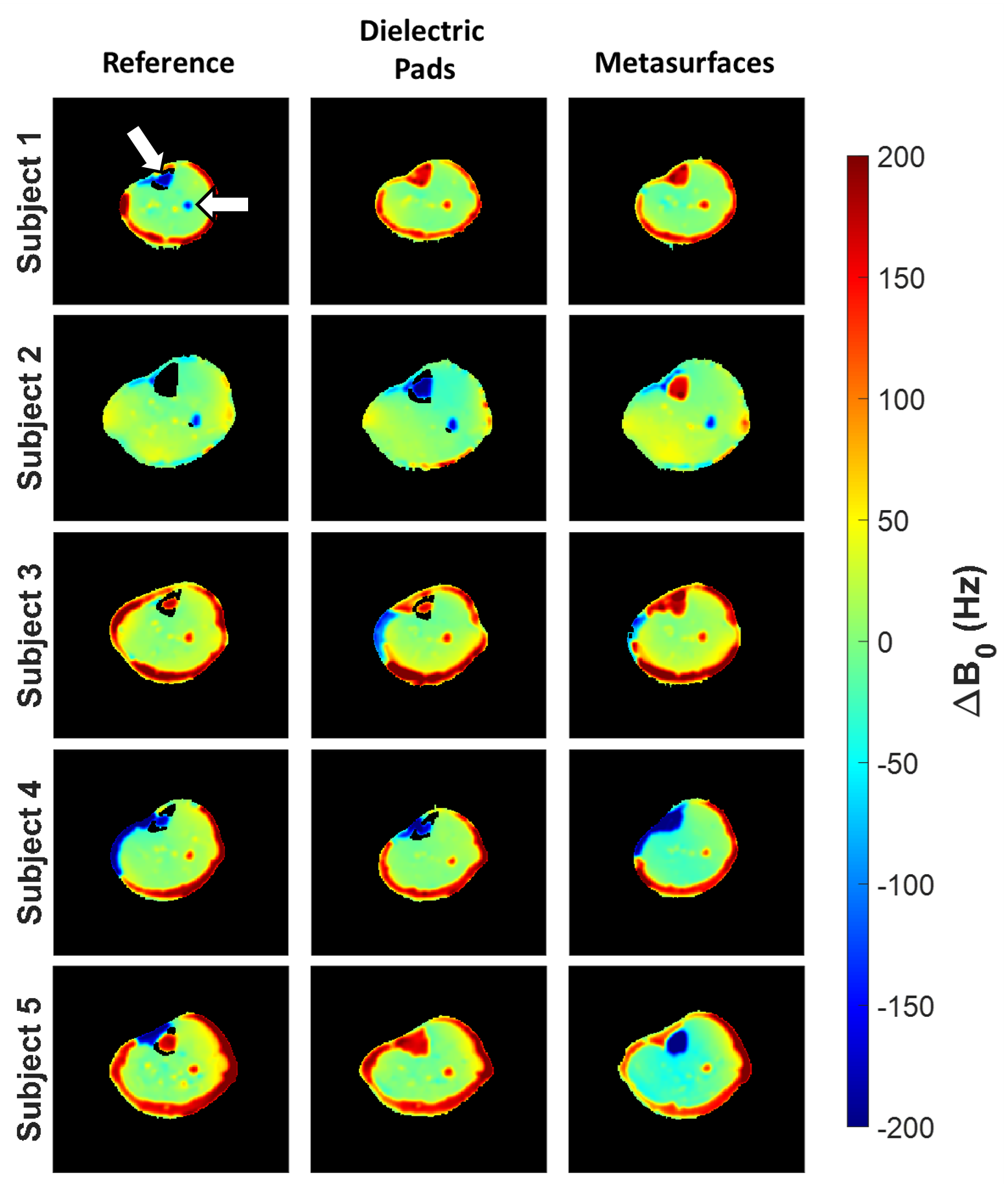
Metasurface optimization data can be seen in Figure 2a, 2b, and 2c which show that a capacitance of 11pF produced the maximal amount of enhancement. The complete set of in vivo axial GRE images were acquired using the optimized metasurface (Figure 1d) and can be seen in Figure 3, showing that the metasurfaces increased the relative SNR value within the calf regions in comparison to both the reference and dielectric pad cases. Specifically, the average relative SNR gain across all five subjects when the metasurfaces were used was 126.5% in comparison to 61.8% when the dielectric pads were used. The metasurfaces also increased the average transmit efficiency across all subjects in the calf region by 27.6% in comparison to 8.9% in the case of the dielectric pad (Figure 4). Lastly, B0 deviation maps, seen in Figure 5, showed no major differences when compared to the other cases, as expected.Discussion
Out study shows that optimized flexible 7T metasurfaces can be used to correct for B1+ inhomogeneities at 7T. Previous studies have used a similar design with a capacitance value of 40pF to correct for B1+ inhomogeneities in abdominal images at 3T, with a high degree of success1. One limitation of the current work lies in the constant capacitance that was used across the metasurface, which may be further optimized in the future to account for asymmetric central unit cell loading, thereby improving the design6.Conclusion
Our results showed that an optimized flexible metasurface design was able to substantially increase the relative SNR of anatomical GRE images of the calf at 7T by increasing the local transmit efficiency. They also address the limitations of dielectric pads by being light in weight and not having a fixed shelf life. Furthermore, these metasurfaces outperformed conventional calcium titanate dielectric padding by nearly double. This work offers a novel approach towards correcting B1+ inhomogeneities and warrants further developments for use in neuroimaging applications at 7T.Acknowledgements
Research reported in this work was supported by the National Institutes of Biomedical Imaging and Bioengineering of the National Institutes of health under award number P41EB029460 and by the National Institute of Aging of the National Institutes of Health under award number R01AG071725.References
1. Vorobyev V, Shchelokova A, Efimtcev A, et al. Improving B1+ homogeneity in abdominal imaging at 3 T with light, flexible, and compact metasurface. Magn Reson Med. Jan 2022;87(1):496-508. doi:10.1002/mrm.28946
2. Slobozhanyuk AP, Poddubny AN, Raaijmakers AJ, et al. Enhancement of Magnetic Resonance Imaging with Metasurfaces. Adv Mater. Mar 2 2016;28(9):1832-8. doi:10.1002/adma.201504270
3. Schmidt R, Slobozhanyuk A, Belov P, Webb A. Flexible and compact hybrid metasurfaces for enhanced ultra high field in vivo magnetic resonance imaging. Sci Rep. May 10 2017;7(1):1678. doi:10.1038/s41598-017-01932-9
4. Volz S, Noth U, Rotarska-Jagiela A, Deichmann R. A fast B1-mapping method for the correction and normalization of magnetization transfer ratio maps at 3 T. Neuroimage. Feb 15 2010;49(4):3015-26. doi:10.1016/j.neuroimage.2009.11.054
5. Teeuwisse WM, Brink WM, Webb AG. Quantitative assessment of the effects of high-permittivity pads in 7 Tesla MRI of the brain. Magn Reson Med. May 2012;67(5):1285-93. doi:10.1002/mrm.23108
6. Brizi D, Monorchio A. An Analytical Approach for the Arbitrary Control of Magnetic Metasurfaces Frequency Response. Ieee Antenn Wirel Pr. Jun 2021;20(6):1003-1007. doi:10.1109/Lawp.2021.3069571
Figures