1220
Preliminary Results on Torso PNS Thresholds at the Ultrasonic Driving Frequency of 20 kHz using a Whole-Body Gradient Coil1Radiology, University Medical Center Utrecht, Utrecht, Netherlands, 2Spinoza Center for Neuroimaging, Amsterdam, Netherlands
Synopsis
Keywords: Safety, Bioeffects & Magnetic Fields
Motivation: For silent whole-body MRI using ultrasonic encoding at 20kHz, very high slew rates will be experienced by body parts such as the torso with PNS risk. However, PNS thresholds for these body parts at ultrasonic frequencies are not well known.
Goal(s): To estimate PNS thresholds for the torso at 20kHz.
Approach: We test volunteers in a whole-body gradient coil driven at 20kHz to determine when PNS occurs and compare to simulations and current IEC guidelines.
Results: PNS occurs at much higher dB/dt values than predicted by IEC guidelines at 20kHz, where we found a mean threshold value of 1316T/s for the torso.
Impact: PNS is a concern for body MRI using ultrasonic encoding. Here, we show much higher measured PNS thresholds than IEC guideline predictions for 20kHz gradient switching, suggesting that whole-body silent MRI at 20kHz is possible with reduced risk of PNS.
Introduction/Background
Peripheral nerve stimulation is induced by changes in the magnetic field (dB/dt) from switching gradient coils and can be experienced as muscle contractions or paresthesia1. Subsequently, dB/dt values act as an indicator for PNS chance. Reduced gradient switching is often employed to lower slew rates, and therefore dB/t, to avoid PNS. However, this can result in extended scan times and reduced spatial resolution. PNS thresholds are used as a safety limit in MRI to ensure PNS is minimised and avoid cardiac stimulation.PNS is also a concern for ultrasonic encoding with switching frequencies above 20 kHz2-3, especially when scaling to a whole-body ultrasonic gradient. The IEC guidelines4 predict that PNS thresholds increase at very high frequencies, which has been observed in volunteer data (above 101kHz)5. Yet, to our knowledge, no direct determination of a PNS threshold has been made at 20 kHz, leaving uncertainty of PNS thresholds for a whole-body ultrasonic gradient coil. We previously presented a whole-body gradient coil at 20kHz for PNS threshold determination6. This work presents an updated version of this coil and preliminary results on PNS thresholds for seven volunteers. We also compare these results to simulations and IEC guideline predictions.
Methods
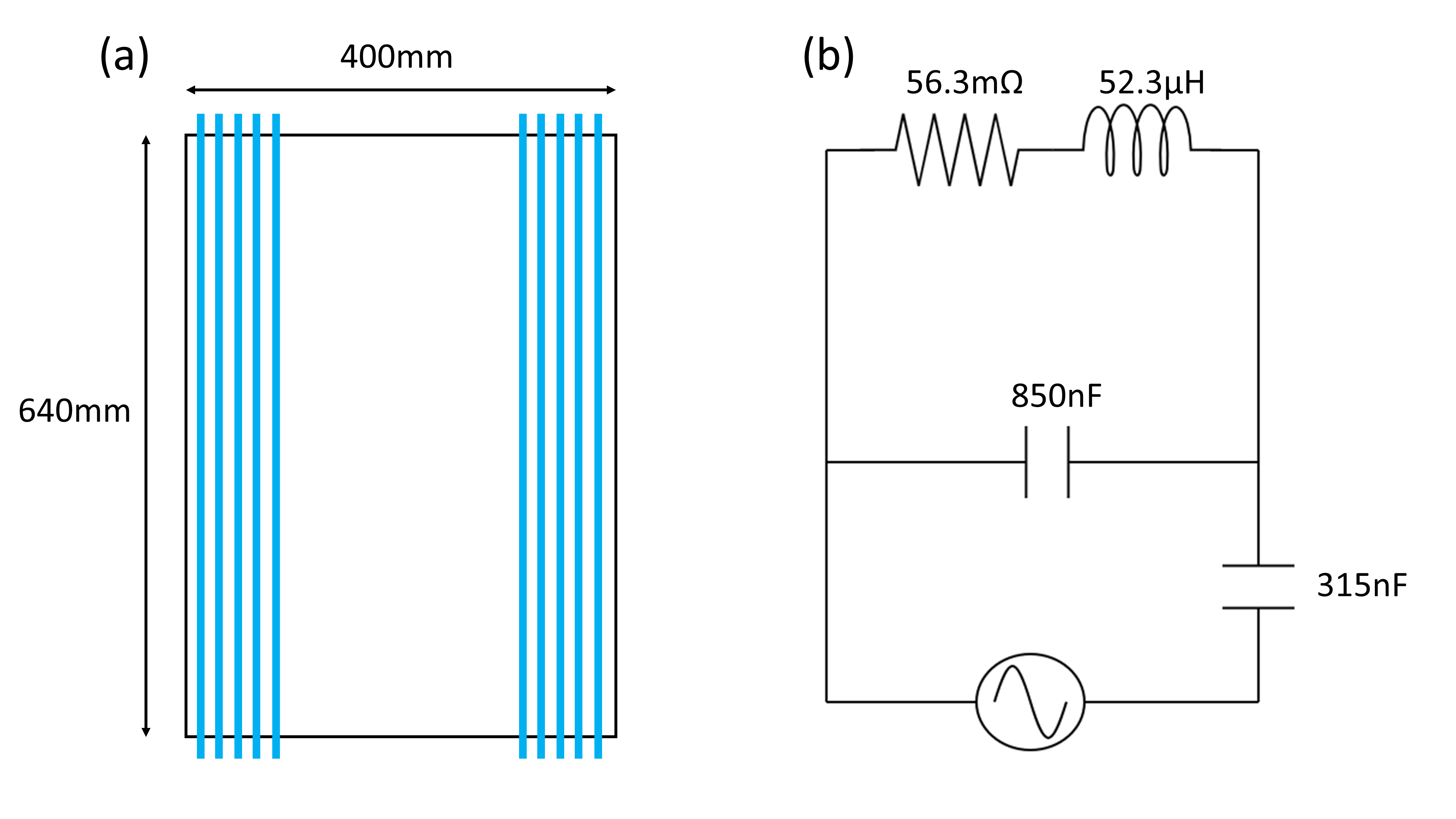
The whole-body gradient coil is a z-axis coil with a length of 40cm and a diameter of 64cm(Figure 1a). The coil design6 was updated with a new capacitor bank achieving a resonance frequency of 20.2kHz matched to 1.1Ω. The measured inductance and resistance of the coil were 52.3µH and 56.3mΩ, respectively(Figure 1b). The capacitor bank can withstand a maximum voltage of 8820V, allowing a current in the coil to induce a maximum field strength of 32mT corresponding to a dB/dt >= 4000T/s for 20.2kHz switching.Magnetic field simulations were performed in Sim4Life (Sim4Life, ZMT, Switzerland) with matching coil parameters to the physical coil(Figure 1) on the Duke body model. Using these values, a comparison to the predicted dB/dt threshold values was made to determine if simulation predictions matched the measured field when PNS was experienced in the volunteers.
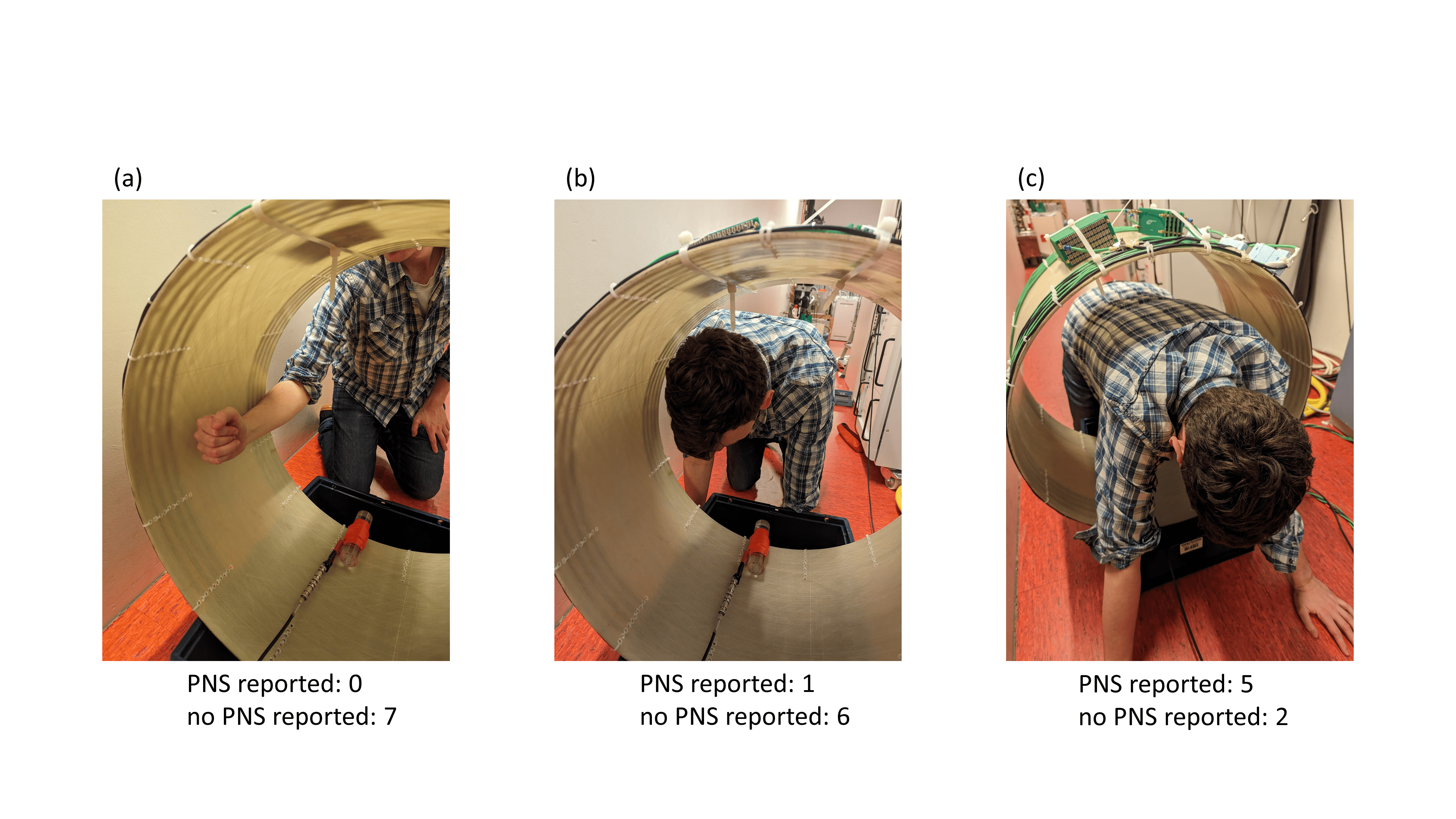
Seven healthy male volunteers were included in this study. PNS testing was completed on the volunteer’s arms, heads and torso(Figure 2). These body parts were assessed at different positions within the coil, corresponding to different dB/dt values. Each measurement involved a 1ms pulse with a current of 332A from the gradient coil. Here, we reduced the current through the coil until no PNS was experienced, allowing PNS threshold determination. A probe solenoid was placed in the gradient coil at the different positions from which we verified the dB/dt from the measured voltage matched dB/dt calculations from Sim4Life(Figure 2).
Results
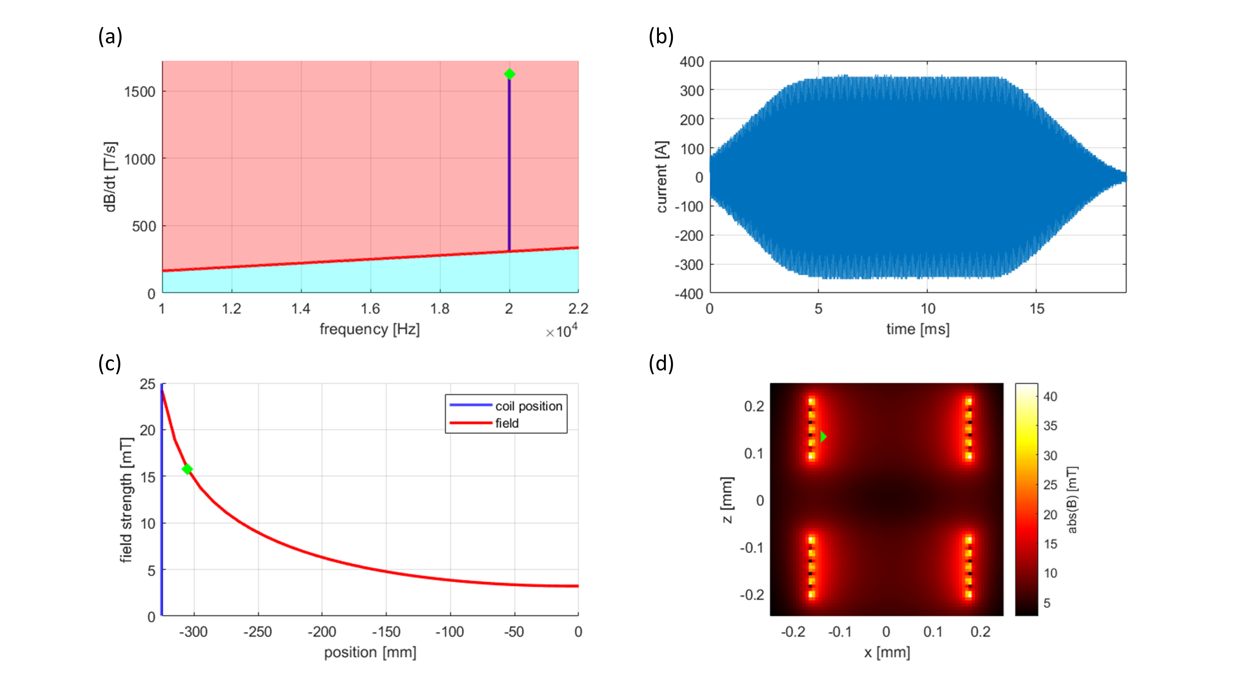
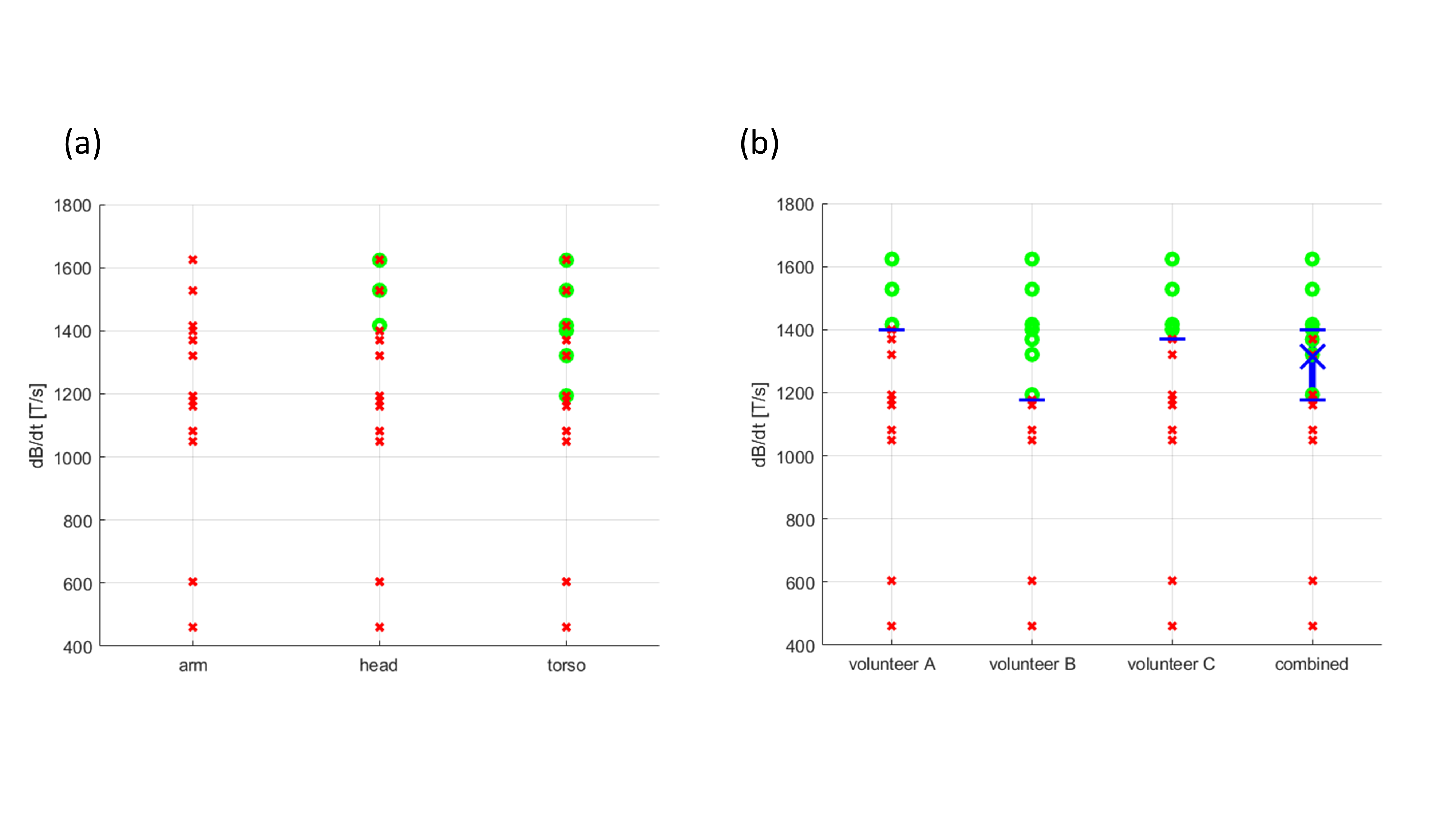
PNS was experienced by six volunteers in this study. PNS was mainly reported (5/7 volunteers) in the torso for the positions closest to the coil conductors, corresponding to the maximum dB/dt; this was experienced as a muscle twitch on the side of the torso. PNS was reported in the head of one volunteer but not in any of the seven volunteers' arms(Figure 2). The volunteer who experienced head PNS did not experience torso PNS.In the position closest to the coils with a current of 332A, the solenoid voltage was measured as 20V. This corresponds to a dB/dt of 1592T/s(Figure 3). At 20.2kHz, the IEC guidelines predict the threshold for PNS to be 311T/s for the torso. Meanwhile, even in the centre of the coil, a measured dB/dt of 481.5T/s did not result in any experienced PNS(Figure 4a).
Head PNS corresponded to a measured dB/dt of 1573T/s. For three volunteers chosen for preliminary torso PNS threshold determination, a mean threshold of 1316T/s was found(Figure 4b), which is 323% greater than the threshold based on IEC calculations. At this threshold, the gradient strength is 34.9mT/m with a slew rate of 4383T/m/s.
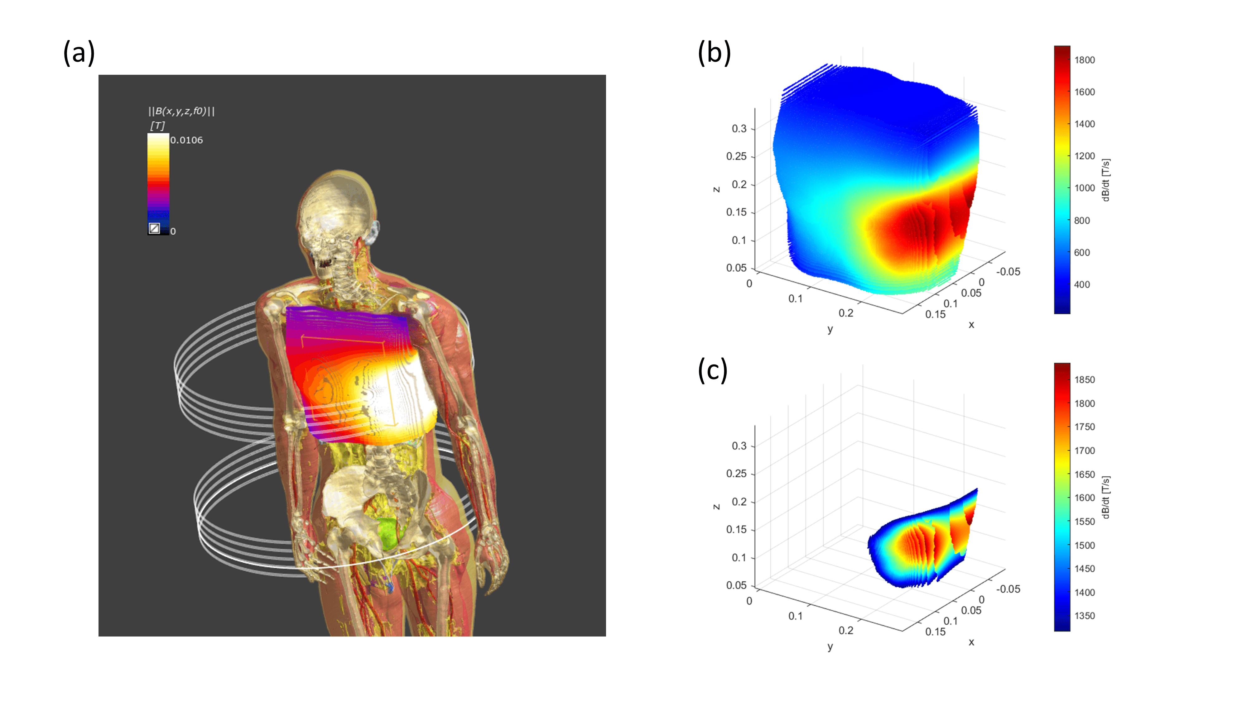
Sim4Life simulations at 332A also demonstrated a large region of the torso to experience dB/dt well above the mean threshold. This indicates the measured preliminary torso PNS threshold is more accurate than the IEC-based predictions(Figure 5).
Conclusion
We presented a setup that can be used to assess PNS thresholds for whole-body gradients at 20kHz. With this setup, we determined a preliminary torso PNS threshold in healthy volunteers that is much greater than the IEC guideline predictions. This result suggests that much greater gradient switching frequencies are possible without inducing PNS. The threshold value can be improved with increased volunteer participation. We hope that with this result, we can determine safe slew rate limits to achieve ultrasonic encoding for future silent whole-body MRI.Acknowledgements
This work has been financed by NWO grant number 18951.References
1. Ham CL, Engels JM, van de Wiel GT, Machielsen A. Peripheral nerve stimulation during MRI: effects of high gradient amplitudes and switching rates. J Magn Reson Imaging. 1997 Sep-Oct;7(5):933-9372. Versteeg E, Klomp DWJ, Siero JCW. A silent gradient axis for soundless spatial encoding to enable fast and quiet brain imaging. Magn Reson Med. 2021;00:1–12
3. Versteeg E, van der Velden TA, van Leeuwen CC, Borgo M, Huijing ER, Hendriks AD, Hendrikse J, Klomp DWJ, Siero JCW. A plug-and-play, lightweight, single-axis gradient insert design for increasing spatiotemporal resolution in echo planar imaging-based brain imaging. NMR Biomed. 2021 Jun;34(6):e4499
4. IEC. Medical electrical equipment – Part 2-33: Particular requirements for the basic safety and essential performance of magnetic resonance equipment for medical diagnosis. Geneva: International Electrotechnical Commissioin 60601-2-33 Edition 4.0; 2022
5. Weinberg IN, Stepanov PY, Fricke ST, et al. Increasing the oscillation frequency of strong magnetic fields above 101 kHz significantly raises peripheral nerve excitation thresholds. Med Phys. 2012; 39(5):2578-83
6. McGrory MJB, Versteeg E, Siero JCW, Klomp DWJ, A novel PNS measurement setup for body gradient coils at an ultrasonic driving frequency of 20kHz, Proc. Intl. Soc. Mag. Reson. Med. 31 (2023) 2876
Figures