1219
Temperature Changes in the Brain due to External Heat Sources: an MR Thermometry Study1Center for Image Sciences - Computational Imaging Group, UMC Utrecht, Utrecht, Netherlands, 2Biomedical Engineering - Medical Imaging Analysis, Eindhoven University of Technology, Eindhoven, Netherlands, 3Tesla Dynamic Coils, Zaltbommel, Netherlands
Synopsis
Keywords: Safety, Safety
Motivation: RF-induced temperature rise is considered a safety risk for MRI.
Goal(s): To study temperature changes in the brain due to (generally considered safe) heat or cool pads placed on a subject's forehead for comparison to RF-induced temperature elevations.
Approach: Using the Projection onto Dipole Fields method, susceptibility and drift induced field changes are separated from temperature effects to enable assessment of temperature rise in the brain.
Results: Up to 2.5 °C temperature rise was measured at the brain edge in 2 volunteers. Similarly, a 2.0 °C decrease was observed at the brain edge when a coolpad was placed on the forehead.
Impact: RF-induced temperature changes in the brain can be considered modest compared to temperatures induced by (generally considered safe) external heating/cooling sources. The temperature increase due to a heatpad on the forehead is much larger than previously measured RF-induced temperature increases.
Introduction
The use of MR thermometry (MRT) to measure RF-induced temperature rise in the brain for normal MRI scans bears great potential for validation of thermal modelling and verification of current RF safety constraints.1,2 Previous studies have measured temperature rises up to 0.3 °C in the brain for head coils operated at maximal SAR.3,4 In this study, we aim to measure temperature rises in the brain for external heat sources. For this purpose, we have applied the Projection onto Dipole Fields (PDF) method,5 commonly used in Quantitive Susceptibly Mapping,6 to correct for susceptibility-induced B0 field disturbances. The resulting method was applied to measure the temperature change in a subject’s brain when a heatpad/coolpad is placed on a subject’s forehead.Methods
MRT was performed using the proton resonance frequency shift (PRFS) method.7 The presence of a heatpad/coolpad considerably changes the temperature in the skull; therefore altering its magnetic susceptibility.8 This induces a local B0 field change which is characterized as the susceptibility difference convoluted with a dipole. Therefore, phase shift between two dynamics can be modelled as: $$\Delta\phi_{PRFS}=D*\Delta\chi+\Delta\phi_{T}+\Delta\phi_{drift}+\Delta\phi_{motion},$$ with $$$\Delta\phi_{PRFS}$$$ the uncorrected PRFS signal, $$$D$$$ a dipole kernel, and $$$\Delta\chi$$$ the change in magnetic susceptibility. Furthermore, $$$\Delta\phi_{T}$$$, $$$\Delta\phi_{drift}$$$, and $$$\Delta\phi_{motion}$$$ are the phase changes due to temperature, drift, and motion, respectively. A method commonly used to separate susceptibility-induced field variations is PDF.9 This method estimates susceptibility sources by solving the following optimization problem: $$ \Delta\chi^*=\underset{\Delta\chi\in\Omega_{skull}}{\arg\min}||\Delta\phi_{PRFS}-\Delta\phi_{motion}-D*\Delta\chi||_2.$$ Here susceptibility sources can only be placed inside the skull ($$$\Omega_{skull}$$$). By correcting the PRFS signal for motion,3 and by assuming that the PDF method also captures drift fields, the reconstructed temperature-induced phase shift is given by: $$ \Delta\phi_{T}=\Delta\phi_{PRFS}-\Delta\phi_{motion}-D*\Delta\chi^*.$$ An overview of the method is visualized in figure 1. It is first verified through simulations and subsequently applied to in vivo data.Simulations
RF-induced temperature rise distributions were simulated using Sim4Life (ZMT, Zurich, Switzerland). Here, the heatpad was implemented as a Dirichlet boundary condition on the model Duke’s forehead.10 The simulated temperature was used as input to the B0 computation algorithm11 to simulate the effect of susceptibility-induced changes in B0. Additionally, a drift field was added. This data was used to create synthetic PRFS data (figure 2).12
Measurements
MR scans were performed at 7T (Achieva, Philips Healthcare, Best, NL) using a Nova 2TxRx head coil (Nova Medical, Wilmington, USA). Receiver elements were excluded from the conventional setup to provide space for placement of a headpad/coolpad. Brain imaging was performed in 2 subjects (2 male, age 24 and 44) and include 16-echo spoiled-GRE MRT scans (TE1=1.2 ms, TEspacing=1.2 ms, TR=30 ms, 5x5x8 mm3 resolution, 220x200x8 mm3 FOV, flip angle 11°, multi-shot, cardiac triggered13) in 3 orthogonal slices with negligible SAR. However, only a single echo was used for temperature measurements (TE=2.4 ms). During MRT scans, the volunteers placed and/or removed the pad on/from their forehead. The heatpad was heated by submerging in hot tapwater for several minutes; the coolpad was placed in the freezer overnight. During scanning, fiber-optic probe measurements (Opsens, Québec, Canada) were performed at the skin-pad interface. After MRT scans, motion-calibration scans were acquired in which volunteers were asked to deliberately move their head.3
Results
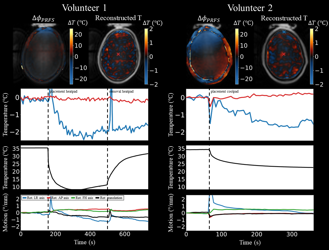
Tests on synthetic data show that the proposed PDF method successfully separates temperature from susceptibility and drift in synthetic PRFS data (figure 3). However, ground truth temperatures are underestimated as $$$D*\Delta\chi^*$$$ also captures temperature effects. The two additional experiments demonstrate that the method is insensitive to the magnitude of susceptibility and drift. Measurements also identify temperature increase in peripheral brain regions neighboring the heatpad (figure 4). Strikingly, when a heatpad is placed a second time–even though the skin already cooled down and susceptibility effects are present in the input data–the brain temperature does not increase anymore. Similarly, a temperature decrease is observed when a coolpad is used (figure 5).Discussion and conclusion
PDF is effective at separating temperature from other effects present in PRFS-based measurements. Although, particularly for more deeply located brain tissue, this method may underestimate temperature changes, it correctly predicts maximum temperature changes in the brain. Note that placement of a heatpad/coolpad on a human’s forehead is considered a safe procedure: coolpads are frequently used for head injuries in sports and a heatpad will not heat more than a hot shower. Here it is shown that these pads induce considerably larger temperature changes in the brain compared to what we can expect from RF-induced heating. However, it should be noted that the pad measurements heat or cool the brain periphery while RF-induced heating for MRI measurements heats up the center of the brain.Acknowledgements
This publication is part of the project "Finding the hotspot: AI unravels tissue heating in MRI" with project number 19995 of the Open Technology Program which is (partly) financed by the Dutch Research Council (NWO).References
- Schaefer DJ, Shellock FG. Health effects and safety of radiofrequency power deposition associated with magnetic resonance procedures. In: Magnetic Resonance Procedures: Health Effects and Safety. ; 2001:55-74.
- Kikken MWI, Steensma BR, van den Berg CAT, Raaijmakers AJE. Multi-echo MR thermometry in the upper leg at 7 T using near-harmonic 2D reconstruction for initialization. Magn Reson Med. 2023;89(6):2347-2360.
- Kikken MWI, Steensma BR, van den Berg CAT, Raaijmakers AJE. MR thermometry of RF heating in the human brain at 7T using the harmonical initialized multi-echo model with SVD-based motion correction. Proceedings of the 2023 ISMRM & SMRT Annual Meeting and Exhibition; 0612.
- Le Ster C, Mauconduit F, Mirkes C, Vignaud A, Boulant N. Measuring radiofrequency field-induced temperature variations in brain MRI exams with motion compensated MR thermometry and field monitoring. Magn Reson Med. 2022;87(3):1390-1400.
- Liu T, Khalidov I, de Rochefort L, et al. A novel background field removal method for MRI using projection onto dipole fields (PDF). NMR Biomed. 2011;24(9):1129-1136.
- Wang Y, Liu T. Quantitative susceptibility mapping (QSM): Decoding MRI data for a tissue magnetic biomarker. Magn Reson Med. 2015;73(1):82-101.
- Rieke V, Pauly KB. MR thermometry. Journal of Magnetic Resonance Imaging. 2008;27(2):376-390.
- Sprinkhuizen SM, Konings MK, Van Der Bom MJ, Viergever MA, Bakker CJG, Bartels LW. Temperature-induced tissue susceptibility changes lead to significant temperature errors in PRFS-based MR thermometry during thermal interventions. Magn Reson Med. 2010;64(5):1360-1372.
- Wu M, Mulder HT, Baron P, et al. Correction of motion-induced susceptibility artifacts and B0 drift during proton resonance frequency shift-based MR thermometry in the pelvis with background field removal methods. Magn Reson Med. 2020;84(5):2495-2511.
- Christ A, Kainz W, Hahn EG, et al. The Virtual Family - Development of surface-based anatomical models of two adults and two children for dosimetric simulations. Phys Med Biol. 2010;55(2).
- Boegle R, MacLaren J, Zaitsev M. Combining prospective motion correction and distortion correction for EPI: Towards a comprehensive correction of motion and susceptibility-induced artifacts. Magnetic Resonance Materials in Physics, Biology and Medicine. 2010;23(4):263-273.
- Poorman ME, Braškutė I, Bartels LW, Grissom WA. Multi‐echo MR thermometry using iterative separation of baseline water and fat images. Magn Reson Med. 2019;81(4):2385-2398.
- Steensma BR, van den Berg CAT, Raaijmakers AJE. Towards high precision thermal based RF safety assessment with cardiac triggered MR thermometry. Proceedings of the 2021 ISMRM & SMRT Annual Meeting and Exhibition; 1260.
- Dymerska B, Eckstein K, Bachrata B, et al. Phase unwrapping with a rapid opensource minimum spanning tree algorithm (ROMEO). Magn Reson Med. 2021;85(4):2294-2308.
Figures
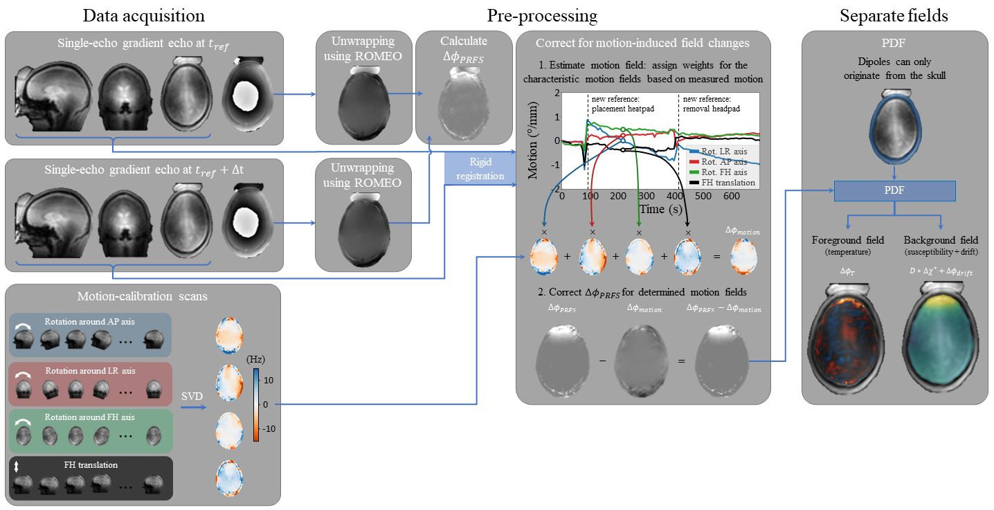
Figure 1: Proposed method to separate temperature from other PRFS effects: drift, motion, and susceptibility. ΔφPRFS is calculated from the unwrapped (using ROMEO14) transverse plane phase data with respect to a reference (e.g., dynamic after placement of heatpad). ΔφPRFS is subsequently corrected for motion-induced field changes using measured motion parameters (determined by registration) and the characteristic motion-induced fields (from the motion-calibration scans). PDF is finally used to separate temperature from susceptibility-induced field changes.
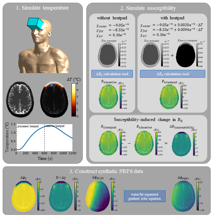
Figure 2: Synthetic PRFS data generation. Temperature changes due to a heatpad are simulated in Sim4Life. Here, we first simulate a steady state (3600 s), followed by heatpad heating implemented through a 45 °C Dirichlet boundary condition (600 s), and finally a cooldown period in which the boundary condition is removed (600 s). The resulting temperatures are subsequently used to simulate how local temperature rise alters susceptibility and therefore disturbs the B0 field. Changes in B0 field due to temperature, susceptibility, and drift are used to construct synthetic PRFS data.
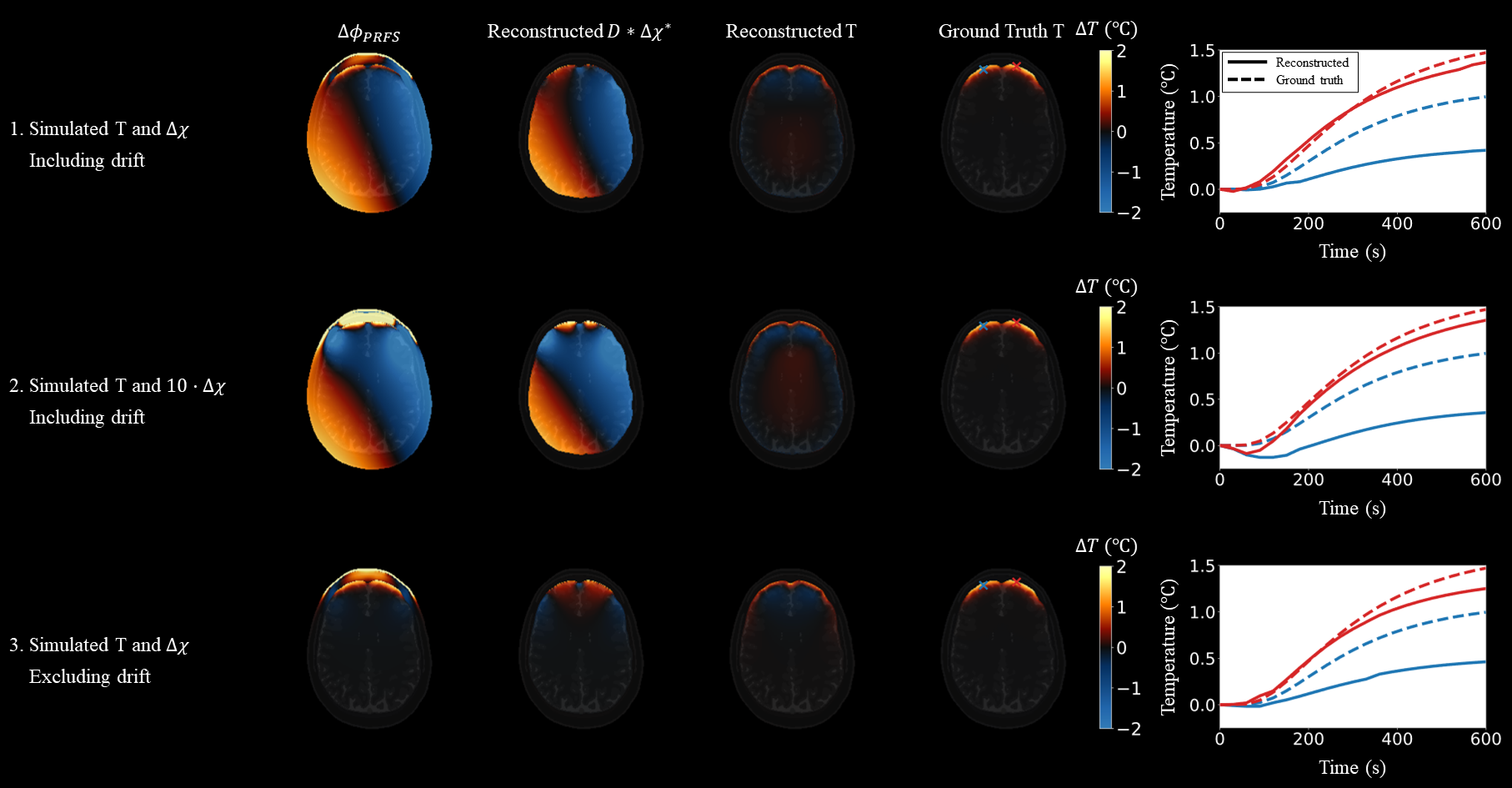
Figure 3: Performance of the PDF algorithm on synthetic PRFS data. Three scenarios are evaluated: (1) default scenario with parameters as shown in figure 2, (2) scenario with 10 times as large B0 field due to susceptibility (more similar to measured data, as can be seen in figure 4), and (3) scenario without B0 field drift. For every scenario, the input, reconstructed susceptibility, reconstructed temperature, and ground truth temperature are provided. Also, the temperature over time is provided for two voxels: exactly at the edge of the brain (red) and slightly more inwards (blue).
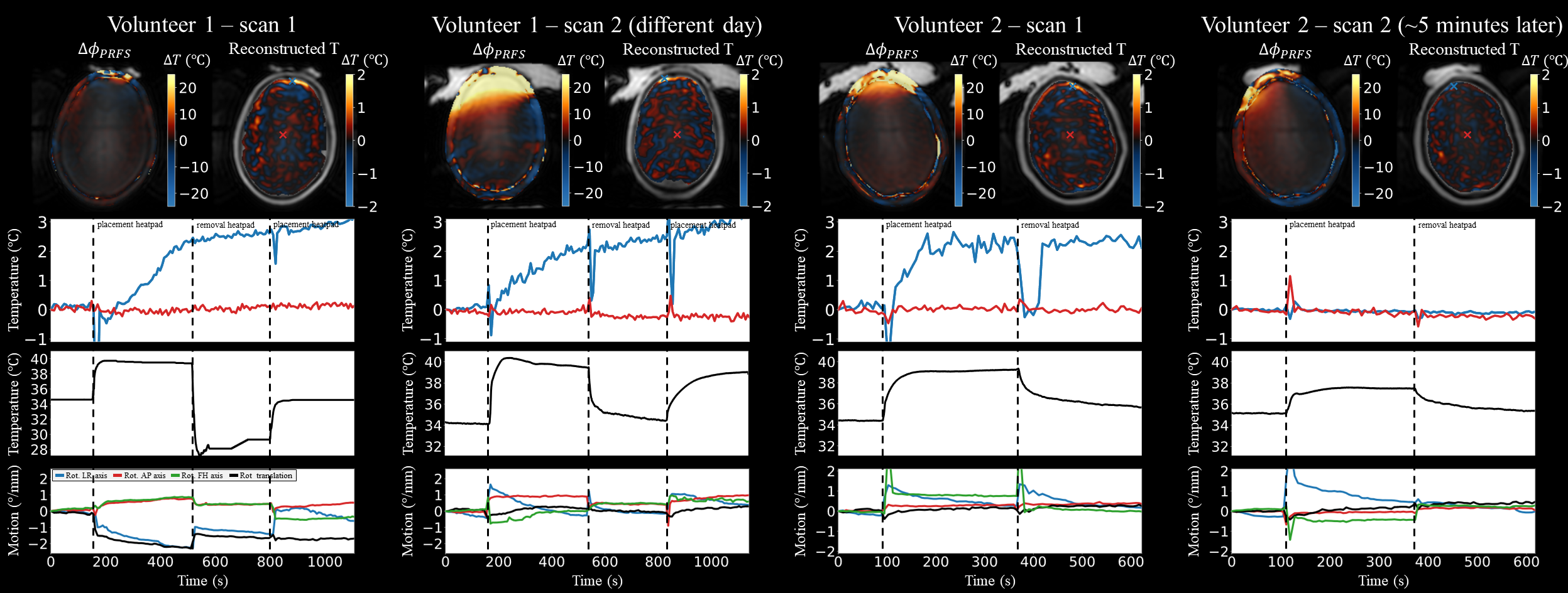
Figure 4: Temperature reconstruction for placement of a heatpad during dynamic scanning in 2 volunteers. The input (ΔφPRFS) to the PDF algorithm clearly shows susceptibility effects. PDF removes these effects, as reconstructed temperatures only show temperature changes at the brain edge (except for the repeated measurement for subject 2). For every scan, the reconstructed temperature in 2 voxels, the probed temperature at the skin surface (black line), and the motion parameters are plotted over time. Dotted lines indicate when the heatpad is placed/removed on/from the forehead.