1218
Evaluation of Specific Absorption Rate of Tight-Fit Array Coils for Human Head MRI at 9.4T in Presence of EEG Caps1High-field MR Center, Max Planck Institute for Biological Cybernetics, Tuebingen, Germany, 2Department of Biomedical Magnetic Resonance, University of Tübingen, Tuebingen, Germany
Synopsis
Keywords: Safety, Safety, Ultra-High-Field, Electroencephalography (EEG)
Motivation: The electroencephalography (EEG) in combination with MRI allows performing multi-modal imaging. The presence of EEG-caps can increase SAR of tight-fit transceiver RF-arrays at Ultra-High-Field.
Goal(s): To numerically evaluate SAR generated by a tight-fit array at 9.4T in the presence of EEG-caps.
Approach: Numerical models of 8-channel 9.4T transceiver arrays with EEG-electrodes were constructed. B1+ and SAR were simulated for the human head voxel models using CST Studio.
Results: In this work, we numerically showed that EEG-caps don’t significantly change B1+ and SAR of the arrays at 9.4T. Furthermore, the created models of the caps can be used in future simulations.
Impact: We numerically showed that EEG caps don’t significantly change B1+ and SAR of the arrays at 9.4T. The developed cap models can be used in future simulations
Introduction
The combined EEG-fMRI at ultra-high field (UHF, B0>7T) brain imaging is a powerful multimodal method that has gained popularity in the last decade[1,2]. However, EEG wires, electrodes, and conductive gel near the human tissue may lead to local heating[3] commonly accessed by evaluating the peak of the local Specific Absorption Rate (SAR). Therefore, the setup combining RF-coil and EEG caps must be evaluated to minimize potential harm to volunteers[4]. To predict SAR performance different numerical simulation software packages can be used. Recently, such evaluation was performed at 7T[5].In this work, we numerically evaluated the alteration of B1+ and pSAR10g for eight-element tight-fit loop[6] and dipole[7] arrays at 9.4T in the presence of EEG caps with 31 and 61 electrodes (EasyCap, Wörthsee, Germany)[8].
Methods
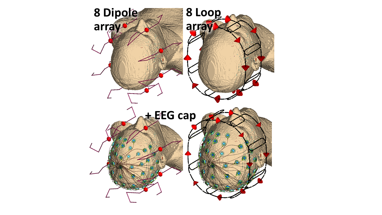
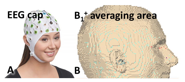
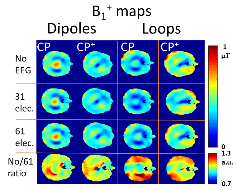
First, two numerical models of EEG caps with 31 and 61 electrodes were created in CST Studio 2022. Then, both models were combined with two transceiver (TxRx) arrays, i.e. eight folded-end dipoles and eight overlapped loops. Numerical models of 8-element arrays without the EEG cap are presented in Fig. 1. Fig. 2A shows a commercial picture as an example of the EEG cap.All array models were loaded by a human voxel model (“Duke”)[9]. In simulations, we used the Duke model with 2-mm and 1-mm isotropic resolutions. All simulations were performed using FIT-TD[10] solver. SAR10g was calculated using the CST Legacy averaging method. After tuning and matching of all array elements the CP- (45-deg phase shift between adjacent elements) and CP+- (90-deg phase shift) modes were calculated. The mean value of B1+ was averaged over the 120-mm transversal slab (Fig.2B) that covers the majority of the brain.
Finally, to demonstrate a change in the B1+ field, the ratio between B1+ without and with EEG caps (61 electrodes) was calculated in MatLab.
Results and Discussion
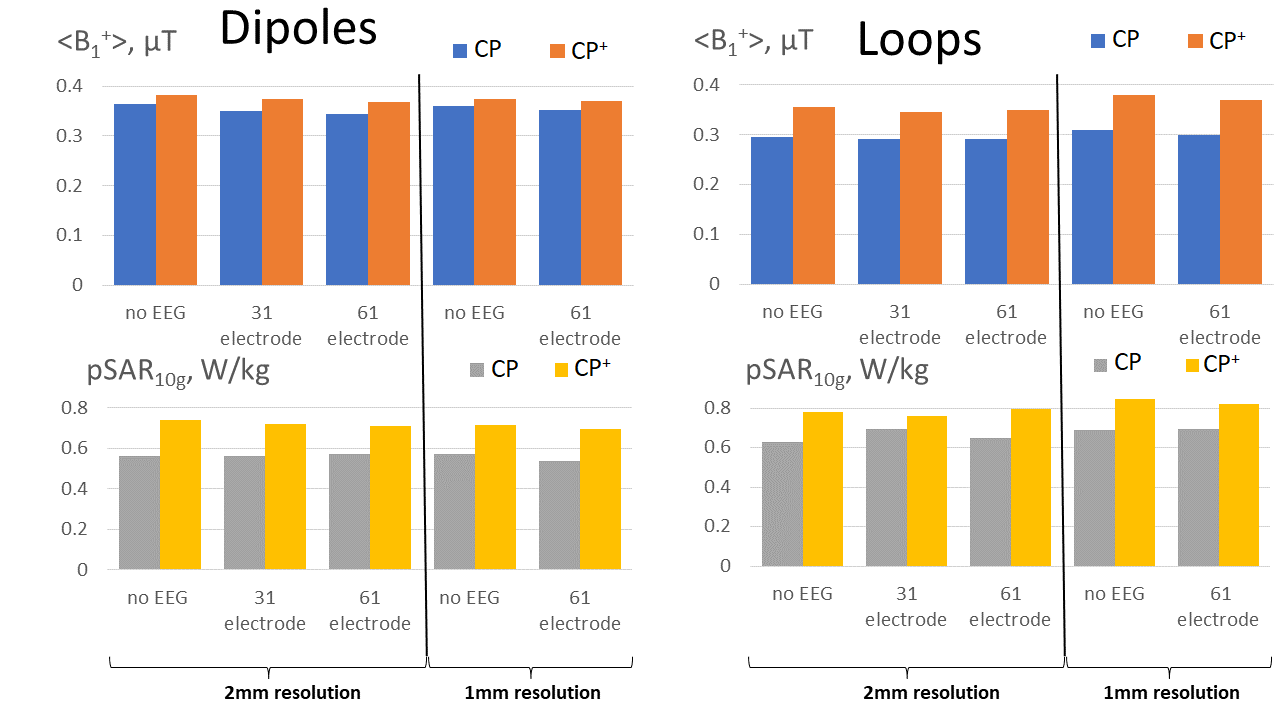
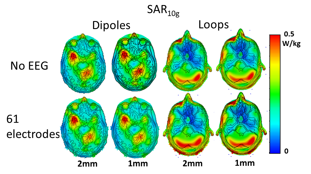
Fig. 3 presents bar plots of the mean B1+ and pSAR10g values for all simulated setups. Fig. 4 shows SAR10g maps in transversal planes cut through pSAR10g locations. Fig. 5 presents central transversal B1+ maps obtained using both dipole and loop arrays without and with EEG caps (both 31 and 61 caps). Fig. 5 also shows ratios of the corresponding B1+ maps demonstrating alterations of the field due to the presence of the EEG cap with 61 electrodes.As seen in Fig. 3, pSAR slightly changes in the presence of EEG caps for both arrays (loops or dipoles) and voxel model resolutions. For example, for the 1-mm voxel model and the EEG cap with 61 electrodes, the pSAR value decreased by 6% and increased by 1% for the dipole and loop arrays, respectively, in presence of EEG caps. For the 2-mm voxel model and the EEG cap with 61 electrodes, presence of the cap led to an increase in the pSAR value by 2% and 4% for the dipole and loop arrays, respectively. Array with 31 electrodes was simulated only for the 2-mm voxel model. In this case, presence of the EEG cap led to an increase of pSAR increase by less than 1% and 3% for the dipole and loop arrays, respectively. Resolution of the voxel model also influenced the alteration of the mean B1+ value, which decreased by 4% (dipoles, 31 electrodes), 2% (dipoles, 61 electrodes), 3% (loops, 31 electrodes), and 2% (loops, 61 electrodes). As seen from these data, the quantitative evaluation depends on the voxel model resolution, e.g. pSAR value calculated for the dipole array in presence of 61 electrodes decreased by 6% and increased by 2% for the 1-mm and 2-mm resolutions, respectively. More importantly, all these changes are very small for all simulated models.
Conclusion
According to the results of our simulations, the presence of EEG caps led only to small drops of the B1+ values and a small increment of the pSAR10g value implying the safe use of eight-channel tight-fit arrays combined with EEG caps.Acknowledgements
No acknowledgement found.References
[1] Goldman, Robin I., et al. "Simultaneous EEG and fMRI of the alpha rhythm." Neuroreport 13.18 (2002): 2487.
[2] Neuner, Irene, et al. "Simultaneous EEG–fMRI acquisition at low, high and ultra-high magnetic fields up to 9.4 T: perspectives and challenges." Neuroimage 102 (2014): 71-79.
[3] Hoffmann J, Henning A, Giapitzakis IA, Scheffler K, Shajan G, Pohmann R, Avdievich NI. Safety testing and operational procedures for self-developed radiofrequency coils. NMR Biomed 2016;29(9):1131-1144.
[4] Kumar, V., Buckenmaier, K., Warbrick, T., Wehrle, R., Pohmann, R., & Scheffler, K. (2020). EEG-fMRI at 9.4T: Safety assessment and effect on B0, B1 and fMRI scans in a phantom. Poster presented at 2020 ISMRM & SMRT Virtual Conference & Exhibition.
[5] Lê, Thanh Phong, et al. "Segmenting electroencephalography wires reduces radiofrequency shielding artifacts in simultaneous electroencephalography and functional magnetic resonance imaging at 7 T." Magnetic resonance in medicine 88.3 (2022): 1450-1464.
[6] Avdievich NI, Giapitzakis IA, Pfrommer A, Henning A. Decoupling of a tight-fit transceiver phased array for human brain imaging at 9.4T: Loop overlapping rediscovered. Magn Reson Med 2018;79(2):1200-1211.
[7] Avdievich NI, Solomakha G, Ruhm L, Henning A, Scheffler K. Unshielded bent folded-end dipole 9.4 T human head transceiver array decoupled using modified passive dipoles. Magn Reson Med 2021;86(1):581-597.
[8] https://www.brainproducts.com/solutions/braincap-mr/
[9] Christ, Andreas, et al. "The Virtual Family—development of surface-based anatomical models of two adults and two children for dosimetric simulations." Physics in Medicine & Biology 55.2 (2009): N23.
[10] T. Weiland: A Discretization Method for the Solution of Maxwell’s Equations for Six-Component Fields, Electronics and Communications AEUE, vol. 31, no. 3, pp. 116–120, 1977.
Figures